Chinese Journal of Tissue Engineering Research ›› 2024, Vol. 28 ›› Issue (28): 4469-4476.doi: 10.12307/2024.340
Previous Articles Next Articles
Metformin pretreatment induces cardiac autophagy to reduce myocardial injury in septic mice
Tian Yong1, 2, Zhou Ying1, Gu Yongxiang1, Yang Guohui3
- 1Guizhou Medical University, Guiyang 550004, Guizhou Province, China; 2Intensive Care Unit, Tongren City People’s Hospital, Tongren 554300, Guizhou Province, China; 3Affiliated Hospital of Guizhou Medical University, Guiyang 550004, Guizhou Province, China
-
Received:2023-04-18Accepted:2023-06-01Online:2024-10-08Published:2023-11-27 -
Contact:Yang Guohui, Professor, Chief physician, Affiliated Hospital of Guizhou Medical University, Guiyang 550004, Guizhou Province, China -
About author:Tian Yong, Master candidate, Guizhou Medical University, Guiyang 550004, Guizhou Province, China; Intensive Care Unit, Tongren City People’s Hospital, Tongren 554300, Guizhou Province, China -
Supported by:Culture Program for the Affiliated Hospital of Guizhou Medical University, The National Natural Science Foundation of China, No. gyfynsfc-2021-54 (to YGH); Guizhou Provincial Health Commission Science and Technology Fund Project, No. gzwjkj2020-1-026 (to YGH)
CLC Number:
Cite this article
Tian Yong, Zhou Ying, Gu Yongxiang, Yang Guohui. Metformin pretreatment induces cardiac autophagy to reduce myocardial injury in septic mice[J]. Chinese Journal of Tissue Engineering Research, 2024, 28(28): 4469-4476.
share this article
Add to citation manager EndNote|Reference Manager|ProCite|BibTeX|RefWorks

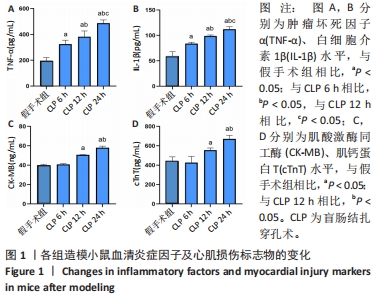
2.1 造模小鼠的一般情况分析 假手术组10 只小鼠及CLP 6 h组10 只小鼠全部存活,假手术组小鼠术后24 h精神、活动及进食正常,无腹泻、畏寒、排泄物异常增多等情况;CLP 6 h组小鼠精神稍萎靡,活动基本正常,无腹泻;CLP 12 h组在术后12 h内死亡1 只小鼠,余存活9 只小鼠出现畏寒、腹泻,进食明显减少,活动明显减少,精神萎靡;CLP 24 h组死亡4 只小鼠,存活6 只小鼠出现嗜睡、拒食、腹泻、竖毛、呼吸急促、眼部有较多分泌物、运动迟缓、不能直立。 2.2 造模小鼠炎症因子及心肌损伤标志物的变化 ELISA检测各组小鼠血清TNF-α、IL-1β、CK-MB及cTnT水平,如图1所示,与假手术组相比,CLP各组炎症因子TNF-α、IL-1β水平均升高(P < 0.05),以CLP 24 h组最为显著;与假手术组相比,CLP 12 h组和CLP 24 h组CK-MB、cTnT水平均显著升高(P < 0.05),以CLP 24 h组更显著,而CLP 6 h组与假手术组相比无统计学差异(P > 0.05),表明在CLP 12 h脓毒症小鼠心肌损伤模型构建成功,且CLP 24 h组较CLP 12 h组心肌损伤更重。"

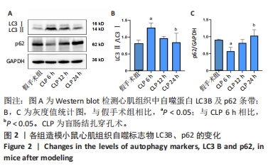
2.3 造模小鼠心肌组织中自噬水平 Western blot检测各组造模小鼠心肌组织自噬标志物(LC3B、p62)的表达水平,如图2所示,与假手术组相比,CLP 6 h组LC3Ⅱ/LC3Ⅰ显著升高(P < 0.05),之后逐渐降低,在CLP 24 h降至基础水平;与假手术组相比,CLP 6 h组p62表达水平显著降低(P < 0.05),之后逐渐恢复,在CLP 24 h已恢复至基础水平,CLP 24 h组与假手术组相比无统计学差异(P > 0.05),这提示在脓毒症早期,自噬处于高表达水平,但在脓毒症心肌损伤中自噬处于抑制状态,在CLP 24 h脓毒症小鼠中自噬处于基础水平,且心肌损伤较CLP 12 h严重,故此研究选择脓毒症心肌损伤中自噬处于显著抑制的时间点(24 h)进行后续研究。"
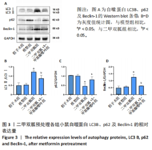
2.4 二甲双胍预处理可诱导脓毒症小鼠心肌损伤中自噬水平 Western blot检测心肌组织中LC3B、Beclin-1及p62的表达情况,如图3所示,与模型组相比,二甲双胍组自噬标志物(LC3Ⅱ/LC3Ⅰ、Beclin-1)显著升高(P < 0.05),且自噬底物p62显著降低(P < 0.05);加用自噬抑制剂3-甲基腺嘌呤对自噬进行抑制后,与二甲双胍组相比,二甲双胍+3-甲基腺嘌呤组LC3Ⅱ/LC3Ⅰ及Beclin-1降低且p62升高(P < 0.05),这表明二甲双胍预处理可增强脓毒症小鼠心肌损伤中自噬水平,且可被自噬抑制剂3-甲基腺嘌呤部分抑制。"

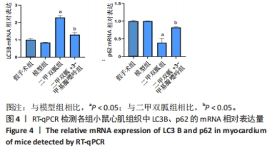
2.5 各组小鼠心肌组织中LC3B、p62的mRNA表达变化 RT-qPCR检测心肌组织中自噬标志物(LC3B、p62)的总mRNA表达水平,如图4所示,与模型组相比,二甲双胍组LC3B mRNA表达水平显著升高且p62 表达水平降低(P < 0.05),与二甲双胍组相比,二甲双胍+3-甲基腺嘌呤组LC3B降低且p62升高(P < 0.05),该结果与Western blot检测结果一致,这表明二甲双胍预处理可增强脓毒症小鼠心肌损伤中自噬水平。"


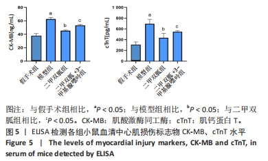
2.6 二甲双胍预处理可减轻脓毒症小鼠心肌损伤 用ELISA试剂盒检测各组小鼠血清中心肌损伤标志物(CK-MB、cTnT)水平,如图5所示,与假手术组相比,模型组心肌损伤标志物CK-MB、cTnT均显著升高(P < 0.05);与模型组相比,二甲双胍组心肌损伤标志物(CK-MB、cTnT)显著降低(P < 0.05),表明二甲双胍预处理可减轻脓毒症心肌损伤;与二甲双胍组相比,二甲双胍+3-甲基腺嘌呤组CK-MB及cTnT均升高(P < 0.05),二甲双胍对脓毒症心肌损伤的保护作用被自噬抑制剂3-甲基腺嘌呤部分消除,这表明二甲双胍预处理增强自噬在脓毒症小鼠心肌损伤的保护作用中扮演重要角色。"
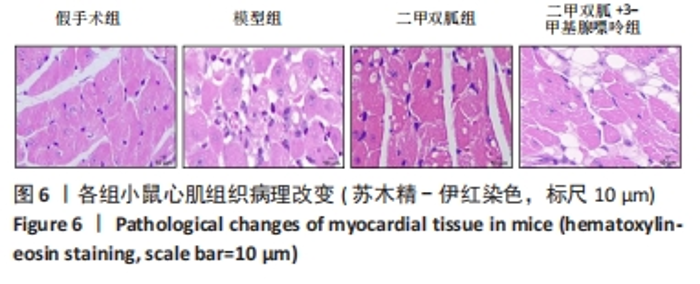
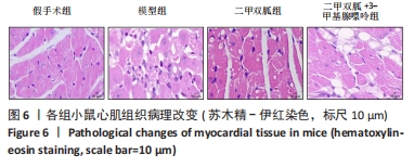
2.7 苏木精-伊红染色评估小鼠心肌组织病理改变 如图6所示,假手术组光镜下心肌纤维细胞分界清楚,无紊乱及坏死,未见炎症细胞浸润;模型组心肌纤维排列紊乱,间质水肿,少量红细胞渗出,可见大量炎症细胞浸润;二甲双胍组心肌纤维稍水肿,局部见少量空泡状改变;二甲双胍+3-甲基腺嘌呤组心肌纤维排列稍紊乱,空泡状改变较二甲双胍组明显增多,见少量炎症细胞浸润,提示二甲双胍预处理对脓毒症心肌组织损伤具有一定的保护作用,这种保护作用可被3-甲基腺嘌呤部分逆转。 "

2.8 二甲双胍预处理诱导自噬减轻脓毒症心肌损伤中的炎症反应 用ELISA检测各组血清中炎症因子TNF-α、IL-6水平,如图7所示,与假手术组相比,模型组TNF-α、IL-6水平均显著升高(P < 0.05),与模型组相比,二甲双胍组TNF-α、IL-6水平均显著降低(P < 0.05),加用3-甲基腺嘌呤对自噬进行抑制后,炎症因子(TNF-α、IL-6)水平升高(P < 0.05),这表明二甲双胍预处理可通过增强自噬减少脓毒症小鼠炎症因子的释放。"


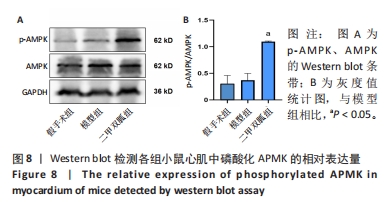
2.9 二甲双胍预处理心肌中磷酸化AMPK的表达变化 用Western blot检测AMPK信号通路中p-AMPK、AMPK的表达,如图8所示,模型组与假手术组相比p-AMPK/AMPK无统计学差异(P > 0.05),与模型组相比,二甲双胍组p-AMPK/AMPK显著升高(P < 0.05),二甲双胍可通过依赖性及非依赖性AMPK信号通路发挥药理作用,这结果表明二甲双胍可能主要通过激活AMPK信号通路来诱导脓毒症心肌损伤中的自噬水平。"
| [1] ZHANG YY, NING BT. Signaling pathways and intervention therapies in sepsis. Signal Transduct Target Ther. 2021;6(1):407. [2] ZHOU B, ZHANG J, CHEN Y, et al. Puerarin protects against sepsis-induced myocardial injury through AMPK-mediated ferroptosis signaling. Aging (Albany NY). 2022;14(8):3617-3632. [3] LI Y, ZHANG L, ZHANG P, et al. Dehydrocorydaline Protects Against Sepsis-Induced Myocardial Injury Through Modulating the TRAF6/NF-κB Pathway. Front Pharmacol. 2021;12:709604. [4] BI CF, LIU J, YANG LS, et al. Research Progress on the Mechanism of Sepsis Induced Myocardial Injury. J Inflamm Res. 2022;15:4275-4290. [5] SUN Y, CAI Y, ZANG QS. Cardiac Autophagy in Sepsis. Cells. 2019; 8(2):141. [6] ISMAIL HASSAN F, DIDARI T, KHAN F, et al. A Review on The Protective Effects of Metformin in Sepsis-Induced Organ Failure. Cell J. 2020;21(4): 363-370. [7] KOLAHDOUZMOHAMMADI M, PAHLAVAN S, SOTOODEHNEJADNEMATALAHI F, et al. Activation of AMPK promotes cardiac differentiation by stimulating the autophagy pathway. J Cell Commun Signal. 2023 Apr 11. doi: 10.1007/s12079-023-00744-z. [8] LI Q, WANG L, JI D, et al. Metformin attenuates cadmium-induced degeneration of spiral ganglion neuron via restoring autophagic flux in primary culture. J Inorg Biochem. 2022;234:111901. [9] LI W, HE P, HUANG Y, et al. Selective autophagy of intracellular organelles: recent research advances. Theranostics. 2021;11(1):222-256. [10] HUANG M, CAI S, SU J. The Pathogenesis of Sepsis and Potential Therapeutic Targets. Int J Mol Sci. 2019;20(21):5376. [11] GROSS AS, GRAEF M. Mechanisms of Autophagy in Metabolic Stress Response. J Mol Biol. 2020;432(1):28-52. [12] LIU C, LIU Y, CHEN H, et al. Myocardial injury: where inflammation and autophagy meet. Burns Trauma. 2023;11:tkac062. [13] 蒋大军,蒋万威,周颖,等.NLRC4炎症小体介导自噬反应在脓毒症心肌功能障碍小鼠中的作用[J].中国组织工程研究,2023,27(23): 3667-3673. [14] JINPIAO Z, ZONGZE Z, QIUYUE Y, et al. Metformin attenuates sevoflurane-induced neurocognitive impairment through AMPK-ULK1-dependent autophagy in aged mice. Brain Res Bull. 2020;157:18-25. [15] LI T, CHEN Y, LI Y, et al. FAM134B-mediated endoplasmic reticulum autophagy protects against sepsis myocardial injury in mice. Aging (Albany NY). 2021;13(10):13535-13547. [16] EVANS L, RHODES A, ALHAZZANI W, et al. Surviving sepsis campaign: international guidelines for management of sepsis and septic shock 2021. Intensive Care Med. 2021;47(11):1181-1247. [17] PÓVOA P, COELHO L, DAL-PIZZOL F, et al. How to use biomarkers of infection or sepsis at the bedside: guide to clinicians. Intensive Care Med. 2023;49(2):142-153. [18] LIU C, ZOU Q, TANG H, et al. Melanin nanoparticles alleviate sepsis-induced myocardial injury by suppressing ferroptosis and inflammation. Bioact Mater. 2022;24:313-321. [19] 杭成文,崔鸣.脓毒症致心肌损伤模型的研究进展[J].中国心血管杂志,2020,25(5):485-488. [20] HO J, YU J, WONG SH, et al. Autophagy in sepsis: Degradation into exhaustion? Autophagy. 2016;12(7):1073-1082. [21] ALA M, ALA M. Metformin for Cardiovascular Protection, Inflammatory Bowel Disease, Osteoporosis, Periodontitis, Polycystic Ovarian Syndrome, Neurodegeneration, Cancer, Inflammation and Senescence: What Is Next? ACS Pharmacol Transl Sci. 2021;4(6):1747-1770. [22] CEJUELA M, MARTIN-CASTILLO B, MENENDEZ JA, et al. Metformin and Breast Cancer: Where Are We Now? Int J Mol Sci. 2022;23(5):2705. [23] LIU G, WU K, ZHANG L, et al. Metformin attenuated endotoxin-induced acute myocarditis via activating AMPK. Int Immunopharmacol. 2017;47:166-172. [24] ZHANG M, SUN W, DU J, et al. Protective Effect of Metformin on Sepsis Myocarditis in Zebrafish. Dose Response. 2020;18(3): 1559325820938543. [25] LIANG H, SONG H, ZHANG X, et al. Metformin attenuated sepsis-related liver injury by modulating gut microbiota. Emerg Microbes Infect. 2022;11(1):815-828. [26] ZHAO H, LYU Y, ZHAI R, et al. Metformin Mitigates Sepsis-Related Neuroinflammation via Modulating Gut Microbiota and Metabolites. Front Immunol. 2022;13:797312. [27] GHAVIMI H, SHEIDAEI S, VAEZ H, et al. Metformin-attenuated sepsis-induced oxidative damages: a novel role for metformin. Iran J Basic Med Sci. 2018;21(5):469-475. [28] TANG G, YANG H, CHEN J, et al. Metformin ameliorates sepsis-induced brain injury by inhibiting apoptosis, oxidative stress and neuroinflammation via the PI3K/Akt signaling pathway. Oncotarget. 2017;8(58):97977-97989. [29] 高婷,陈忠.二甲双胍对脂多糖诱导的大鼠心肌细胞H9C2损伤的保护机制[J].南京医科大学学报(自然科学版),2019,39(9):1298-1303. [30] LI M, GOU Y, YU H, et al. Mechanism of Metformin on LPS-Induced Bacterial Myocarditis. Dose Response. 2019;17(2):1559325819847409. [31] WU B, SONG H, FAN M, et al. Luteolin attenuates sepsis‑induced myocardial injury by enhancing autophagy in mice. Int J Mol Med. 2020;45(5):1477-1487. [32] TANG R, JIA L, LI Y, et al. Narciclasine attenuates sepsis-induced myocardial injury by modulating autophagy. Aging (Albany NY). 2021; 13(11):15151-15163. [33] WANG X, XIE D, DAI H, et al. Clemastine protects against sepsis-induced myocardial injury in vivo and in vitro. Bioengineered. 2022;13(3):7134-7146. [34] WU S, ZHANG H, CHEN N, et al. Metformin protects cardiomyocytes against oxygen-glucose deprivation injury by promoting autophagic flux through AMPK pathway. J Drug Target. 2021;29(5):551-561. [35] WEN X, XIE B, YUAN S, et al. The “Self-Sacrifice” of ImmuneCells in Sepsis. Front Immunol. 2022;13:833479. [36] QIU P, LIU Y, ZHANG J. Review: the Role and Mechanisms of Macrophage Autophagy in Sepsis. Inflammation. 2019;42(1):6-19. [37] LU G, WU Z, SHANG J, et al. The effects of metformin on autophagy. Biomed Pharmacother. 2021;137:111286. [38] HUANG KY, QUE JQ, HU ZS, et al. Metformin suppresses inflammation and apoptosis of myocardiocytes by inhibiting autophagy in a model of ischemia-reperfusion injury. Int J Biol Sci. 2020;16(14):2559-2579. [39] WANG Y, YANG Z, ZHENG G, et al. Metformin promotes autophagy in ischemia/reperfusion myocardium via cytoplasmic AMPKα1 and nuclear AMPKα2 pathways. Life Sci. 2019;225:64-71. [40] WANG Y, AN H, LIU T, et al. Metformin Improves Mitochondrial Respiratory Activity through Activation of AMPK. Cell Rep. 2019;29(6): 1511-1523.e5. [41] BU Y, PENG M, TANG X, et al. Protective effects of metformin in various cardiovascular diseases: Clinical evidence and AMPK-dependent mechanisms. J Cell Mol Med. 2022;26(19):4886-4903. [42] TURCO E, FRACCHIOLLA D, MARTENS S. Recruitment and Activation of the ULK1/Atg1 Kinase Complex in Selective Autophagy. J Mol Biol. 2020;432(1):123-134. [43] LI SX, LI C, PANG XR, et al. Metformin Attenuates Silica-Induced Pulmonary Fibrosis by Activating Autophagy via the AMPK-mTOR Signaling Pathway. Front Pharmacol. 2021;12:719589. [44] SONG H, ZHANG X, ZHAI R, et al. Metformin attenuated sepsis-associated liver injury and inflammatory response in aged mice. Bioengineered. 2022;13(2):4598-4609. [45] LI Y, CHEN Y. AMPK and Autophagy. Adv Exp Med Biol. 2019;1206:85-108. [46] LAMOIA TE, SHULMAN GI. Cellular and Molecular Mechanisms of Metformin Action. Endocr Rev. 2021;42(1):77-96. [47] TZANAVARI T, VARELA A, THEOCHARIS S, et al. Metformin protects against infection-induced myocardial dysfunction. Metabolism. 2016; 65(10):1447-1458. [48] DI MAURO S, FILIPPELLO A, SCAMPORRINO A, et al. Metformin: When Should We Fear Lactic Acidosis? Int J Mol Sci. 2022;23(15):8320. [49] L’HEUREUX M, STERNBERG M, BRATH L, et al. Sepsis-Induced Cardiomyopathy: a Comprehensive Review. Curr Cardiol Rep. 2020; 22(5):35. [50] CHEN P, AN Q, HUANG Y, et al. Prevention of endotoxin-induced cardiomyopathy using sodium tanshinone IIA sulfonate: Involvement of augmented autophagy and NLRP3 inflammasome suppression. Eur J Pharmacol. 2021;909:174438. [51] 迪丽热巴•吐尔逊,杨春波,王毅,等.脓毒性心肌损伤机制及治疗的研究进展[J].中华危重病急救医学,2022,34(10):1112-1115. [52] NIKOUEE A, KIM M, DING X, et al. Beclin-1-Dependent Autophagy Improves Outcomes of Pneumonia-Induced Sepsis. Front Cell Infect Microbiol. 2021;11:706637. |
| [1] | Wang Ji, Zhang Min, Li Wenbo, Yang Zhongya, Zhang Long. Effect of aerobic exercise on glycolipid metabolism, skeletal muscle inflammation and autophagy in type 2 diabetic rats [J]. Chinese Journal of Tissue Engineering Research, 2024, 28(8): 1200-1205. |
| [2] | Zhou Bangyu, Li Jie, Ruan Yushang, Geng Funeng, Li Shaobo. Effects of Periplaneta americana powder on motor function and autophagic protein Beclin-1 in rats undergoing spinal cord hemisection [J]. Chinese Journal of Tissue Engineering Research, 2024, 28(8): 1223-1228. |
| [3] | Sheng Siqi, Xie Lin, Zhao Xiangyu, Jiang Yideng, Wu Kai, Xiong Jiantuan, Yang Anning, Hao Yinju, Jiao Yun. Involvement of miR-144-3p in Cbs+/- mouse hepatocyte autophagy induced by high-methionine diet [J]. Chinese Journal of Tissue Engineering Research, 2024, 28(8): 1289-1294. |
| [4] | Pan Xiaolong, Fan Feiyan, Ying Chunmiao, Liu Feixiang, Zhang Yunke. Effect and mechanism of traditional Chinese medicine on inhibiting the aging of mesenchymal stem cells [J]. Chinese Journal of Tissue Engineering Research, 2024, 28(7): 1091-1098. |
| [5] | Huang Yuxin, Liang Wenzi, Chen Xiuwen, Ni Na, Zhao Yinglin, Lin Changmin. Role of autophagy in hair regeneration [J]. Chinese Journal of Tissue Engineering Research, 2024, 28(7): 1112-1117. |
| [6] | Liu Qiwei, Zhang Junhui, Yang Yuan, Wang Jinjuan. Role and mechanism of umbilical cord mesenchymal stem cells on polycystic ovary syndrome [J]. Chinese Journal of Tissue Engineering Research, 2024, 28(7): 1015-1020. |
| [7] | Xue Jingwen, Wang Fangfang, Zhang Xin, Pang Ruifeng, Wang Xiaoye, Ma Xiaoru. Effect of ganoderma spore on mitochondrial autophagy and apoptosis in testicular tissue of diabetic rats [J]. Chinese Journal of Tissue Engineering Research, 2024, 28(4): 562-568. |
| [8] | Yang Yuqing, Chen Zhiyu. Role and application of early transient presence of M1 macrophages in bone tissue engineering [J]. Chinese Journal of Tissue Engineering Research, 2024, 28(4): 594-601. |
| [9] | Yan Binghan, Li Zhichao, Su Hui, Xue Haipeng, Xu Zhanwang, Tan Guoqing. Mechanisms of traditional Chinese medicine monomers in the treatment of osteoarthritis by targeting autophagy [J]. Chinese Journal of Tissue Engineering Research, 2024, 28(4): 627-632. |
| [10] | Zhai Sheng, Aikeremujiang • Aerken, Zhang Zheng, Rixiati • Paerhati, Hao Feihu. CircCDR1as/miR-7-5p/RAF1 axis promotes autophagy levels in steroid-induced necrosis of the femoral head [J]. Chinese Journal of Tissue Engineering Research, 2024, 28(28): 4455-4460. |
| [11] | Chen Ying, Xia Tianqin, Hua Jianlin, Yin Jinzhu, Song Lijuan, Wang Qing, Yu Jiezhong, Huang Jianjun, Ma Cungen. The role and mechanism of TLRs/MyD88/NF-κB signaling pathway in multiple sclerosis [J]. Chinese Journal of Tissue Engineering Research, 2024, 28(28): 4578-4585. |
| [12] | Liu Ying, Liu Yalei, Liu Yu. Screening and analysis of differentially expressed long non-coding RNAs in adriamycin-induced myocardial injury antagonized with daisy leaf gentianone [J]. Chinese Journal of Tissue Engineering Research, 2024, 28(26): 4121-4128. |
| [13] | Ma Lingju, Wang Lexin, Chi Hongyang, Zhang Jingwen, Peng Hongjian, Gao Chunlan, Jiang Yideng, Huang Hui, Yang Li, Ma Shengchao. Role of METTL3 in homocysteine-induced autophagy in mouse islet beta cells [J]. Chinese Journal of Tissue Engineering Research, 2024, 28(26): 4221-4225. |
| [14] | Zhu Yongzhao, Fang Chao, Zhao Fang, Zhang Qing, Zhao Dan. Mechanism by which lycium barbarum polysaccharides inhibit keratinocyte apoptosis in burn wounds via autophagy [J]. Chinese Journal of Tissue Engineering Research, 2024, 28(23): 3686-3691. |
| [15] | Liang Xiaolong, Zheng Kai, Geng Dechun, Xu Yaozeng. Novel programmed cell death in periprosthetic osteolysis [J]. Chinese Journal of Tissue Engineering Research, 2024, 28(21): 3393-3399. |
| Viewed | ||||||
|
Full text |
|
|||||
|
Abstract |
|
|||||