Chinese Journal of Tissue Engineering Research ›› 2024, Vol. 28 ›› Issue (19): 2953-2959.doi: 10.12307/2024.156
Bone marrow mesenchymal stem cell exosomes combined with epigallocatechin-3-gallate in treatment of spinal cord ischemia/reperfusion injury in rats
Long Zhisheng, Gong Feipeng, Wen Jiabin, Min Huan, Shu Yang, Lai Zhuoxi, Chen Gang
- Department of Orthopedics, First Affiliated Hospital, Nanchang Medical College, Jiangxi Provincial People’s Hospital, Nanchang 330006, Jiangxi Province, China
-
Received:2023-04-22Accepted:2023-05-25Online:2024-07-08Published:2023-09-25 -
About author:Long Zhisheng, Associate chief physician, MD, Department of Orthopedics, First Affiliated Hospital, Nanchang Medical College, Jiangxi Provincial People’s Hospital, Nanchang 330006, Jiangxi Province, China -
Supported by:National Natural Science Foundation of China, No. 32060222 (to LZS); Project of Jiangxi Provincial Administration of Traditional Chinese Medicine, No. 2021B018 (to LZS); Project of Jiangxi Provincial Health Commission, No. 202310014 (to LZS)
CLC Number:
Cite this article
Long Zhisheng, Gong Feipeng, Wen Jiabin, Min Huan, Shu Yang, Lai Zhuoxi, Chen Gang. Bone marrow mesenchymal stem cell exosomes combined with epigallocatechin-3-gallate in treatment of spinal cord ischemia/reperfusion injury in rats[J]. Chinese Journal of Tissue Engineering Research, 2024, 28(19): 2953-2959.
share this article
Add to citation manager EndNote|Reference Manager|ProCite|BibTeX|RefWorks
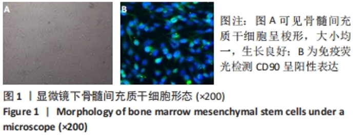
2.1 骨髓间充质干细胞形态 第3代骨髓间充质干细胞呈梭形改变,生长状态良好,见图1A。免疫荧光检测特异标记物对间充质干细胞进行鉴定,可见其CD90表达明显,见图1B。"

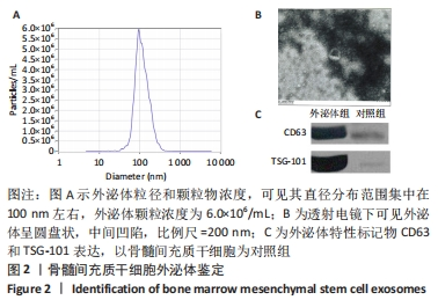
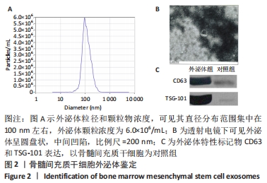
2.2 BMSCs-Exo鉴定结果 BMSCs-Exo颗粒直径大小为100 nm左右,外泌体颗粒物浓度为6.0×10⁶个/mL,透射电镜下呈圆盘状,中间凹陷,与对照组比较,外泌体的特异性标记物CD63和TSG-101表达显著升高,见图2。"
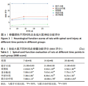
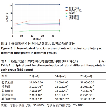
2.3 实验动物数量分析 实验共纳入50只SD大鼠,每组10只,中途因出现麻醉过量、出血、术后感染及原因不明等造成8只大鼠死亡,重新补充大鼠进行造模及相应的干预。 2.4 神经功能评分 术后7 d,假手术组大鼠神经功能正常,而模型组、外泌体组、茶多酚组及联合治疗组有不同程度的神经功能障碍,术后7,14,28 d,假手术组BBB评分均优于其余4组(P < 0.05);7 d评分:联合治疗组优于模型组(P < 0.05);14 d评分:联合治疗组优于模型组、茶多酚组、外泌体组(P < 0.05);28 d评分:联合治疗组优于模型组,茶多酚组(P < 0.05),见图3,表1。"
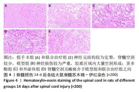
2.5 苏木精-伊红染色结果 脊髓损伤后14 d,假手术组神经元形态正常,脊髓中央灰质区域内见大量神经元细胞,无明显空洞及瘢痕组织生成;模型组神经元数量少于假手术组,同时神经元周围可见大量瘢痕组织和空泡状坏死区域;外泌体组、茶多酚组神经元数量和坏死区域较模型组有所改善,联合治疗组神经元结构较为完整,损伤恢复良好,胶质空洞坏死区域较少,见图4。"


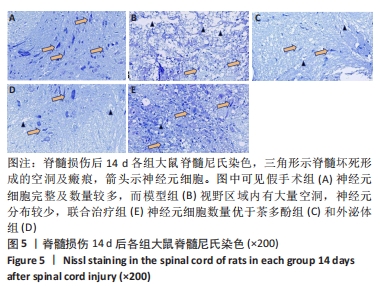
2.6 尼氏染色结果 脊髓损伤14 d,假手术组有大量正常神经元分布,而模型组神经损伤较为严重,观察区域内神经元分布最少,联合治疗组神经元分布优于茶多酚组及外泌体组,见图5。"
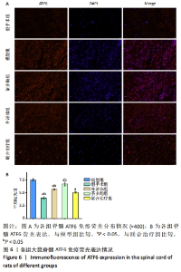
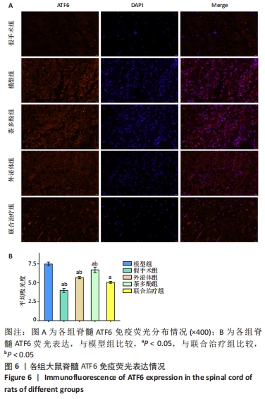
2.7 ATF6免疫荧光染色结果 各组均可见ATF6表达,其中模型组ATF6表达最高,模型组与其余4组比较差异有显著性意义(P < 0.05),说明脊髓损伤后内质网应激明显增强,经茶多酚、外泌体、联合治疗等干预,ATF6表达有不同程度下降,其中联合治疗组ATF6表达最低,其与茶多酚组、外泌体组比较,差异有显著性意义(P < 0.05),结果说明联合治疗组抑制内质网应激最显著,见图6。"
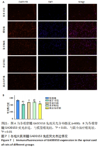
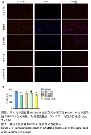
2.8 GADD153免疫荧光染色结果 各组均可见GADD153表达,其中模型组GADD153表达最高,假手术组GADD153表达最少,与模型组比较,其余4组GADD153表达下降(P < 0.05)。联合治疗组GADD153低于茶多酚组(P < 0.05)。结果说明茶多酚、外泌体及两者联合治疗能够降低GADD153表达,均能够降低内质网应激所致的神经元损伤,联合治疗组抑制内质网应激最显著,见图7。"
| [1] LIU WZ, MA ZJ, LI JR, et al. Mesenchymal stem cell-derived exosomes: therapeutic opportunities and challenges for spinal cord injury. Stem Cell Res Ther. 2021;12(1):102. [2] 陈平波,王晶,孙勇,等.京尼平交联音猬因子复合纤维蛋白支架修复大鼠脊髓损伤[J].中国组织工程研究,2022,26(21):3345-3350. [3] 龙智生,马泽民,吕国华,等.大鼠急性脊髓损伤后内质网应激相关蛋白CHOP的表达及其意义[J].中国脊柱脊髓杂志,2009,19(6): 458-463. [4] YAO M, PU PM, LI ZY, et al. Melatonin restores endoplasmic reticulum homeostasis to protect injured neurons in a rat model of chronic cervical cord compression. J Pineal Res. 2023;74(4):e12859. [5] 韩晓光,田伟,刘波,等.表没食子儿茶素没食子酸酯对大鼠脊髓损伤后神经功能恢复的影响[J].中国脊柱脊髓杂志,2013,23(11): 998-1005. [6] ZHOU Y, WEN LL, LI YF, et al. Exosomes derived from bone marrow mesenchymal stem cells protect the injured spinal cord by inhibiting pericyte pyroptosis. Neural Regen Res. 2022;17(1):194-202. [7] 谢兴奇,胡伟,屠冠军.骨髓间充质干细胞来源外泌体与硫酸软骨素酶ABC联合治疗大鼠脊髓损伤[J].中国组织工程研究,2022, 26(1):20-26. [8] WEI X, ZHOU Z, LI L, et al. Intrathecal Injection of 3-Methyladenine Reduces Neuronal Damage and Promotes Functional Recovery via Autophagy Attenuation after Spinal Cord Ischemia/Reperfusion Injury in Rats. Biol Pharm Bull. 2016;39(5):665-673. [9] SUNG SE, SEO MS, KIM YI, et al. Human Epidural AD-MSC Exosomes Improve Function Recovery after Spinal Cord Injury in Rats. Biomedicines. 2022;10(3):678. [10] KANCHANAPALLY R, DESHMUKH SK, CHAVVA SR, et al. Drug-loaded exosomal preparations from different cell types exhibit distinctive loading capability, yield, and antitumor efficacies: a comparative analysis. Int J Nanomedicine. 2019;14:531-541. [11] REN Z, QI Y, SUN S, et al. Mesenchymal Stem Cell-Derived Exosomes: Hope for Spinal Cord Injury Repair. Stem Cells Dev. 2020;29(23):1467-1478. [12] CAMBRIA RP, DAVISON JK, ZANNETTI S, et al. Clinical experience with epidural cooling for spinal cord protection during thoracic and thoracoabdominal aneurysm repair. J Vasc Surg. 1997;25(2):234-241. [13] LI GL, FAROOQUE M, OLSSON Y. Changes of Fas and Fas ligand immunoreactivity after compression trauma to rat spinal cord. Acta Neuropathol. 2000;100(1):75-81. [14] XU D, CUI S, SUN Y, et al. Overexpression of glucose-regulated protein 94 after spinal cord injury in rats. J Neurol Sci. 2011;309(1-2):141-147. [15] YONG C, ARNOLD PM, ZOUBINE MN, et al. Apoptosis in cellular compartments of rat spinal cord after severe contusion injury. J Neurotrauma. 1998;15(7):459-472. [16] PAHL HL. Signal transduction from the endoplasmic reticulum to the cell nucleus. Physiol Rev. 1999;79(3):683-701. [17] IIDA K, LI Y, MCGRATH BC, et al. PERK eIF2 alpha kinase is required to regulate the viability of the exocrine pancreas in mice. BMC Cell Biol. 2007;8:38. [18] BO N, YILIN H, CHAOYUE Y, et al. Acrylamide induces NLRP3 inflammasome activation via oxidative stress- and endoplasmic reticulum stress-mediated MAPK pathway in HepG2 cells. Food Chem Toxicol. 2020;145:111679. [19] GUO Y, LU Y, WANG J, et al. Dysregulated ion channels and transporters activate endoplasmic reticulum stress in rat kidney of fetal growth restriction. Life Sci. 2020;259:118276. [20] LI Y, YI M, WANG D, et al. LncRNA KCNQ1OT1 Regulates Endoplasmic Reticulum Stress to Affect Cerebral Ischemia-Reperfusion Injury Through Targeting miR-30b/GRP78. Inflammation. 2020;43(6):2264-2275. [21] 龙智生,马泽民,邓幼文,等.甲泼尼龙对大鼠急性脊髓损伤后内质网应激相关蛋白CHOP表达的影响[J].南昌大学学报(医学版), 2013,53(1):16-20. [22] ZHANG L, YAN R, WU Z. Metagenomics analysis of intestinal flora modulatory effect of green tea polyphenols by a circadian rhythm dysfunction mouse model. J Food Biochem. 2020 Aug 10:e13430. doi: 10.1111/jfbc.13430. [23] AHADI S, ZARGARI M, KHALATBARY AR. Assessment of the neuroprotective effects of (-)-epigallocatechin-3-gallate on spinal cord ischemia-reperfusion injury in rats. J Spinal Cord Med. 2021;44(5): 725-732. [24] ZHANG S, CAO M, FANG F. The Role of Epigallocatechin-3-Gallate in Autophagy and Endoplasmic Reticulum Stress (ERS)-Induced Apoptosis of Human Diseases. Med Sci Monit. 2020;26:e924558. [25] TIAN W, HAN XG, LIU YJ, et al. Intrathecal epigallocatechin gallate treatment improves functional recovery after spinal cord injury by upregulating the expression of BDNF and GDNF. Neurochem Res. 2013;38(4):772-729. [26] RENNO WM, BENOV L, KHAN KM. Possible role of antioxidative capacity of (-)-epigallocatechin-3-gallate treatment in morphological and neurobehavioral recovery after sciatic nerve crush injury. J Neurosurg Spine. 2017;27(5):593-613. [27] STEINMANN J, BUER J, PIETSCHMANN T, et al. Anti-infective properties of epigallocatechin-3-gallate (EGCG), a component of green tea. Br J Pharmacol. 2013;168(5):1059-1073. [28] LI L, ZHANG Y, MU J, et al. Transplantation of Human Mesenchymal Stem-Cell-Derived Exosomes Immobilized in an Adhesive Hydrogel for Effective Treatment of Spinal Cord Injury. Nano Lett. 2020;20(6): 4298-4305. [29] TRAN PHL, XIANG D, TRAN TTD, et al. Exosomes and Nanoengineering: A Match Made for Precision Therapeutics. Adv Mater. 2020;32(18): e1904040. [30] HAZRATI A, MALEKPOUR K, SOUDI S, et al. Mesenchymal stromal/stem cells spheroid culture effect on the therapeutic efficacy of these cells and their exosomes: A new strategy to overcome cell therapy limitations. Biomed Pharmacother. 2022;152:113211. [31] YU W, LI S, GUAN X, et al. Higher yield and enhanced therapeutic effects of exosomes derived from MSCs in hydrogel-assisted 3D culture system for bone regeneration. Biomater Adv. 2022;133:112646. [32] FAN L, LIU C, CHEN X, et al. Exosomes-Loaded Electroconductive Hydrogel Synergistically Promotes Tissue Repair after Spinal Cord Injury via Immunoregulation and Enhancement of Myelinated Axon Growth. Adv Sci (Weinh). 2022;9(13):e2105586. [33] RAN N, LI W, ZHANG R, et al. Autologous exosome facilitates load and target delivery of bioactive peptides to repair spinal cord injury. Bioact Mater. 2022;25:766-782. |
| [1] | Chen Kaijia, Liu Jingyun, Cao Ning, Sun Jianbo, Zhou Yan, Mei Jianguo, Ren Qiang. Application and prospect of tissue engineering in treatment of osteonecrosis of the femoral head [J]. Chinese Journal of Tissue Engineering Research, 2024, 28(9): 1450-1456. |
| [2] | Yang Yifeng, Ye Nan, Wang Lin, Guo Shuaicheng, Huang Jian. Signaling pathway of dexmedetomidine against ischemia-reperfusion injury [J]. Chinese Journal of Tissue Engineering Research, 2024, 28(9): 1464-1469. |
| [3] | Yue Yun, Wang Peipei, Yuan Zhaohe, He Shengcun, Jia Xusheng, Liu Qian, Li Zhantao, Fu Huiling, Song Fei, Jia Menghui. Effects of croton cream on JNK/p38 MAPK signaling pathway and neuronal apoptosis in cerebral ischemia-reperfusion injury rats [J]. Chinese Journal of Tissue Engineering Research, 2024, 28(8): 1186-1192. |
| [4] | Zhou Bangyu, Li Jie, Ruan Yushang, Geng Funeng, Li Shaobo. Effects of Periplaneta americana powder on motor function and autophagic protein Beclin-1 in rats undergoing spinal cord hemisection [J]. Chinese Journal of Tissue Engineering Research, 2024, 28(8): 1223-1228. |
| [5] | Liu Hanfeng, Wang Jingjing, Yu Yunsheng. Artificial exosomes in treatment of myocardial infarction: current status and prospects [J]. Chinese Journal of Tissue Engineering Research, 2024, 28(7): 1118-1123. |
| [6] | Ma Shuwei, He Sheng, Han Bing, Zhang Liaoyun. Exosomes derived from mesenchymal stem cells in treatment of animals with acute liver failure: a meta-analysis [J]. Chinese Journal of Tissue Engineering Research, 2024, 28(7): 1137-1142. |
| [7] | Feng Ruiqin, Han Na, Zhang Meng, Gu Xinyi, Zhang Fengshi. Combination of 1% platelet-rich plasma and bone marrow mesenchymal stem cells improves the recovery of peripheral nerve injury [J]. Chinese Journal of Tissue Engineering Research, 2024, 28(7): 985-992. |
| [8] | Qiu Xiaoyan, Li Bixin, Li Jingdi, Fan Chuiqin, Ma Lian, Wang Hongwu. Differentiation of insulin-producing cells from human umbilical cord mesenchymal stem cells infected by MAFA-PDX1 overexpressed lentivirus [J]. Chinese Journal of Tissue Engineering Research, 2024, 28(7): 1000-1006. |
| [9] | Zeng Fanzhuo, Li Yuxin, Sun Jiachen, Gu Xinyang, Wen Shan, Tian He, Mei Xifan. Efficient strategies for microglia replacement in spinal cord injury models [J]. Chinese Journal of Tissue Engineering Research, 2024, 28(7): 1007-1014. |
| [10] | Liu Qiwei, Zhang Junhui, Yang Yuan, Wang Jinjuan. Role and mechanism of umbilical cord mesenchymal stem cells on polycystic ovary syndrome [J]. Chinese Journal of Tissue Engineering Research, 2024, 28(7): 1015-1020. |
| [11] | Liu Tao, Zhang Wenkai, Ma Ziqian, Zhang Yan, Chen Xueming. Riluzole interferes with the activation of NLRP3 inflammasome in microglia of rats with spinal cord injury [J]. Chinese Journal of Tissue Engineering Research, 2024, 28(7): 1036-1042. |
| [12] | Pan Xiaolong, Fan Feiyan, Ying Chunmiao, Liu Feixiang, Zhang Yunke. Effect and mechanism of traditional Chinese medicine on inhibiting the aging of mesenchymal stem cells [J]. Chinese Journal of Tissue Engineering Research, 2024, 28(7): 1091-1098. |
| [13] | Zhang Kefan, Shi Hui. Research status and application prospect of cytokine therapy for osteoarthritis [J]. Chinese Journal of Tissue Engineering Research, 2024, 28(6): 961-967. |
| [14] | Shen Ziqing, Xia Tian, Shan Yibo, Zhu Ruijun, Wan Haoxin, Ding Hao, Pan Shu, Zhao Jun. Vascularized tracheal substitutes constructed by exosome-load hydrogel-modified 3D printed scaffolds [J]. Chinese Journal of Tissue Engineering Research, 2024, 28(5): 697-705. |
| [15] | Wei Yuanxun, Chen Feng, Lin Zonghan, Zhang Chi, Pan Chengzhen, Wei Zongbo. The mechanism of Notch signaling pathway in osteoporosis and its prevention and treatment with traditional Chinese medicine [J]. Chinese Journal of Tissue Engineering Research, 2024, 28(4): 587-593. |
| Viewed | ||||||
|
Full text |
|
|||||
|
Abstract |
|
|||||