Chinese Journal of Tissue Engineering Research ›› 2023, Vol. 27 ›› Issue (31): 4965-4970.doi: 10.12307/2023.705
Previous Articles Next Articles
Quantitative evaluation of bone structure changes in femoroacetabular impingement syndrome by magnetic resonance imaging
Li Xiaojuan1, Zhang Yuanzhi2, Yang Xiaoguang3, Gao Yang3, Wu Qiong3
- 1Health Management Service Center of Inner Mongolia Autonomous Region, Hohhot 010010, Inner Mongolia Autonomous Region, China; 2Department of Orthopedics, 3Department of Radiology, The Affiliated Hospital of Inner Mongolia Medical University, Hohhot 010059, Inner Mongolia Autonomous Region, China
-
Received:2022-09-05Accepted:2022-11-08Online:2023-11-08Published:2023-01-31 -
Contact:Zhang Yuanzhi, MD, Chief physician, Department of Orthopedics, The Affiliated Hospital of Inner Mongolia Medical University, Hohhot 010059, Inner Mongolia Autonomous Region, China Wu Qiong, MD, Chief physician, Department of Radiology, The Affiliated Hospital of Inner Mongolia Medical University, Hohhot 010059, Inner Mongolia Autonomous Region, China -
About author:Li Xiaojuan, Master, Associate chief technician, Health Management Service Center of Inner Mongolia Autonomous Region, Hohhot 010010, Inner Mongolia Autonomous Region, China -
Supported by:Scientific and Technological Achievements Transformation Project of Inner Mongolia Autonomous Region, No. CGZH 2018148 (to ZYZ); Science and Technology Planning Project of Inner Mongolia Autonomous Region, No. 201802157 (to ZYZ); “Zhiyuan” Talent Fund Project of Inner Mongolia Medical University, No. ZY0120011 (to ZYZ): Science and Technology Million Project of Inner Mongolia Medical University, No. YKD2017KJBW(LH)031 (to WQ)
CLC Number:
Cite this article
Li Xiaojuan, Zhang Yuanzhi, Yang Xiaoguang, Gao Yang, Wu Qiong. Quantitative evaluation of bone structure changes in femoroacetabular impingement syndrome by magnetic resonance imaging[J]. Chinese Journal of Tissue Engineering Research, 2023, 27(31): 4965-4970.
share this article
Add to citation manager EndNote|Reference Manager|ProCite|BibTeX|RefWorks

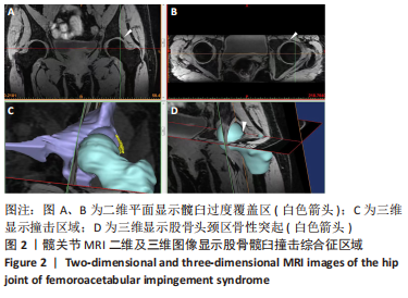
2.1 患者一般资料分析 通过回顾性分析的方式,选取2021年1-12月期间符合标准的双侧股骨髋臼撞击综合征患者36例,其中男性18例,女性18例。男性患者年龄36-50岁,平均(44.61±4.02)岁;身高170-190 cm,平均(177.89±5.31) cm;体质量74-86 kg,平均(79.22±3.42) kg。 女性患者年龄37-53岁,平均(43.11±3.89)岁;身高158-170 cm,平均(164.11±4.01) cm;体质量61-79 kg,平均(71.11±5.09) kg。所有数据呈正态分布。在纳入的36例患者中,根据股骨髋臼撞击综合征分型,有25例患者为混合型,7例患者为凸轮型,4例患者为钳夹型。 2.2 主要观察指标分析 通过对患者的髋关节MRI图像进行三维可视化分析,可直观地显示患髋的三维结构形态,在完整保留髋关节软组织的同时也能其清楚地显示骨性结构,研究以黄色表示撞击区域,患髋髋臼和股骨头形态发生改变,髋臼存在过度覆盖或突起,股骨头呈现非球型,部分可见明显的骨性突起。患髋股骨头骨性突起6例,髋臼过度覆盖9例,均伴有不同程度的髋臼缘增生硬化或游离骨赘,见 图2。"
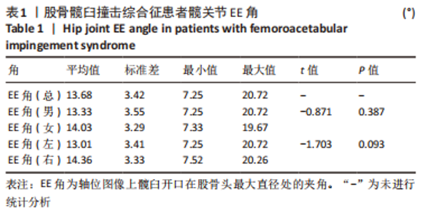
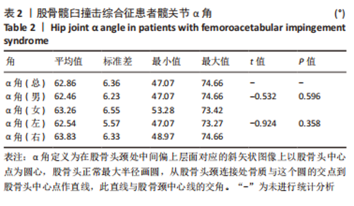
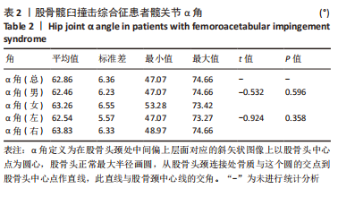
选定轴位图像上股骨头直径为最大时测量患髋EE角度,结果为7.25°-20.72°,平均(13.68±3.42)°,见表1;在髋关节MRI三维重建图像上调整至斜矢状,并可在二维和三维图像上直观地显示α角度,结果为47.07°-74.66°,平均(62.86±6.36)°,见表2,所有患者EE角和α角在男女之间、左右髋关节之间差异均无显著性意义(P > 0.05)。"
"
| [1] 李亚超,王伟强,李韶平.髋关节撞击综合征影像学研究进展[J].安徽医学,2017,38(2):261-264. [2] ALSHAMEERI Z, KHANDUJA V. The effect of femoro-acetabular impingement on the kinematics and kinetics of the hip joint. Int Orthop. 2014;38(8):1615-1620. [3] JOHNSON KA. Impingement of the lesser trochanter on the ischial ramus after total hip arthroplasty. Report of three cases. J Bone Joint Surg Am. 1977;59(2):268-269. [4] TOSUN O, ALGIN O, YALCIN N, et al. Ischiofemoral impingement: evaluation with new MRI parameters and assessment of their reliability. Skeletal Radiol. 2012;41(5):575-587. [5] BREDELLA MA, AZEVEDO DC, OLIVEIRA AL, et al. Pelvic morphology in ischiofemoral impingement. Skeletal Radiol. 2015;44(2):249-253. [6] TANNAST M, GORICKI D, BECK M, et al. Hip damage occurs at the zone of femoroacetabular impingement. Clin Orthop Relat Res. 2008; 466(2):273-280. [7] TANNAST M, SIEBENROCK KA, ANDERSON SE. Femoroacetabular impingement: radiographic diagnosis--what the radiologist should know. AJR Am J Roentgenol. 2007;188(6):1540-1552. [8] MASCARENHAS VV, CAETANO A, DANTAS P, et al. Advances in FAI Imaging: a Focused Review. Curr Rev Musculoskelet Med. 2020;13(5): 622-640. [9] OMAR IM, BLOUNT KJ. Magnetic Resonance Imaging of the Hip. Top Magn Reson Imaging. 2015;24(4):165-181. [10] KIM T, MIN BH, YOON SH, et al. An in vitro comparative study of T2 and T2* mappings of human articular cartilage at 3-Tesla MRI using histology as the standard of reference. Skeletal Radiol. 2014;43(7):947-954. [11] 吴琼,高阳,牛广明,等.基于磁共振图像的膝关节软骨可视化初步研究[J].中国医药导报,2017,14(5):103-105. [12] NÖTZLI HP, WYSS TF, STOECKLIN CH, et al. The contour of the femoral head-neck junction as a predictor for the risk of anterior impingement. J Bone Joint Surg Br. 2002;84(4):556-560. [13] YOU T, YANG B, ZHANG XT, et al. Are “normal hips” being labeled as femoroacetabular impingement due to EE angle? Medicine (Baltimore). 2017;96(13):e6410. [14] DIAZ-LEDEZMA C, NOVACK T, MARIN-PEÑA O, et al. The relevance of the radiological signs of acetabular retroversion among patients with femoroacetabular impingement. Bone Joint J. 2013;95-B(7):893-899. [15] SUTTER R, DIETRICH TJ, ZINGG PO, et al. Femoral antetorsion: comparing asymptomatic volunteers and patients with femoroacetabular impingement. Radiology. 2012;263(2):475-483. [16] WERNER CM, COPELAND CE, STROMBERG J, et al. Correlation of the cross-over ratio of the cross-over sign on conventional pelvic radiographs with computed tomography retroversion measurements. Skeletal Radiol. 2010;39(7):655-660. [17] 陈焱君,刘波,卢建烨,等. MSCT对髋关节撞击综合征的影像学研究[J].中国CT和MRI杂志,2013,11(3):98-102. [18] COOKE WR, GILL HS, MURRAY DW, et al. Discrete mineralisation of the acetabular labrum: a novel marker of femoroacetabular impingement? Br J Radiol. 2013;86(1021):20120182. [19] SIEBENROCK KA, FERNER F, NOBLE PC, et al. The cam-type deformity of the proximal femur arises in childhood in response to vigorous sporting activity. Clin Orthop Relat Res. 2011;469(11):3229-3240. [20] SUTTER R, DIETRICH TJ, ZINGG PO, et al. How useful is the alpha angle for discriminating between symptomatic patients with cam-type femoroacetabular impingement and asymptomatic volunteers? Radiology. 2012;264(2):514-521. [21] 孙钢,李敏,姜庆军,等.凸轮型髋臼撞击综合征影像学表现[J].实用放射学杂志,2010,26(5):695-698. [22] PANZER S, AUGAT P, ESCH U. CT assessment of herniation pits: prevalence, characteristics, and potential association with morphological predictors of femoroacetabular impingement. Eur Radiol. 2008;18(9): 1869-1875. [23] APRATO A, MASSÈ A, FALETTI C, et al. Magnetic resonance arthrography for femoroacetabular impingement surgery: is it reliable? J Orthop Traumatol. 2013;14(3):201-206. [24] SMITH TO, HILTON G, TOMS AP, et al. The diagnostic accuracy of acetabular labral tears using magnetic resonance imaging and magnetic resonance arthrography: a meta-analysis. Eur Radiol. 2011; 21(4):863-874. [25] LATTANZI R, PETCHPRAPA C, ASCANI D, et al. Detection of cartilage damage in femoroacetabular impingement with standardized dGEMRIC at 3 T. Osteoarthritis Cartilage. 2014;22(3):447-456. [26] ELLERMANN J, ZIEGLER C, NISSI MJ, et al. Acetabular cartilage assessment in patients with femoroacetabular impingement by using T2* mapping with arthroscopic verification. Radiology. 2014;271(2): 512-523. [27] ROBINSON P. Conventional 3-T MRI and 1.5-T MR arthrography of femoroacetabular impingement. AJR Am J Roentgenol. 2012;199(3): 509-515. [28] CRESPO-RODRÍGUEZ AM, DE LUCAS-VILLARRUBIA JC, PASTRANA-LEDESMA M, et al. The diagnostic performance of non-contrast 3-Tesla magnetic resonance imaging (3-T MRI) versus 1.5-Tesla magnetic resonance arthrography (1.5-T MRA) in femoro-acetabular impingement. Eur J Radiol. 2017;88:109-116. [29] 庞智晖,魏秋实,周广全,等.个体股骨头坏死三维有限元模型的建立与应用[J].生物医学工程学杂志,2012,29(4):251-255. [30] STEPHEN JM, CALDER JD, WILLIAMS A, et al. Comparative accuracy of lower limb bone geometry determined using MRI, CT, and direct bone 3D models. J Orthop Res. 2021;39(9):1870-1876. [31] LERCH TD, SCHMARANZER F, HANKE MS, et al. Torsional deformities of the femur in patients with femoroacetabular impingement : Dynamic 3D impingement simulation can be helpful for the planning of surgical hip dislocation and hip arthroscopy. Orthopade. 2020;49(6):471-481. [32] YAN K, XI Y, SASIPONGANAN C, et al. Does 3DMR provide equivalent information as 3DCT for the pre-operative evaluation of adult Hip pain conditions of femoroacetabular impingement and Hip dysplasia? Br J Radiol. 2018;91(1092):20180474. [33] LI W, ABRAM F, BEAUDOIN G, et al. Human hip joint cartilage: MRI quantitative thickness and volume measurements discriminating acetabulum and femoral head. IEEE Trans Biomed Eng. 2008;55(12): 2731-2740. [34] NARDI C, DE FALCO L, CARACCHINI G, et al. A three-dimensional measurement method on MR arthrography of the hip to classify femoro-acetabular impingement. Jpn J Radiol. 2021;39(12):1175-1185. [35] SCHAUWECKER N, XI Y, SLEPICKA C, et al. Quantifying differences in femoral head and neck asphericity in CAM type femoroacetabular impingement and hip dysplasia versus controls using radial 3DCT imaging and volumetric segmentation. Br J Radiol. 2020;93(1110):20190039. [36] SCHMARANZER F, HELFENSTEIN R, ZENG G, et al. Automatic MRI-based Three-dimensional Models of Hip Cartilage Provide Improved Morphologic and Biochemical Analysis. Clin Orthop Relat Res. 2019; 477(5):1036-1052. [37] DENIZ CM, XIANG S, HALLYBURTON RS, et al. Segmentation of the Proximal Femur from MR Images using Deep Convolutional Neural Networks. Sci Rep. 2018;8(1):16485. [38] CHENG CT, HO TY, LEE TY, et al. Application of a deep learning algorithm for detection and visualization of hip fractures on plain pelvic radiographs. Eur Radiol. 2019;29(10):5469-5477. [39] ZENG G, SCHMARANZER F, DEGONDA C, et al. MRI-based 3D models of the hip joint enables radiation-free computer-assisted planning of periacetabular osteotomy for treatment of hip dysplasia using deep learning for automatic segmentation. Eur J Radiol Open. 2020;8: 100303. [40] GUIRGUIS A, POLSTER J, KARIM W, et al. Interchangeability of CT and 3D “pseudo-CT” MRI for preoperative planning in patients with femoroacetabular impingement. Skeletal Radiol. 2020;49(7):1073-1080. |
| [1] | Du Xueting, Zhang Xiaodong, Chen Yanjun, Wang Mei, Chen Wubiao, Huang Wenhua. Application of compressed sensing technology in two-dimensional magnetic resonance imaging of the ankle joint [J]. Chinese Journal of Tissue Engineering Research, 2023, 27(9): 1396-1402. |
| [2] | Liu Guangluan, Guo Zonglei, Ge Jin, Huang Dong, Wang Yehua. Anatomic risk factors for medial meniscus posterior root tears combined with anterior cruciate ligament injuries [J]. Chinese Journal of Tissue Engineering Research, 2023, 27(5): 663-668. |
| [3] | Guo Yingqi, Gong Xianxu, Zhang Yan, Xiao Han, Wang Ye, Gu Wenguang. Meniscus extrusion and patellofemoral joint cartilage injury and bone marrow lesions: MRI semi-quantitative score [J]. Chinese Journal of Tissue Engineering Research, 2023, 27(4): 600-605. |
| [4] | Liu Hao, Yang Hongsheng, Zeng Zhimou, Wang Liping, Yang Kunhai, Hu Yongrong, Qu Bo. Lumbar MRI vertebral bone quality score to evaluate the severity of osteoporosis in postmenopausal women [J]. Chinese Journal of Tissue Engineering Research, 2023, 27(4): 606-611. |
| [5] | Wu Tong, Yin Caiyun, Zhao Mingzhe, Zhu Yishen. Application of functional peptides for biomedical diagnosis [J]. Chinese Journal of Tissue Engineering Research, 2023, 27(3): 478-485. |
| [6] | Li Panpan, Qing Haomiao, Ren Sixie, Zhang Yuanyuan. Correlation of medial and lateral posterior tibial slope and their differences with anterior cruciate ligament injury [J]. Chinese Journal of Tissue Engineering Research, 2023, 27(27): 4379-4384. |
| [7] | Gao Yue, Fu Ziwei, Wu Yanbo, Pan Shinong, Lu Zhao. Clinical and imaging features of slipped capital femoral epiphysis [J]. Chinese Journal of Tissue Engineering Research, 2023, 27(27): 4421-4428. |
| [8] | Zhang Jinsheng, Tian Li, Li Sanqiang, Zhang Xixian. Chinese medicine promotes post-stroke brain function remodeling based on visual decoding of functional magnetic resonance imaging [J]. Chinese Journal of Tissue Engineering Research, 2023, 27(23): 3747-3754. |
| [9] | Chen Hao, Wang Rui, Jiang Shaowei, Wu Lei. Correlation of MRI quantitative measurement of medial meniscus extrusion and medial meniscus injury pattern with cartilage damage [J]. Chinese Journal of Tissue Engineering Research, 2023, 27(22): 3567-3572. |
| [10] | Wang Suping, Qiu Demei, Fan Zhonghe, Hu Bo. Bibliometrics and visual analysis of research in the field of rehabilitation for femoroacetabular impingement syndrome in the past decade [J]. Chinese Journal of Tissue Engineering Research, 2023, 27(17): 2754-2762. |
| [11] | Zeng Weipeng, Lin Jianping, Zhou Gang, Mao Hanru. Correlation of anterior cruciate ligament injury with patella alta and femoral trochlear dysplasia in adults evaluated by magnetic resonance imaging [J]. Chinese Journal of Tissue Engineering Research, 2023, 27(13): 2071-2075. |
| [12] | Zhao Jing, Liu Xiaobo, Zhang Yue, Zhang Jiaming, Zhong Dongling, Li Juan, Jin Rongjiang. Visualization analysis of neuromuscular electrical stimulation therapy based on CiteSpace: therapeutic effects, hot spots, and developmental trends [J]. Chinese Journal of Tissue Engineering Research, 2022, 26(8): 1234-1241. |
| [13] | Lu Qinxue, Xu Ning, Yang Yinglan, Han Qianqian, Duanmu Xianyu, Guo Yuwei, Han Qing. Femoroacetabular impingement: strength trainings for nerve-muscle, peripheral muscle and core muscle [J]. Chinese Journal of Tissue Engineering Research, 2022, 26(5): 786-791. |
| [14] | Ma Jiang, Zhang Di, Zhao Tianyu, Liu Xiaoxiao, Wang Ju, Lu Li, Wang Ying, Jin Song. Mechanism and application prospects of motor imagery in spinal cord injury [J]. Chinese Journal of Tissue Engineering Research, 2022, 26(36): 5897-5904. |
| [15] | Liu Yang, Zhu Zhiqiang, Zhao Xiaowei, Xiang Yujie, Xiao Jian, Cheng Lifen. Hot topics and international frontiers of electromyography in the field of body movements [J]. Chinese Journal of Tissue Engineering Research, 2022, 26(35): 5707-5715. |
| Viewed | ||||||
|
Full text |
|
|||||
|
Abstract |
|
|||||