Chinese Journal of Tissue Engineering Research ›› 2023, Vol. 27 ›› Issue (27): 4379-4384.doi: 10.12307/2023.610
Previous Articles Next Articles
Correlation of medial and lateral posterior tibial slope and their differences with anterior cruciate ligament injury
Li Panpan1, Qing Haomiao2, Ren Sixie1, Zhang Yuanyuan1
- 1Department of Radiology, Chengdu Second People’s Hospital, Chengdu 610000, Sichuan Province, China; 2Department of Radiology, Sichuan Cancer Hospital, Chengdu 610000, Sichuan Province, China
-
Received:2022-06-15Accepted:2022-07-28Online:2023-09-28Published:2022-11-08 -
Contact:Zhang Yuanyuan, Associate chief physician, Master, Department of Radiology, Chengdu Second People’s Hospital, Chengdu 610000, Sichuan Province, China -
About author:Li Panpan, Master, Attending physician, Department of Radiology, Chengdu Second People’s Hospital, Chengdu 610000, Sichuan Province, China -
Supported by:Sichuan Provincial Department of Science and Technology Fund Project, No. 2019YJ0585 (to QHM); 2021 Special Fund Project of Scientific Research Nursery Program of Chengdu Second People’s Hospital, No. (2020) 27-16 (to ZYY)
CLC Number:
Cite this article
Li Panpan, Qing Haomiao, Ren Sixie, Zhang Yuanyuan. Correlation of medial and lateral posterior tibial slope and their differences with anterior cruciate ligament injury[J]. Chinese Journal of Tissue Engineering Research, 2023, 27(27): 4379-4384.
share this article
Add to citation manager EndNote|Reference Manager|ProCite|BibTeX|RefWorks
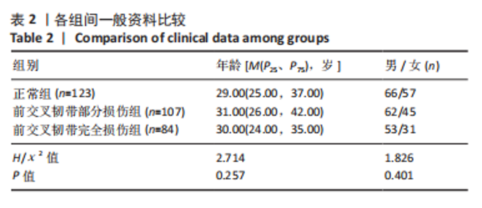
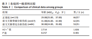
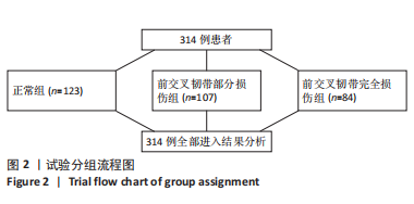
2.1 参与者数量分析 314例患者的MRI影像资料全部进入结果分析。 2.2 各组基线资料比较 3组间的年龄、性别比较差异无显著性意义(P > 0.05),见表2。"
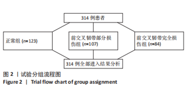
2.3 试验分组流程 见图2。"
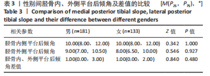
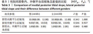
2.4 性别间胫骨内、外侧平台后倾角及差值的比较 总样本314例,其中男181例,女133例,经Kolmogorov-Smirnov Z检验,男性胫骨内、外侧平台后倾角及差值与女性之间比较差异无显著性意义(P > 0.05),见表3。 "
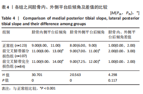
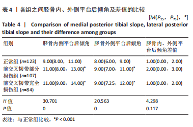
2.5 各组间胫骨内、外侧平台后倾角及差值的比较分析 经Kruskal Wallis H检验,3组之间胫骨内、外侧平台后倾角比较差异有显著性意义(P < 0.001),胫骨内、外侧平台后倾角差值比较差异无显著性意义(P > 0.05);进一步多重比较,前交叉韧带部分损伤组、前交叉韧带完全损伤组胫骨内、外侧平台后倾角均高于正常组(P < 0.001),前交叉韧带部分损伤组与前交叉韧带完全损伤组胫骨内、外侧平台后倾角比较差异无显著性意义(P > 0.05),见表4。"
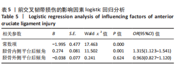
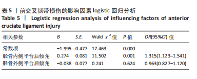
2.6 前交叉韧带损伤的影响因素分析 为探讨前交叉韧带损伤的影响因素,根据前交叉韧带有无损伤分为正常组和损伤组,进行二分类logistic回归分析。结果显示,胫骨内侧平台后倾角是前交叉韧带损伤的独立危险因素[OR=1.315,95%CI(1.123-1.541),P < 0.05],见表5。"
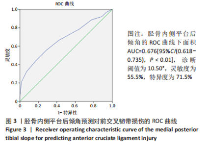
2.7 胫骨内侧平台后倾角的ROC曲线分析 利用ROC曲线分析胫骨内侧平台后倾角对前交叉韧带损伤的预测价值,胫骨内侧平台后倾角的ROC曲线下面积AUC=0.676[95%CI(0.618-0.735),P < 0.01],诊断阈值为10.50°,灵敏度为55.5%,特异度为71.5%,见图3。"

| [1] BAYER S, MEREDITH SJ, WILSON KW, et al. Knee Morphological Risk Factors for Anterior Cruciate Ligament Injury: A Systematic Review. J Bone Joint Surg Am. 2020;102(8):703-718. [2] GIANOTTI SM, MARSHALL SW, HUME PA, et al. Incidence of anterior cruciate ligament injury and other knee ligament injuries: A national population-based study. J Sci Med Sport. 2009;12(6):622-627. [3] EGGER AC, PARIKH SN, WILSON PL, et al. What’s New in the Management of Pediatric Anterior Cruciate Ligament Tears and Tibial Spine Fractures. Instr Course Lect. 2021;70:399-414. [4] LANSDOWN D, MA CB. The Influence of Tibial and Femoral Bone Morphology on Knee Kinematics in the Anterior Cruciate Ligament Injured Knee. Clin Sports Med. 2018;37(1):127-136. [5] NAZZAL EM, ZSIDAI B, PUJOL O, et al. Considerations of the Posterior Tibial Slope in Anterior Cruciate Ligament Reconstruction: a Scoping Review. Curr Rev Musculoske. 2022;15(4):291-299. [6] LIU Z, JIANG J, YI Q, et al. An increased posterior tibial slope is associated with a higher risk of graft failure following ACL reconstruction: a systematic review. Knee Surg Sports Traumatol Arthrosc. 2022;30(7):2377-2387. [7] 龚恒,黄斌,付立功,等.中国汉族人与美国高加索白人膝关节几何形态的比较[J].中国组织工程研究,2020,24(33):5366-5370. [8] BUCHER C, LAMY D, DEBATY G, et al. Validity of the lever sign test for the clinical diagnosis of anterior cruciate ligament tears: Assessments in ski resorts. Orthop Traumatol Surg Res. 2022;108(3):103254. [9] 常丽鹏,赵敏,龚国龄,等.MRI在膝关节半月板损伤,前交叉韧带损伤诊断中的应用价值研究[J].中国CT和MRI杂志,2020,18(8):164-167. [10] ADHIKARI V, JOSHI A, SINGH N, et al. Predictive Accuracy of Blumensaat Line Angle and Its Apex along with Anterior Cruciate Ligament Tear with Abundant Remnant. J Nepal Health Res Counc. 2021;18(4):604-609. [11] GUNAYDIN B, SAHIN GG, SARI A, et al. A new method for diagnosis of anterior cruciate ligament tear: MRI with maximum flexion of knee in the prone position: A case control study. Int J Surg. 2019;68(2):142-147. [12] 翁习生,卫小春,主译.膝关节创伤及疾病的MRI图谱[M].北京:中国协和医科大学出版社,2005:29-31. [13] HUDEK R, FUCHS B, REGENFELDER F, et al. Is noncontact ACL injury associated with the posterior tibial and meniscal slope? Clin Orthop Relat Res. 2011;469(8): 2377-2384. [14] 王善安,杨友刚,戚世鹏,等. 前交叉韧带损伤合并内侧胫骨平台骨性关节炎患者临床特点分析[J]. 成都医学院学报,2021,42(4):371-374. [15] KAPLAN JT, RAMSAY JW, CAMERON SE, et al. Association Between Knee Anatomic Metrics and Biomechanics for Male Soldiers Landing With Load. Am J Sport Med. 2020;48(7):1389-1397. [16] 敖英芳,刘宝戈,龚熹,主译.骨科运动医学原理与实践[M].北京:北京大学出版社,2005:1065-1066. [17] BLAHA JD, MANCINELLI CA, SIMONS WH, et al. Kinematics of human knee using an open chain cadaver model. Clin Orthop Relat Res. 2003;(410):25-34. [18] ALAIA MJ, KAPLAN DJ, MANNINO BJ, et al. Tibial Sagittal Slope in Anterior Cruciate Ligament Injury and Treatment. J Am Acad Orthop Surg. 2021;29(21): e1045-e1056. [19] MAROUANE H, SHIRAZI-ADL A, HASHEMI J. Quantification of the role of tibial posterior slope in knee joint mechanics and ACL force in simulated gait. J Biomech. 2015;10(48): 1899-1905. [20] MEIER M, JANSSEN D, KOECK FX, et al. Variations in medial and lateral slope and medial proximal tibial angle. Knee Surg Sports Traumatol Arthrosc. 2021;29(3): 939-946. [21] ALJUHANI WS, QASIM SS, ALRASHEED A, et al. The effect of gender, age, and body mass index on the medial anda lateral posterior tibial slopes: a magnetic resonance imaging study. Knee Surg Relat Res. 2021;33(1):12-30. [22] 佟志忠,蒋雯,程晓光,等.利用EOS X线影像系统评估前交叉韧带损伤患者的胫骨平台后倾角[J].骨科临床与研究杂志,2020,5(5):268-271. [23] KACMAZ IE, TOPKAYA Y, BASA CD, et al. Posterior tibial slope of the knee measured on X-rays in a Turkish population. Surg Radiol Anat. 2020;42(6):673-679. [24] BERNHARDSON AS, AMAN ZS, DORNAN GJ, et al. Tibial Slope and Its Effect on Force in Anterior Cruciate Ligament Grafts: Anterior Cruciate Ligament Force Increases Linearly as Posterior Tibial Slope Increases. Am J Sport Med. 2019; 47(2):296-302. [25] NUNLEY RM, NAM D, JOHNSON SR, et al. Extreme Variability in Posterior Slope of the Proximal Tibia: Measurements on 2395 CT Scans of Patients Undergoing UKA? J Arthroplasty. 2014;29(8):1677-1680. [26] RAJA B, MARATHE N, DESAI J, et al. Evaluation of anatomic risk factors using magnetic resonance imaging in non-contact anterior cruciate ligament injury. J Clin Orthop Trauma. 2019;10(4):710-715. [27] KUMAR PANIGRAHI T, DAS A, MOHANTY T, et al. Study of relationship of posterior tibial slope in anterior cruciate ligament injury. J Orthop. 2020;21:487-490. [28] HOHMANN E, TETSWORTH K, GLATT V, et al. Medial and Lateral Posterior Tibial Slope Are Independent Risk Factors for Noncontact ACL Injury in Both Men and Women. Orthop J Sports Med. 2021;9(8):227-239. [29] GOLDSTEIN SA, WILSON DL, SONGTEGARD DA, et al. The mechanical properties of human tibial trabecular bone as a function of metaphyseal location. J Biomech. 1983;16(12):965-969. [30] 郑亮亮,周志鹏.非接触性前交叉韧带损伤风险因素的循证研究进展[J].中国运动医学杂志,2014,33(6):596-604. [31] TODD MS, LALLISS S, GARCIA E, et al. The relationship between posterior tibial slope and anterior cruciate ligament injuries. Am J Sport Med. 2010;38(1):63-67. [32] VYAS S, VAN ECK CF, VYAS N, et al. Increased medial tibial slope in teenage pediatric population with open physes and anterior cruciate ligament injuries. Knee Surg Sports Traumatol Arthrosc. 2010;19(3):372-377. [33] OKAZAKI Y, FURUMATSU T, HIRANAKA T, et al. Steep posterior slope of the medial tibial plateau is associated with ramp lesions of the medial meniscus and a concomitant anterior cruciate ligament injury. Asia Pac J Sports Med Arthrosc Rehabil Technol. 2021;24(3):23-28. [34] WILLINGER L, BALENDRA G, PAI V, et al. Medial meniscal ramp lesions in ACL-injured elite athletes are strongly associated with medial collateral ligament injuries and medial tibial bone bruising on MRI. Knee Surg Sports Traumatol Arthrosc. 2022;30(5):1502-1510. [35] SALMON LJ, HEATH E, AKRAWI H, et al. 20 year outcomes of ACL reconstruction with hamstring tendon autograft. The catastrophic effect of age and posterior tibial slope. Am J Sport Med. 2018;46(3):531-543. [36] LIU C, GE J, HUANG C, et al. A radiographic model predicting the status of the anterior cruciate ligament in varus knee with osteoarthritis. Bmc Musculoskelet Disord. 2022;23(1):603-612. [37] ZEE MJM, KEIZER MNJ, DIJKERMAN L, et al. The correlation between posterior tibial slope and dynamic anterior tibial translation and dynamic range of tibial rotation. J Exp Orthop. 2021;8(1):71-78. [38] HOHMANN E, TETSWORTH K, GLATT V, et al. The posterior horn of the medial and lateral meniscus both reduce the effective posterior tibial slope: a radiographic MRI study. Surg Radiol Anat. 2021;43(7):1123-1130. [39] JIANG J, LIU Z, WANG X, et al. Increased Posterior Tibial Slope and Meniscal Slope Could Be Risk Factors for Meniscal Injuries: A Systematic Review. Arthroscopy. 2022;38(7):2331-2341. [40] SMEETS A, WILLEMS M, GILSON L, et al. Neuromuscular and biomechanical landing alterations persist in athletes returning to sport after anterior cruciate ligament reconstruction. Knee. 2021;33:305-317. [41] HUANG M, LI Y, LI H, et al. Correlation between knee anatomical angles and anterior cruciate ligament injury in males. Radiol Med. 2021;(10):1201-1206. |
| [1] | Du Xueting, Zhang Xiaodong, Chen Yanjun, Wang Mei, Chen Wubiao, Huang Wenhua. Application of compressed sensing technology in two-dimensional magnetic resonance imaging of the ankle joint [J]. Chinese Journal of Tissue Engineering Research, 2023, 27(9): 1396-1402. |
| [2] | Bi Gengchao, Zhang Yanlong, Li Qiuyue, Hu Longwei, Zhang Yu. Knee joint mechanics and activation characteristics of surrounding muscles during deep jumps at different heights and distances [J]. Chinese Journal of Tissue Engineering Research, 2023, 27(8): 1211-1218. |
| [3] | Xiong Bohan, Yu Yang, Lu Xiaojun, Wang Xu, Yang Tengyun, Zhang Yaozhang, Liao Xinyu, Zhou Xiaoxiang, He Lu, Li Yanlin. Research progress in promoting tendon to bone healing during anterior cruciate ligament reconstruction [J]. Chinese Journal of Tissue Engineering Research, 2023, 27(5): 779-786. |
| [4] | Liu Guangluan, Guo Zonglei, Ge Jin, Huang Dong, Wang Yehua. Anatomic risk factors for medial meniscus posterior root tears combined with anterior cruciate ligament injuries [J]. Chinese Journal of Tissue Engineering Research, 2023, 27(5): 663-668. |
| [5] | Wei Bo, Yao Qingqiang, Tang Cheng, Li Xuxiang, Xu Yan, Wang Liming. Advantage of medial pivot prosthesis in total knee arthroplasty via medial subvastus approach [J]. Chinese Journal of Tissue Engineering Research, 2023, 27(4): 552-557. |
| [6] | Guo Yingqi, Gong Xianxu, Zhang Yan, Xiao Han, Wang Ye, Gu Wenguang. Meniscus extrusion and patellofemoral joint cartilage injury and bone marrow lesions: MRI semi-quantitative score [J]. Chinese Journal of Tissue Engineering Research, 2023, 27(4): 600-605. |
| [7] | Liu Hao, Yang Hongsheng, Zeng Zhimou, Wang Liping, Yang Kunhai, Hu Yongrong, Qu Bo. Lumbar MRI vertebral bone quality score to evaluate the severity of osteoporosis in postmenopausal women [J]. Chinese Journal of Tissue Engineering Research, 2023, 27(4): 606-611. |
| [8] | Li Shihao, Li Qi, Li Zhen, Zhang Yuanyuan, Liu Miaomiao, Ouyang Yi, Xu Weiguo. Plantar pressure and gait analysis in patients with anterior cruciate ligament injury and reconstruction [J]. Chinese Journal of Tissue Engineering Research, 2023, 27(4): 626-631. |
| [9] | Wu Tong, Yin Caiyun, Zhao Mingzhe, Zhu Yishen. Application of functional peptides for biomedical diagnosis [J]. Chinese Journal of Tissue Engineering Research, 2023, 27(3): 478-485. |
| [10] | Chen Hao, Wang Rui, Jiang Shaowei, Wu Lei. Correlation of MRI quantitative measurement of medial meniscus extrusion and medial meniscus injury pattern with cartilage damage [J]. Chinese Journal of Tissue Engineering Research, 2023, 27(22): 3567-3572. |
| [11] | Wang Jiawei, Liu Ye. Hip and knee coordination patterns in relation to lower limb muscle activity during drop jumps at different heights [J]. Chinese Journal of Tissue Engineering Research, 2023, 27(2): 276-281. |
| [12] | Zhang Xiaohui, Zhang Mingjun, Wang Jianping, Qu Haijun, Chai Le, Li Yu, Zhang Xinmin. Constructing digital model of knee joint and surrounding soft tissue surface by three-dimensional image registration technology [J]. Chinese Journal of Tissue Engineering Research, 2023, 27(18): 2814-2819. |
| [13] | Xu Zhi, Li Yuwan, Jin Ying, Liu Yi. Effect of partial posterior root tear of medial meniscus on biomechanics of the knee joint during gait cycle [J]. Chinese Journal of Tissue Engineering Research, 2023, 27(18): 2824-2830. |
| [14] | Zeng Weipeng, Lin Jianping, Zhou Gang, Mao Hanru. Correlation of anterior cruciate ligament injury with patella alta and femoral trochlear dysplasia in adults evaluated by magnetic resonance imaging [J]. Chinese Journal of Tissue Engineering Research, 2023, 27(13): 2071-2075. |
| [15] | Fang Run, Ning Rende, Liu Yulong, Bi Chenghao, Zhou Daobin. Significance of X-ray measurement of the anatomical relationship between adult lateral tibial plateau and lateral femoral condyle in fracture reduction [J]. Chinese Journal of Tissue Engineering Research, 2023, 27(13): 2076-2080. |
| Viewed | ||||||
|
Full text |
|
|||||
|
Abstract |
|
|||||