[1] 徐飞, 李彦林, 王国梁, 等. 内减张技术在辅助前交叉韧带重建中的研究进展[J]. 中国修复重建外科杂志,2021,35(12):1630-1636.
[2] Chan CX, Wong KL, Toh SJ, et al. Epidemiology of patients with anterior cruciate ligament injuries undergoing reconstruction surgery in a multi-ethnic Asian population. Res Sports Med. 2021;29(1):12-24.
[3] Tseng HJ, Lo HL, Lin YC, et al. Analyze the Differential Rates of Anterior Cruciate Ligament Injuries Between Men and Women by Biomechanical Study of Single-Leg Landing in Badminton. Indian J Orthop. 2021;55(Suppl 2):409-417.
[4] Huang YL, Jung J, Mulligan CMS, et al. A majority of anterior cruciate ligament injuries can be prevented by injury prevention programs: a systematic review of randomized controlled trials and cluster-randomized controlled trials with meta-analysis. Am J Sports Med. 2020;48(6):1505-1515.
[5] Basukala B, Joshi A, Pradhan I. The effect of the intercondylar notch shape and notch width index on anterior cruciate ligament injuries. J Nepal Health Res Counc. 2020;17(4):532-536.
[6] Hosseinzadeh S, Kiapour AM. Sex Differences in Anatomic Features Linked to Anterior Cruciate Ligament Injuries During Skeletal Growth and Maturation. Am J Sports Med. 2020;48(9):2205-2212.
[7] 黄碧滢, 邓文宇, 李韬, 等. 前交叉韧带重建术中股骨隧道定位方法研究进展[J]. 中国修复重建外科杂志,2021,35(1):118-123.
[8] Migliorini F, Maffulli N, Eschweiler J, et al. Lateral retinacular release combined with MPFL reconstruction for patellofemoral instability: a systematic review. Arch Orthop Trauma Surg. 2021;141(2): 283-292.
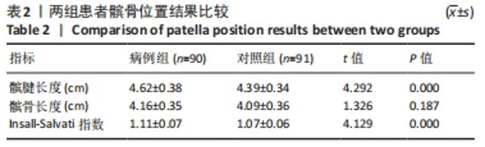
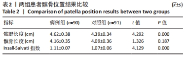
[9] Degnan AJ, Maldjian C, Adam RJ, et al. Comparison of Insall-Salvati ratios in children with an acute anterior cruciateligament tear and a matched control population. AJR Am J Roentgenol. 2015;204(1):161-166.
[10] Insall J, Salvati E. Patella position in the normal knee joint. Radiology. 1971;101(1):101-104.
[11] Hiemstra LA, Kerslake S, Loewen M, et al. Effect of Trochlear Dysplasia on Outcomes After Isolated Soft Tissue Stabilization for Patellar Instability. Am J Sports Med. 2016;44(6):1515-1523.
[12] Botchu R, Obaid H, Rennie WJ. Correlation between trochlear dysplasia and the notch index. J Orthop Surg (Hong Kong). 2013;21(3): 290-293.
[13] Dejour D, Le Coultre B. Osteotomies in Patello-Femoral Instabilities. Sports Med Arthrosc Rev. 2018;26(1):8-15.
[14] Montalvo AM, Schneider DK, Webster KE, et al. Anterior Cruciate Ligament Injury Risk in Sport: A Systematic Review and Meta-Analysis of Injury Incidence by Sex and Sport Classification. J Athl Train. 2019;54(5):472-482.
[15] DE Carli A, Koverech G, Gaj E, et al. Anterior cruciate ligament injury in elite football players: video analysis of 128 cases. J Sports Med Phys Fitness. 2022;62(2):222-228.
[16] Webster KE, Hewett TE. Anterior Cruciate Ligament Injury and Knee Osteoarthritis: An Umbrella Systematic Review and Meta-analysis. Clin J Sport Med. 2022;32(2):145-152.
[17] Kingery MT, Anil U, Berlinberg EJ, et al. Changes in the Synovial Fluid Cytokine Profile of the Knee Between an Acute Anterior Cruciate Ligament Injury and Surgical Reconstruction. Am J Sports Med. 2022; 50(2):451-460.
[18] Snoeker B, Turkiewicz A, Magnusson K, et al. Risk of knee osteoarthritis after different types of knee injuries in young adults: a population-based cohort study. Br J Sports Med. 2020;54(12):725-730.
[19] Poulsen E, Goncalves GH, Bricca A, et al. Knee osteoarthritis risk is increased 4-6 fold after knee injury - a systematic review and meta-analysis. Br J Sports Med. 2019;53(23):1454-1463.
[20] Ekås GR, Laane MM, Larmo A, et al. Knee Pathology in Young Adults After Pediatric Anterior Cruciate Ligament Injury: A Prospective Case Series of 47 Patients with a Mean 9.5-Year Follow-up. Am J Sports Med. 2019;47(7):1557-1566.
[21] Zadehmohammad A, Grillari J, Stevanovic V, et al. Osteoarthritis Progression after ACL Reconstruction Was Significantly Higher Than That of the Healthy Contralateral Knees: Long-Term Follow Up Study of Mean 16.4 Years. J Clin Med. 2022;11(3):775.
[22] van Yperen DT, Reijman M, van Es EM, et al. Twenty-Year Follow-up Study Comparing Operative Versus Nonoperative Treatment of Anterior Cruciate Ligament Ruptures in High-Level Athletes. Am J Sports Med. 2018;46(5):1129-1136.
[23] Rodriguez K, Soni M, Joshi PK, et al. Anterior Cruciate Ligament Injury: Conservative Versus Surgical Treatment. Cureus. 2021;13(12): e20206.
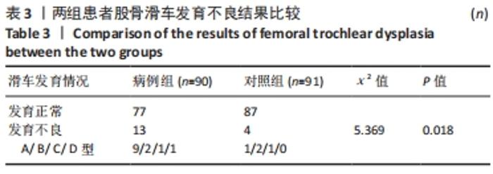
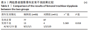
[24] Kwak YH, Nam JH, Koh YG, et al. Correlation of Femoral Trochlear Dysplasia With Anterior Cruciate Ligament Injury in Skeletally Immature Patients. Orthop J Sports Med. 2021;9(9):23259671211022690.
[25] Chen M, Qin L, Li M, et al. Correlation analysis between femoral trochlear dysplasia and anterior cruciate ligament injury based on CT measurement. Quant Imaging Med Surg. 2020;10(4):847-852.
[26] Botchu R, Obaid H, Rennie WJ. Correlation between trochlear dysplasia and anterior cruciate ligament injury. J Orthop Surg (Hong Kong). 2013;21(2):185-188.
[27] Pfeifer CE, Beattie PF, Sacko RS, et al. Risk factors associated with non-contact anterior cruciate ligament injury: a systematic review. Int J Sports Phys Ther. 2018;13(4):575-587.
[28] Akgün AS, Agirman M. Associations between anterior cruciate ligament injuries and patella alta and trochlear dysplasia in adults using magnetic resonance imaging. J Knee Surg. 2021;34(11):1220-1226.
[29] Kwak YH, Nam JH, Koh YG, et al. Femoral trochlear morphology is associated with anterior cruciate ligament injury in skeletally immature patients. Knee Surg Sports Traumatol Arthrosc. 2020;28(12):3969-3977.
[30] Van Ginckel A, Verdonk P, Witvrouw E. Cartilage adaptation after anterior cruciate ligament injury and reconstruction: implications for clinical management and research? A systematic review of longitudinal MRI studies. Osteoarthritis Cartilage. 2013;21(8):1009-1024.
[31] Macri EM, Culvenor AG, Morris HG, et al. Lateral displacement, sulcus angle and trochlear angle are associated with early patellofemoral osteoarthritis following anterior cruciate ligament reconstruction. Knee Surg Sports Traumatol Arthrosc. 2018;26(9):2622-2629.
[32] Ntagiopoulos PG, Bonin N, Sonnery-Cottet B, et al. The incidence of trochlear dysplasia in anterior cruciate ligament tears. Int Orthop. 2014;38(6):1269-1275.
|