Chinese Journal of Tissue Engineering Research ›› 2023, Vol. 27 ›› Issue (22): 3567-3572.doi: 10.12307/2023.393
Previous Articles Next Articles
Correlation of MRI quantitative measurement of medial meniscus extrusion and medial meniscus injury pattern with cartilage damage
Chen Hao, Wang Rui, Jiang Shaowei, Wu Lei
- Department of Sports Trauma and Arthroscopic Surgery, First Affiliated Hospital of Anhui Medical University, Hefei 230022, Anhui Province, China
-
Received:2022-06-02Accepted:2022-07-14Online:2023-08-08Published:2022-11-02 -
Contact:Wang Rui, Doctoral candidate, Associate chief physician, Department of Sports Trauma and Arthroscopic Surgery, First Affiliated Hospital of Anhui Medical University, Hefei 230022, Anhui Province, China -
About author:Chen Hao, Master candidate, Physician, Department of Sports Trauma and Arthroscopic Surgery, First Affiliated Hospital of Anhui Medical University, Hefei 230022, Anhui Province, China
CLC Number:
Cite this article
Chen Hao, Wang Rui, Jiang Shaowei, Wu Lei. Correlation of MRI quantitative measurement of medial meniscus extrusion and medial meniscus injury pattern with cartilage damage[J]. Chinese Journal of Tissue Engineering Research, 2023, 27(22): 3567-3572.
share this article
Add to citation manager EndNote|Reference Manager|ProCite|BibTeX|RefWorks
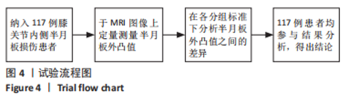
2.1 参与者数量分析 纳入膝关节内侧半月板损伤患者117例,全部进入结果分析,无脱落。 2.2 试验流程图 见图4。"
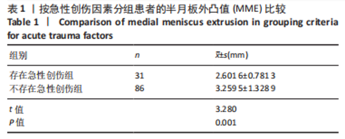
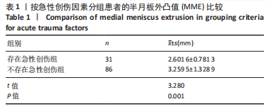
2.3 创伤因素相关组MME值的比较 病史中存在可明确回忆的急性创伤患者有31例(26.5%),无明确回忆的急性创伤患者有86例(73.5%),两组间MME值比较差异有显著性意义(t=3.280,P=0.001),见表1。"
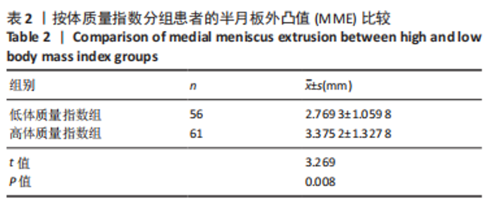
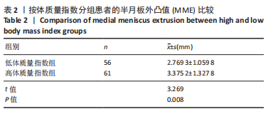
2.4 不同体质量指数分组MME值的比较 根据体质量指数分组,低体质量指数组(体质量指数≤25 kg/m2) 56例(47.9%),高体质量指数组(体质量指数 > 25 kg/m2) 61例(52.1%),两组间MME值比较差异有显著性意义(t=3.269,P=0.008),见表2。"
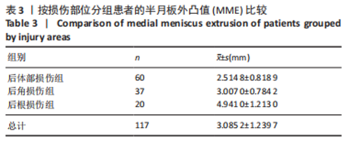
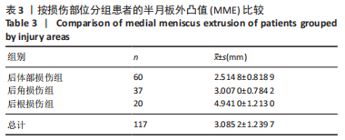
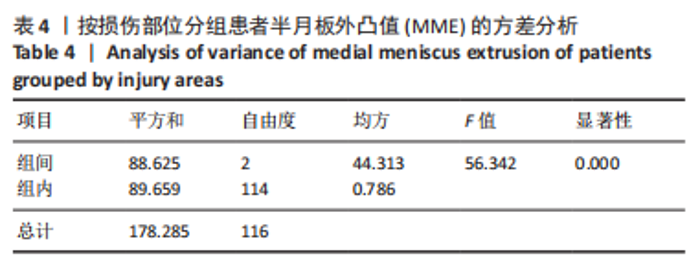
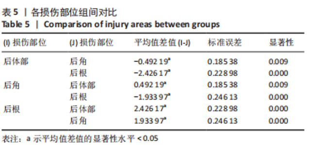
2.5 半月板损伤部位组间的MME值比较 半月板后体部损伤组60例(51.2%),后角损伤组37例(31.6%),后根断裂组20例(17.1%),3组间MME值比较采用单因素方差分析,差异有显著性学意义(总体F=56.342,P=0.000,P < 0.05);各组间两两对比差异均有显著性意义(P < 0.05),见表3-5。"
"
"
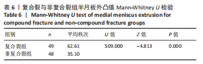
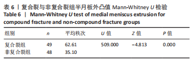
2.6 复合裂与非复合裂组的MME值比较 按照术中关节镜下证实的损伤情况,将非内侧半月板后根断裂的患者分为半月板复合裂组和非复合裂组(复合裂组MME平均秩62.61,非复合裂组MME平均秩35.10),两组间采用非参数检验,差异有显著性意义(P=0.000),见表6及图5。"
"

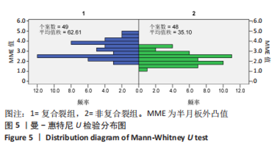
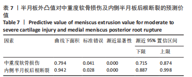
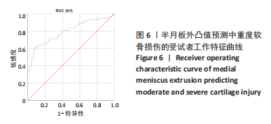
2.7 MME值对中重度软骨损伤的预测价值 将关节镜下证实的Outerbridge 3级与4级软骨损伤认为是中重度的软骨损伤,绘制了MME预测中重度软骨损伤的ROC曲线,其AUC为0.794(95%CI:0.715-0.874,P=0.000),有一定的预测意义,见表7及图6。"

"
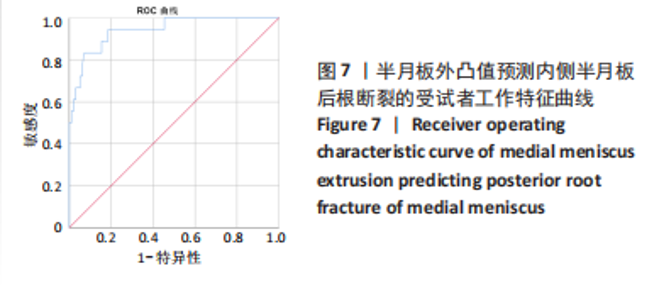
2.8 MME值 对内侧半月板后根断裂的预测价值 绘制了MME预测内侧半月板后根断裂的ROC曲线,其AUC为0.942 (95%CI:0.887-0.998,P=0.000),有较强的预测意义。通过计算约登指数,当选择cuff-off值 > 2.555 0时,诊断内侧半月板后根断裂的敏感度为94.4%,特异度为54.6%,见表7及图7。"
| [1] THOMPSON WO, THAETE FL, FU FH, et al. Tibial meniscal dynamics using three-dimensional reconstruction of magnetic resonance images. Am J Sports Med. 1991;19(3):210-215; discussion 215-216. [2] DASZKIEWICZ K, LUCZKIEWICZ P. Biomechanics of the medial meniscus in the osteoarthritic knee joint. Peer J. 2021;9:27. [3] NHA KW, LEE YS, HWANG DH, et al. Second-Look Arthroscopic Findings After Open-Wedge High Tibia Osteotomy Focusing on the Posterior Root Tears of the Medial Meniscus. Arthroscopy. 2013;29(2):226-231. [4] LEAFBLAD ND, SMITH PA, STUART MJ, et al. Arthroscopic Centralization of the Extruded Medial Meniscus. Arthrosc Tech. 2021;10(1):e43-e48. [5] SHARIF B, ASHRAF T, SAIFUDDIN A. Magnetic resonance imaging of the meniscal roots. Skeletal Radiol. 2020;49(5):661-676. [6] KRYCH AJ, HEVESI M, LELAND DP, et al. Meniscal Root Injuries. J Am Acad Orthop Surg. 2020;28(12):491-499. [7] BHATIA S, LAPRADE CM, ELLMAN MB, et al. Meniscal Root Tears Significance, Diagnosis, and Treatment. Am J Sports Med. 2014;42(12): 3016-3030. [8] SWAMY N, WADHWA V, BAJAJ G, et al. Medial meniscal extrusion: Detection, evaluation and clinical implications. Eur J Radiol. 2018;102: 115-124. [9] OZEKI N, SEIL R, KRYCH AJ, et al. Surgical treatment of complex meniscus tear and disease: state of the art. J ISAKOS. 2021;6(1):35-45. [10] FUJISAWA T, CHOE H, KUSABA Y, et al. Medial meniscus extrusion and stage are related to the size of spontaneous osteonecrosis of the knee in patients who underwent high tibial osteotomy. Knee. 2022;36:72-79. [11] 江佩师, 陈志伟, 方玉基, 等. 602例膝关节半月板损伤流行病学调查[J]. 中南医学科学杂志,2020,48(2):160-163. [12] GUILAK F, RATCLIFFE A, LANE N, et al. Mechanical and biochemical changes in the superficial zone of articular cartilage in canine experimental osteoarthritis. J Orthop Res. 1994;12(4):474-484. [13] BRUNS K, SVENSSON F, TURKIEWICZ A, et al. Meniscus body position and its change over four years in asymptomatic adults: a cohort study using data from the Osteoarthritis Initiative (OAI). BMC Musculoskelet Disord. 2014;15:32. [14] STENSBY JD, PRINGLE LC, CRIM J. MRI of the Meniscus. Clin in Sports Med. 2021;40(4):641-655. [15] HISASHI K, MUNETA T, KOHNO Y, et al. MRI study of medial meniscus degeneration of osteoarthritic knees with or without posterior root tear. J Exp Orthop. 2022;9(1):38. [16] HAJEK PC, GYLYS-MORIN VM, BAKER LL, et al. The high signal intensity meniscus of the knee. Magnetic resonance evaluation and in vivo correlation. Invest Radiol. 1987;22(11):883-890. [17] ISHII Y, ISHIKAWA M, HAYASHI S, et al. The correlation between osteoarthritis stage and the effect of the lateral wedge insole for 3 months on medial meniscus extrusion in the knee joint. Knee. 2021; 28:110-116. [18] COSTA CR, MORRISON WB, CARRINO JA. Medial meniscus extrusion on knee MRI: Is extent associated with severity of degeneration or type of tear? Am J Roentgenol. 2004;183(1):17-23. [19] KOZAKI T, FUKUI D, YAMAMOTO E, et al. Medial meniscus extrusion and varus tilt of joint line convergence angle increase stress in the medial compartment of the knee joint in the knee extension position -finite element analysis. J Exp Orthop. 2022;9(1):49. [20] ZHANG F, BIERMA-ZEINSTRA SM, OEI EHG, et al. Factors associated with meniscal body extrusion on knee MRI in overweight and obese women. Osteoarthritis Cartilage. 2017;25(5):694-699. [21] MUNUGODA IP, BEAVERS DP, WIRTH W, et al. The effect of weight loss on the progression of meniscal extrusion and size in knee osteoarthritis: a post-hoc analysis of the Intensive Diet and Exercise for Arthritis (IDEA) trial. Osteoarthritis Cartilage. 2020;28(4):410-417. [22] SHARMA K, ECKSTEIN F, WIRTH W, et al. Meniscus position and size in knees with versus without structural knee osteoarthritis progression - data from the osteoarthritis initiative. Osteoarthritis Cartilage. 2019; 27:S467-S468. [23] KIM C, BIN SI, LEE BS, et al. Volumetric assessment of extrusion in medial meniscus posterior root tears through semi-automatic segmentation on 3-tesla magnetic resonance images. Orthop Traumatol Surg Res. 2020;106(5):963-968. [24] CHIBA D, SASAKI T, ISHIBASHI Y. Greater medial meniscus extrusion seen on ultrasonography indicates the risk of MRI-detected complete medial meniscus posterior root tear in a Japanese population with knee pain. Sci Rep. 2022;12(1):4756. [25] KRYCH AJ, BERNARD CD, LELAND DP, et al. Isolated meniscus extrusion associated with meniscotibial ligament abnormality. Knee Surg Sports Traumatol Arthrosc. 2020;28(11):3599-3605. [26] KOGA H, NAKAMURA T, NAKAGAWA Y, et al. Arthroscopic Centralization Using Knotless Anchors for Extruded Medial Meniscus. Arthrosc Tech. 2021;10(3):e639-e645. [27] CHERNCHUJIT B, AGRAWAL S. Arthroscopic All-Inside Medial Meniscus Extrusion Reduction. Arthrosc Tech. 2019;8(5):e495-e501. [28] HIRANAKA T, FURUMATSU T, MIYAZAWA S, et al. Transtibial pullout repair techniques using two simple stitches for medial meniscus posterior root tear can prevent the progression of medial meniscus extrusion and obtain successful outcomes. Eur J Orthop Surg Traumatol. 2022;32(5):795-802. [29] WU TY. Arthroscopic Medial Meniscus Posterior Root Repair with Centralization Using Knotless Suture Anchors. Arthrosc Tech. 2022; 11(4):e661-e668. [30] CLIFTON WILLIMON S, BUSCH MT, MURATA A, et al. Transosseous Meniscus Root Repair in Pediatric Patients and Association With Durable Midterm Outcomes and High Rates of Return to Sports. Am J Sports Med. 2022;50(8):2070-2074. [31] CHUNG KS, HA JK, RA HJ, et al. Preoperative varus alignment and postoperative meniscus extrusion are the main long-term predictive factors of clinical failure of meniscal root repair. Knee Surg Sports Traumatol Arthrosc. 2021;29(12):4122-4130. |
| [1] | Li Xiaomin, Tian Xiangdong, Tan Yetong, Zhu Guangyu, Wang Rongtian, Wang Jian, Xue Zhipeng, Ma Sheng, Hu Yuanyi, Huang Ye, Ding Tiansong. Changes of lower limb force line and knee function after high tibial osteotomy in osteoporotic medial ventricular knee osteoarthritis [J]. Chinese Journal of Tissue Engineering Research, 2023, 27(9): 1325-1329. |
| [2] | Du Xueting, Zhang Xiaodong, Chen Yanjun, Wang Mei, Chen Wubiao, Huang Wenhua. Application of compressed sensing technology in two-dimensional magnetic resonance imaging of the ankle joint [J]. Chinese Journal of Tissue Engineering Research, 2023, 27(9): 1396-1402. |
| [3] | Zheng Bo, Zhang Xiuli, Zhou Hao, He Zebi, Zhou Jin, Zhou Weiyun, Li Peng. Arthroscopy-assisted locking hollow screw fixation and open reduction plate internal fixation in the treatment of Schatzker II-III tibial plateau fractures: early CT evaluation [J]. Chinese Journal of Tissue Engineering Research, 2023, 27(9): 1410-1416. |
| [4] | Liu Guangluan, Guo Zonglei, Ge Jin, Huang Dong, Wang Yehua. Anatomic risk factors for medial meniscus posterior root tears combined with anterior cruciate ligament injuries [J]. Chinese Journal of Tissue Engineering Research, 2023, 27(5): 663-668. |
| [5] | Guo Yingqi, Gong Xianxu, Zhang Yan, Xiao Han, Wang Ye, Gu Wenguang. Meniscus extrusion and patellofemoral joint cartilage injury and bone marrow lesions: MRI semi-quantitative score [J]. Chinese Journal of Tissue Engineering Research, 2023, 27(4): 600-605. |
| [6] | Liu Hao, Yang Hongsheng, Zeng Zhimou, Wang Liping, Yang Kunhai, Hu Yongrong, Qu Bo. Lumbar MRI vertebral bone quality score to evaluate the severity of osteoporosis in postmenopausal women [J]. Chinese Journal of Tissue Engineering Research, 2023, 27(4): 606-611. |
| [7] | Wu Tong, Yin Caiyun, Zhao Mingzhe, Zhu Yishen. Application of functional peptides for biomedical diagnosis [J]. Chinese Journal of Tissue Engineering Research, 2023, 27(3): 478-485. |
| [8] | Zhong Hehe, Jin Ying, Liu Xiuqi, Xiang Kuan, Wu Shuhong, Peng Jiachen, Liu Yi. High tibial osteotomy combined with arthroscopy to treat degenerative tear in the posterior horn of medial meniscus combined with varus deformity of the knee [J]. Chinese Journal of Tissue Engineering Research, 2023, 27(22): 3531-3536. |
| [9] | Wu Mingjie, Li Liang, Zhang Xiaoqiang, Luo Zhiping, Wu Jiachang, Sang Hongxun. Stability of anterior cruciate ligament reconstructed by double-loop titanium plate suspension under arthroscopy [J]. Chinese Journal of Tissue Engineering Research, 2023, 27(18): 2878-2883. |
| [10] | Wang Suping, Qiu Demei, Fan Zhonghe, Hu Bo. Bibliometrics and visual analysis of research in the field of rehabilitation for femoroacetabular impingement syndrome in the past decade [J]. Chinese Journal of Tissue Engineering Research, 2023, 27(17): 2754-2762. |
| [11] | Zeng Guanglong, Xie Qingxiang, Li Yongcong, Su Boyuan. All-arthroscopic repair of anterior talofibular ligament with suture anchors versus InternalBrace for chronic lateral ankle instability [J]. Chinese Journal of Tissue Engineering Research, 2023, 27(13): 2064-2070. |
| [12] | Zeng Weipeng, Lin Jianping, Zhou Gang, Mao Hanru. Correlation of anterior cruciate ligament injury with patella alta and femoral trochlear dysplasia in adults evaluated by magnetic resonance imaging [J]. Chinese Journal of Tissue Engineering Research, 2023, 27(13): 2071-2075. |
| [13] | Yang Yang, Li Naxi, Zhang Jian, Wang Mian, Gong Taifang, Gu Liuwei. Effect of tourniquet combined with exsanguination band use on short-term lower extremity venous thrombosis after knee arthroscopy [J]. Chinese Journal of Tissue Engineering Research, 2022, 26(6): 898-903. |
| [14] | Lu Qinxue, Xu Ning, Yang Yinglan, Han Qianqian, Duanmu Xianyu, Guo Yuwei, Han Qing. Femoroacetabular impingement: strength trainings for nerve-muscle, peripheral muscle and core muscle [J]. Chinese Journal of Tissue Engineering Research, 2022, 26(5): 786-791. |
| [15] | Zhang Tong, Cai Jinchi, Yuan Zhifa, Zhao Haiyan, Han Xingwen, Wang Wenji. Hyaluronic acid-based composite hydrogel in cartilage injury caused by osteoarthritis: application and mechanism [J]. Chinese Journal of Tissue Engineering Research, 2022, 26(4): 617-625. |
| Viewed | ||||||
|
Full text |
|
|||||
|
Abstract |
|
|||||