Chinese Journal of Tissue Engineering Research ›› 2022, Vol. 26 ›› Issue (31): 4982-4987.doi: 10.12307/2022.777
Previous Articles Next Articles
Bax/Bcl-2 protein and apoptosis during adipose-derived stromal cell differentiation into astrocytes
Zhang Lili, Ou Ya, Yuan Xiaodong, Zhang Pingshu
- Department of Neurology, Kailuan General Hospital, Hebei Key Laboratory of Neurobiology, Tangshan 063000, Hebei Province, China
-
Received:2021-11-24Accepted:2022-01-06Online:2022-11-08Published:2022-04-24 -
Contact:Yuan Xiaodong, Professor, Department of Neurology, Kailuan General Hospital, Hebei Key Laboratory of Neurobiology, Tangshan 063000, Hebei Province, China -
About author:Zhang Lili, Master, Attending physician, Department of Neurology, Kailuan General Hospital, Hebei Key Laboratory of Neurobiology, Tangshan 063000, Hebei Province, China -
Supported by:Science and Technology Project of Hebei Province, No. 20567622H (to YXD)
CLC Number:
Cite this article
Zhang Lili, Ou Ya, Yuan Xiaodong, Zhang Pingshu. Bax/Bcl-2 protein and apoptosis during adipose-derived stromal cell differentiation into astrocytes[J]. Chinese Journal of Tissue Engineering Research, 2022, 26(31): 4982-4987.
share this article
Add to citation manager EndNote|Reference Manager|ProCite|BibTeX|RefWorks
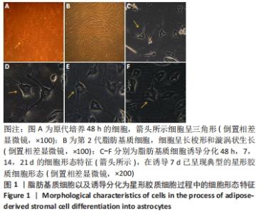
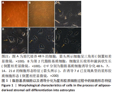
2.1 脂肪基质细胞形态及诱导分化为星形胶质细胞过程中的形态特征 脂肪基质细胞于48 h时可见已完全贴壁,可见细胞呈多种形态,呈类圆形、梭形、不规则形。培养至第7天可见大量长梭形细胞呈漩涡状排列;第10-14天细胞已80%融合,此时可进行细胞传代。应用第3代生长状态良好的脂肪基质细胞进行诱导分化,诱导至48 h时可见细胞呈圆形、多边形、不规则形,细胞周围可见光晕,部分细胞的胞体周围伸出细长突起,并有多个分支,细胞质均匀,细胞核较大,呈卵圆形或圆形,核仁清晰可见;诱导7 d时,可见分化细胞呈现典型的星形胶质细胞形态,细胞体呈圆形或椭圆形,有细长的突起和分支较多,并交织成网状,细胞质均匀,细胞核大,且多偏于细胞体一侧,呈卵圆形或圆形,核仁清晰可见;诱导14 d时,细胞形态与7 d时无明显变化;诱导21 d时,存活的细胞数量明显减少,细胞体呈三角形或不规则形,细胞的突起较诱导14 d时变短、变少,细胞核与诱导14 d时相比无明显变化,见图1。"

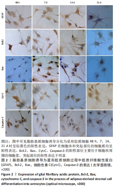
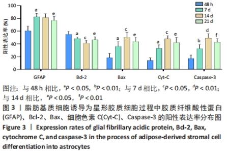
2.2 免疫细胞化学法检测脂肪基质细胞诱导分化为星形胶质细胞过程中胶质纤维酸性蛋白、Bcl-2、Bax、细胞色素C、Caspase-3的表达 胶质纤维酸性蛋白的阳性表达率随诱导时间的延长逐渐升高,在诱导至7 d时达到高峰(P < 0.05),诱导7 d与14 d差异无显著性意义(P > 0.05)。Bcl-2的细胞阳性表达率随诱导时间的延长逐渐降低,至诱导14 d时达到最低(P < 0.05)。Bax、细胞色素C的阳性表达率随诱导时间的延长逐渐升高,诱导至14 d时达到高峰(P < 0.05)。Caspase-3的阳性表达率随诱导时间的延长逐渐升高,诱导至14 d时达到高峰(P < 0.01)。胶质纤维酸性蛋白在诱导48 h及7,14,21 d均可见阳性表达细胞,阳性表达部位主要在细胞体和突起部位。Bcl-2、Bax、细胞色素C、Caspase-3在诱导48 h及7,14,21 d均可见阳性表达细胞,而且阳性表达部位均主要位于细胞核周围的细胞浆,而在突起部位表达不明显,见图2,图3。"
"

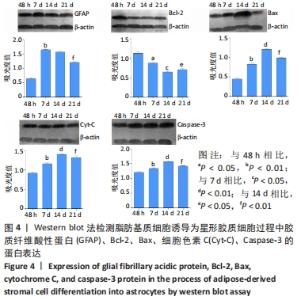
2.3 Western blot检测脂肪基质细胞诱导分化为星形胶质细胞过程中胶质纤维酸性蛋白、Bcl-2、Bax、细胞色素C、Caspase-3 的表达水平 随着诱导时间的延长,胶质纤维酸性蛋白的表达水平逐渐增加,至诱导第7天达到高峰期(P < 0.01),诱导7 d与14 d差异无显著性意义(P > 0.05)。Bcl-2的表达水平随诱导时间的延长逐渐降低,至诱导14 d时达到最低(P < 0.05)。Bax、细胞色素C、Caspase-3的表达水平随诱导时间的延长逐渐升高,在诱导至14 d时达到高峰(P < 0.01),见图4。"

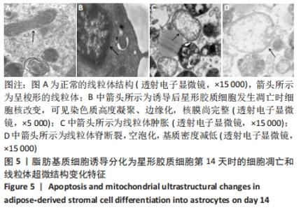
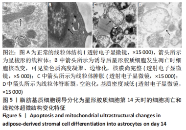
2.4 脂肪基质细胞诱导分化为星形胶质细胞第14天时的细胞和线粒体超微结构变化特征 脂肪基质细胞诱导分化为星形胶质细胞第14天,在透射电子显微镜下可以见到大量呈圆形或椭圆形的线粒体结构。正常线粒体的基质密度高,嵴排列规整。部分线粒体肿胀,体积变大;还可见部分线粒体嵴断裂,甚至空泡化,线粒体基质密度减低。另外,还可见到凋亡细胞,细胞体积缩小,细胞核呈不规则形,染色质浓缩,呈块状,见图5。"
| [1] CHI H, CHANG HY, SANG TK. Neuronal Cell Death Mechanisms in Major Neurodegenerative Diseases. Int J Mol Sci. 2018;19(10):3082. [2] DE GIOIA R, BIELLA F, CITTERIO G, et al. Neural Stem Cell Transplantation for Neurodegenerative Diseases. Int J Mol Sci. 2020; 21(9):3103. [3] YE CQ, YUAN XD, LIU H, et al. Ultrastructure of neuronal-like cells differentiated from adult adipose-derived stromal cells. Neural Regen Res. 2010;5(19):1456-1463. [4] OU Y, YUAN XD, CAI YN, et al. A novel ethanol-based method to induce differentiation of adipose-derived stromal cells into astrocytes. Neural Regen Res. 2011;6(10):738-743. [5] YUAN XD, CAI YN, OU Y, et al. Adult adipose-derived stromal cells differentiate into neurons with normal electrophysiological functions. Neural Regen Res. 2011;6(34):2681-2686. [6] 蔡亚楠,元小冬,欧亚,等.成人脂肪基质细胞体外诱导分化为神经前体细胞的数量达峰时间研究[J].中华行为医学与脑科学杂志, 2011,20(4):302-305. [7] OU Y, YUAN XD, CAI YN, et al. Ultrastructure and electrophysiology of astrocytes differentiated from adult adipose-derived stromal cells. Chin Med J (Engl). 2011;124(17):2656-2660. [8] LU YH, YUAN XD, OU Y, et al. Autophagy and apoptosis during about adult adipose-derived stromal cells differentiation into neuron-like cells in vitro. Neural Regen Res. 2012;7(16):1205-1212. [9] LIN Z, XIE F, TRIVIÑO M, et al. Ectopic Expression of a Self-Incompatibility Module Triggers Growth Arrest and Cell Death in Vegetative Cells. Plant Physiol. 2020;183(4):1765-1779. [10] WON M, LUO Y, LEE DH, et al. BAX is an essential key mediator of AP5M1-induced apoptosis in cervical carcinoma cells. Biochem Biophys Res Commun. 2019;518(2):368-373. [11] HU H, TIAN M, DING C, et al. The C/EBP Homologous Protein (CHOP) Transcription Factor Functions in Endoplasmic Reticulum Stress-Induced Apoptosis and Microbial Infection. Front Immunol. 2019;9:3083. [12] TAN BL, NORHAIZAN ME, CHAN LC. ROS-Mediated Mitochondrial Pathway is Required for Manilkara Zapota (L.) P. Royen Leaf Methanol Extract Inducing Apoptosis in the Modulation of Caspase Activation and EGFR/NF-κB Activities of HeLa Human Cervical Cancer Cells. Evid Based Complement Alternat Med. 2018;2018:6578648. [13] VEZZANI B, CARINCI M, PATERGNANI S, et al. The Dichotomous Role of Inflammation in the CNS: A Mitochondrial Point of View. Biomolecules. 2020;10(10):1437. [14] 郑凯,杨梅桂,闫朝君,等.线粒体动力学与细胞凋亡[J].中国细胞生物学学报,2019,41(8):1465-1476. [15] MARTINIS P, GRANCARA S, KANAMORI Y, et al. Involvement of the biogenic active amine agmatine in mitochondrial membrane permeabilization and release of pro-apoptotic factors. Amino Acids. 2020;52(2):161-169. [16] MACCHIONI L, PETRICCIUOLO M, DAVIDESCU M, et al. Palmitate lipotoxicity in enteric glial cells: Lipid remodeling and mitochondrial ROS are responsible for cyt c release outside mitochondria. Biochim Biophys Acta Mol Cell Biol Lipids. 2018;1863(8):895-908. [17] WANG Q, ZHANG L, YUAN X, et al. The Relationship between the Bcl-2/Bax Proteins and the Mitochondria-Mediated Apoptosis Pathway in the Differentiation of Adipose-Derived Stromal Cells into Neurons. PLoS One. 2016;11(10):e0163327. [18] ANTONSSON B. Bax and other pro-apoptotic Bcl-2 family “killer-proteins” and their victim the mitochondrion. Cell Tissue Res. 2001; 306(3):347-361. [19] 冯健愉,朱玉山,陈佺,等.Bcl-2家族蛋白的生理功能及结构基础[J].中国细胞生物学学报,2019,41(8):1477-1489. [20] RENAULT TT, DEJEAN LM, MANON S. A brewing understanding of the regulation of Bax function by Bcl-xL and Bcl-2. Mech Ageing Dev. 2017;161(Pt B):201-210. [21] MAHDAVI S, KHODARAHMI P, ROODBARI NH. Effects of cadmium on Bcl-2/ Bax expression ratio in rat cortex brain and hippocampus. Hum Exp Toxicol. 2018;37(3):321-328. [22] BERGANDI L, MUNGO E, MORONE R, et al. Hyperglycemia Promotes Chemoresistance Through the Reduction of the Mitochondrial DNA Damage, the Bax/Bcl-2 and Bax/Bcl-XL Ratio, and the Cells in Sub-G1 Phase Due to Antitumoral Drugs Induced-Cytotoxicity in Human Colon Adenocarcinoma Cells. Front Pharmacol. 2018;9:866. [23] HOPE JM, LOPEZ-CAVESTANY M, WANG W, et al. Activation of Piezo1 sensitizes cells to TRAIL-mediated apoptosis through mitochondrial outer membrane permeability. Cell Death Dis. 2019;10(11):837. [24] YANG Z, WANG WE, ZHANG Q. CIAPIN1 siRNA inhibits proliferation, migration and promotes apoptosis of VSMCs by regulating Bcl-2 and Bax. Curr Neurovasc Res. 2013;10(1):4-10. [25] ZHOU P, XIE W, LUO Y, et al. Inhibitory Effects of Ginsenoside Rb1 on Early Atherosclerosis in ApoE-/- Mice via Inhibition of Apoptosis and Enhancing Autophagy. Molecules. 2018;23(11):2912. [26] LIU LS, BAI XQ, GAO Y, et al. PCSK9 Promotes oxLDL-Induced PC12 Cell Apoptosis Through the Bcl-2/Bax-Caspase 9/3 Signaling Pathway. J Alzheimers Dis. 2017;57(3):723-734. [27] RAK M, BÉNIT P, CHRÉTIEN D, et al. Mitochondrial cytochrome c oxidase deficiency. Clin Sci (Lond). 2016;130(6):393-407. [28] CLEETER MW, COOPER JM, DARLEY-USMAR VM, et al. Reversible inhibition of cytochrome c oxidase, the terminal enzyme of the mitochondrial respiratory chain, by nitric oxide. Implications for neurodegenerative diseases. FEBS Lett. 1994;345(1):50-54. [29] TRUMPOWER BL. The protonmotive Q cycle. Energy transduction by coupling of proton translocation to electron transfer by the cytochrome bc1 complex. J Biol Chem. 1990;265(20):11409-11412. [30] MARTINEZ LYONS A, ARDISSONE A, REYES A, et al. COA7 (C1orf163/RESA1) mutations associated with mitochondrial leukoencephalopathy and cytochrome c oxidase deficiency. J Med Genet. 2016;53(12):846-849. [31] OSHIMA Y, VERHOEVEN N, CARTIER E, et al. The OMM-severed and IMM-ubiquitinated mitochondria are intermediates of mitochondrial proteotoxicity-induced autophagy in PRKN/parkin-deficient cells. Autophagy. 2021;17(11):3884-3886. [32] GU J, CHEN Y, TONG L, et al. Astaxanthin-loaded polymer-lipid hybrid nanoparticles (ATX-LPN): assessment of potential otoprotective effects. J Nanobiotechnology. 2020;18(1):53. [33] KISELEVSKY DB. Granzymes and Mitochondria. Biochemistry (Mosc). 2020;85(2):131-139. [34] LAI CI, CHU YL, HO CT, et al. Antcin K, an active triterpenoid from the fruiting bodies of basswood cultivated Antrodia cinnamomea, induces mitochondria and endoplasmic reticulum stress-mediated apoptosis in human hepatoma cells. J Tradit Complement Med. 2015;6(1):48-56. [35] HUANG H, LIU C, FU X, et al. Microcystin-LR Induced Apoptosis in Rat Sertoli Cells via the Mitochondrial Caspase-Dependent Pathway: Role of Reactive Oxygen Species. Front Physiol. 2016;7:397. [36] 王艳杰,邓雯,张鹏飞.细胞色素C与细胞凋亡研究进展[J].动物医学进展,2012,33(7):89-92. [37] BENDER CE, FITZGERALD P, TAIT SW, et al. Mitochondrial pathway of apoptosis is ancestral in metazoans. Proc Natl Acad Sci U S A. 2012; 109(13):4904-4909. [38] 王晓庚,刘林,左健,等.沉默FoxM1通过促进线粒体释放细胞色素C诱导口腔鳞癌细胞凋亡[J].中国病理生理杂志,2019,35(3): 430-435. [39] JEONG SY, SEOL DW. The role of mitochondria in apoptosis. BMB Rep. 2008;41(1):11-22. [40] 朱玉山,卢铁元,王蕊,等.Bcl-2家族蛋白调控线粒体膜通透性和细胞色素C释放的新机制[J].生命科学,2011,23(11):1076-1080. [41] SALIMI A, BAHREINI F, JAMALI Z, et al. Mesalazine Induces Oxidative Stress and Cytochrome c Release in Isolated Rat Heart Mitochondria: An Analysis of Cardiotoxic Effects. Int J Toxicol. 2020;39(3):241-247. [42] WANG L, MA G, ZHANG Y, et al. Effect of mitochondrial cytochrome c release and its redox state on the mitochondrial-dependent apoptotic cascade reaction and tenderization of yak meat during postmortem aging. Food Res Int. 2018;111:488-497. [43] LI L, WANG F, ZHANG J, et al. Typical phthalic acid esters induce apoptosis by regulating the PI3K/Akt/Bcl-2 signaling pathway in rat insulinoma cells. Ecotoxicol Environ Saf. 2021;208:111461. [44] NOMURA M, SHIMIZU S, SUGIYAMA T, et al. 14-3-3 Interacts directly with and negatively regulates pro-apoptotic Bax. J Biol Chem. 2003; 278(3):2058-2065. [45] JEZEK J, CHANG KT, JOSHI AM, et al. Mitochondrial translocation of cyclin C stimulates intrinsic apoptosis through Bax recruitment. EMBO Rep. 2019;20(9):e47425. [46] GIULIANO M, LAURICELLA M, CALVARUSO G, et al. The apoptotic effects and synergistic interaction of sodium butyrate and MG132 in human retinoblastoma Y79 cells. Cancer Res. 1999;59(21):5586-5595. [47] LI H, TONG J, BAO J, et al. Hematoporphyrin monomethyl ether combined with He-Ne laser irradiation-induced apoptosis in canine breast cancer cells through the mitochondrial pathway. J Vet Sci. 2016; 17(2):235-242. [48] GOGVADZE V, ORRENIUS S, ZHIVOTOVSKY B. Multiple pathways of cytochrome c release from mitochondria in apoptosis. Biochim Biophys Acta. 2006;1757(5-6):639-647. [49] WANG X. Molecular mechanism of cytochrome c release from mitochondria during apoptosis. Biochimica et Biophysica Acta (BBA) - Bioenergetics. 2016;1857:e7. |
| [1] | LIU Danni, SUN Guanghua, ZHOU Guijuan, LIU Hongya, ZHOU Jun, TAN Jinqu, HUANG Xiarong, PENG Ting, FENG Wei-bin, LUO Fu. Effect of electroacupuncture on apoptosis of neurons in cerebral cortex of rats with cerebral ischemia-reperfusion injury at "Shuigou" and "Baihui" points [J]. Chinese Journal of Tissue Engineering Research, 2022, 26(在线): 1-6. |
| [2] | Jin Tao, Liu Lin, Zhu Xiaoyan, Shi Yucong, Niu Jianxiong, Zhang Tongtong, Wu Shujin, Yang Qingshan. Osteoarthritis and mitochondrial abnormalities [J]. Chinese Journal of Tissue Engineering Research, 2022, 26(9): 1452-1458. |
| [3] | Li Wei, Zhu Hanmin, Wang Xin, Gao Xue, Cui Jing, Liu Yuxin, Huang Shuming. Effect of Zuogui Wan on bone morphogenetic protein 2 signaling pathway in ovariectomized osteoporosis mice [J]. Chinese Journal of Tissue Engineering Research, 2022, 26(8): 1173-1179. |
| [4] | Wang Shuo, Liu Wenying, Lü Chaofan, Li Jiacong, Geng Yi, Zhao Yungang. Cardioprotective effect of 3-nitro-N-methyl salicylamide on the isolated rat heart under cold ischemia preservation [J]. Chinese Journal of Tissue Engineering Research, 2022, 26(8): 1194-1201. |
| [5] | Yang Shenglin, Pu Xingwei, Luo Chunshan, Yang Jianwen. Neuroprotective effects of tetrandrine preconditioning in rabbits with spinal cord ischemia-reperfusion injury [J]. Chinese Journal of Tissue Engineering Research, 2022, 26(8): 1223-1227. |
| [6] | Hu Wei, Xie Xingqi, Tu Guanjun. Exosomes derived from bone marrow mesenchymal stem cells improve the integrity of the blood-spinal cord barrier after spinal cord injury [J]. Chinese Journal of Tissue Engineering Research, 2022, 26(7): 992-998. |
| [7] | Gao Yujin, Peng Shuanglin, Ma Zhichao, Lu Shi, Cao Huayue, Wang Lang, Xiao Jingang. Osteogenic ability of adipose stem cells in diabetic osteoporosis mice [J]. Chinese Journal of Tissue Engineering Research, 2022, 26(7): 999-1004. |
| [8] | Huang Chenwei, Fei Yankang, Zhu Mengmei, Li Penghao, Yu Bing. Important role of glutathione in stemness and regulation of stem cells [J]. Chinese Journal of Tissue Engineering Research, 2022, 26(7): 1119-1124. |
| [9] | Hui Xiaoshan, Bai Jing, Zhou Siyuan, Wang Jie, Zhang Jinsheng, He Qingyong, Meng Peipei. Theoretical mechanism of traditional Chinese medicine theory on stem cell induced differentiation [J]. Chinese Journal of Tissue Engineering Research, 2022, 26(7): 1125-1129. |
| [10] | An Weizheng, He Xiao, Ren Shuai, Liu Jianyu. Potential of muscle-derived stem cells in peripheral nerve regeneration [J]. Chinese Journal of Tissue Engineering Research, 2022, 26(7): 1130-1136. |
| [11] | Liang Xuezhen, Yang Xi, Li Jiacheng, Luo Di, Xu Bo, Li Gang. Bushen Huoxue capsule regulates osteogenic and adipogenic differentiation of rat bone marrow mesenchymal stem cells via Hedgehog signaling pathway [J]. Chinese Journal of Tissue Engineering Research, 2022, 26(7): 1020-1026. |
| [12] | Liu Feng, Peng Yuhuan, Luo Liangping, Wu Benqing. Plant-derived basic fibroblast growth factor maintains the growth and differentiation of human embryonic stem cells [J]. Chinese Journal of Tissue Engineering Research, 2022, 26(7): 1032-1037. |
| [13] | Wen Dandan, Li Qiang, Shen Caiqi, Ji Zhe, Jin Peisheng. Nocardia rubra cell wall skeleton for extemal use improves the viability of adipogenic mesenchymal stem cells and promotes diabetes wound repair [J]. Chinese Journal of Tissue Engineering Research, 2022, 26(7): 1038-1044. |
| [14] | Luo Xiaoling, Zhang Li, Yang Maohua, Xu Jie, Xu Xiaomei. Effect of naringenin on osteogenic differentiation of human periodontal ligament stem cells [J]. Chinese Journal of Tissue Engineering Research, 2022, 26(7): 1051-1056. |
| [15] | Xiong Tinglin, Ying Menghui, Zhang Lisha, Zhang Xiaogang, Yang Yan. Electrophysiological characteristics of cardiomyocytes differentiated from induced pluripotent stem cells [J]. Chinese Journal of Tissue Engineering Research, 2022, 26(7): 1063-1067. |
| Viewed | ||||||
|
Full text |
|
|||||
|
Abstract |
|
|||||