Chinese Journal of Tissue Engineering Research ›› 2022, Vol. 26 ›› Issue (31): 5020-5025.doi: 10.12307/2022.721
Previous Articles Next Articles
Effects of myeloid-derived growth factor on ventricular remodeling in aging mice
Sun Ying1, Xiang Guangda1, 2, Xu Xiaoli2
- 1Graduate School of Wuhan University of Science and Technology, Wuhan 430081, Hubei Province, China; 2Department of Endocrinology, Central Theater Command General Hospital of Chinese People’s Liberation Army, Wuhan 430070, Hubei Province, China
-
Received:2021-03-25Accepted:2021-05-12Online:2022-11-08Published:2022-04-25 -
Contact:Xiang Guangda, MD, Chief physician, Graduate School of Wuhan University of Science and Technology, Wuhan 430081, Hubei Province, China; Department of Endocrinology, Central Theater Command General Hospital of Chinese People’s Liberation Army, Wuhan 430070, Hubei Province, China -
About author:Sun Ying, Master, Graduate School of Wuhan University of Science and Technology, Wuhan 430081, Hubei Province, China -
Supported by:2018 National Natural Science Foundation of China, No. 81870573 (to XGD); 2015 National Natural Science Foundation of China, No. 81570730 (to XGD)
CLC Number:
Cite this article
Sun Ying, Xiang Guangda, Xu Xiaoli. Effects of myeloid-derived growth factor on ventricular remodeling in aging mice[J]. Chinese Journal of Tissue Engineering Research, 2022, 26(31): 5020-5025.
share this article
Add to citation manager EndNote|Reference Manager|ProCite|BibTeX|RefWorks
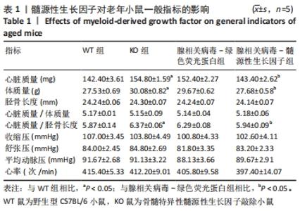
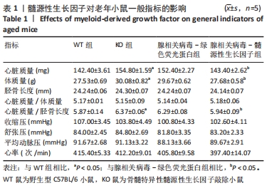
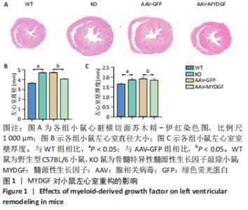
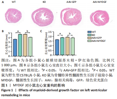
2.1 实验动物数量分析 32只小鼠随机分为4组,即WT组、KO组、AAV-GFP组、AAV-MYDGF组,每组8只。实验期间KO组小鼠死亡3只、AAV-GFP组小鼠死亡1只。最终进入结果分析每组5只,共20只。 2.2 MYDGF缺乏导致老年小鼠左心室内径大小、后壁厚度及心脏质量增加,加重心室重构 结果观察到KO组小鼠心脏大体外观,左心室内径大小及后壁厚度较WT组明显增大(P < 0.05),心脏质量、体质量、心脏质量/胫骨长度等高于WT组小鼠(P < 0.05)。然而,两组小鼠间心脏质量/体质量、胫骨长度、血压和心率相比差异无显著性意义(P > 0.05)。补充MYDGF后,与AAV-GFP组相比,AAV-MYDGF组小鼠心脏质量、体质量、心脏质量/胫骨长度、左心室内径大小及后壁厚度均减小(P < 0.05),心脏质量/体质量、胫骨长度、血压、心率无明显变化(P > 0.05),见表1及图1。"
"
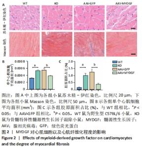
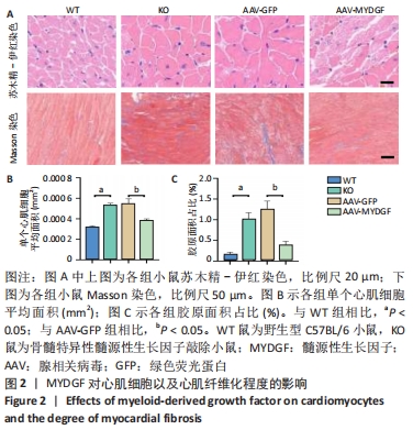
2.3 MYDGF缺乏导致心肌细胞肥大、心肌纤维化 为了进一步研究MYDGF敲除后所引发的小鼠心脏结构的改变,采用相关病理学技术对心脏组织结构进行分析。通过对心脏苏木精-伊红染色横切面观察,KO组小鼠心肌细胞横截面积明显大于WT组(P < 0.05),进一步使用Image-Pro Plus 6.0分析软件计算出单个心肌细胞平均面积(mm2)=心肌细胞总细胞/心肌细胞数量,其结果与镜下观察一致。Masson染色结果显示,心肌胶原纤维和血管显示蓝色,心肌细胞显示粉红色。与WT组小鼠相比,KO组小鼠心肌间质纤维化水平程度加重(P < 0.05)。而补充MYDGF治疗后,发现AAV-MYDGF组小鼠心肌肥大程度较AAV-GFP组小鼠减轻(P < 0.05),心肌间质纤维化水平程度也有所缓解(P < 0.05),见图2。"
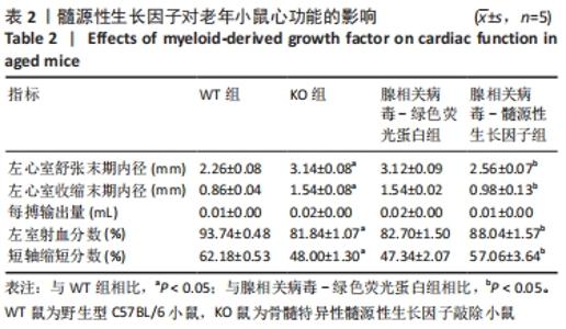
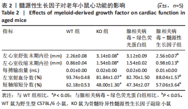
2.4 MYDGF缺失引起心功能下降 超声心动图检查结果显示,与WT组小鼠相比,KO组小鼠左心室舒张末期内径、左心室收缩末期内径均明显增大(P < 0.05),提示左心室腔扩大;且小鼠心脏收缩功能下降,主要表现为射血分数、短轴缩短分数显著降低(P < 0.05)。给予MYDGF处理后则显著缩小了AAV-MYDGF组小鼠左心室舒张末期内径及左心室收缩末期内径(P < 0.05),而射血分数、短轴缩短分数较AAV-GFP组增大(P < 0.05);每搏输出量较前无明显改变(P > 0.05)。以上结果表明恢复MYDGF后小鼠心功能可得到一定改善,见表2。"

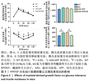
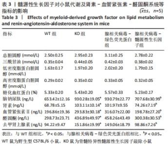
2.5 MYDGF缺乏导致小鼠胰岛素抵抗、交感神经和肾素-血管紧张素-醛固酮系统激活 见图3及表3。"
"
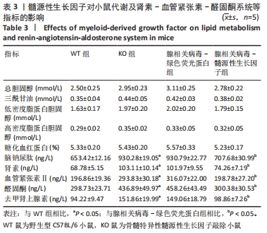

腹腔葡萄糖耐量实验结果显示,腹腔注射葡萄糖后,KO组小鼠血糖升高比例高于WT组,其差异无显著性意义(P > 0.05)。胰岛素耐量实验结果提示,腹腔注射胰岛素后,小鼠血糖开始下降。与WT组相比,KO组小鼠胰岛素敏感性明显下降,15,30,60 min血糖水平均高于WT组(P < 0.05);120 min时KO组小鼠血糖仍高于WT组,但差异无显著性意义(P > 0.05)。此外还发现KO组小鼠血脑钠尿肽、肾素、血管紧张素Ⅱ、醛固酮及去甲肾上腺素水平较WT组升高(P < 0.05)。上述结果表明MYDGF缺乏可导致小鼠发生胰岛素抵抗、肾素-血管紧张素-醛固酮系统激活以及交感神经兴奋。 补充MYDGF后,与AAV-GFP组相比,AAV-MYDGF组小鼠对胰岛素的敏感性增加,血糖下降的比例高于AAV-GFP组(P < 0.05)。腹腔葡萄糖耐量实验结果则提示,两组间差异无显著性意义(P > 0.05)。AAV-MYDGF组小鼠血脑钠尿肽、肾素、血管紧张素Ⅱ、醛固酮及去甲肾上腺素水平较AAV-GFP组下降(P < 0.05)。各组间空腹血糖、糖化血红蛋白、总胆固醇、三酰甘油、低密度脂蛋白胆固醇、高密度脂蛋白胆固醇等指标相比差异仍无显著性意义(P > 0.05)。 "

| [1] ROTH GA, FOROUZANFAR MH, MORAN AE, et al. Demographic and epidemiologic drivers of global cardiovascular mortality. New Eng J Med. 2015;372(14):1333-1341. [2] OBAS V, VASAN R. The aging heart. Clin Sci. 2018;132(13):1367-1382. [3] 李嘉欣,钱欣,赵俊捷,等.衰老对心脏结构和功能的影响与机制的研究[J].心脏杂志,2020,32(2):88-90+103. [4] STEENMAN M, LANDE G. Cardiac aging and heart disease in humans. Biophys Rev. 2017;9(2):131. [5] LOFFREDO F, STEINHAUSER M, JAY S, et al. Growth differentiation factor 11 is a circulating factor that reverses age-related cardiac hypertrophy. Cell. 2013;153(4):828-839. [6] 胡盛寿, 杨跃进, 郑哲,等.《中国心血管病报告2018》概要[J]. 中国循环杂志,2019,34(3):209-220. [7] 陶丽婵, 贾方. 运动训练对心脏衰老的保护机制[J]. 上海大学学报:自然科学版,2017,23(6):828-834. [8] NO MH, HEO JW, YOO SZ, et al. Effects of aging and exercise training on mitochondrial function and apoptosis in the rat heart. Eur J Physiol. 2020; 472(2):179-193. [9] 宋亚男,王烙佩,郑杰,等. 竹节参总皂苷通过调节TGF-β1/Smad3通路改善衰老大鼠心肌纤维化的作用研究[J]. 中国中药杂志,2018,43(22): 155-160. [10] 庞溢媛,秦雪梅,杜冠华,等. 基于衰老假说的黄芩黄酮药理作用及机制研究进展[J]. 中草药,2019,50(13):3207-3216. [11] CHANG WT, FISCH S, DANGWAL S, et al. Angiotensin II blockers improve cardiac coronary flow under hemodynamic pressure overload. Hypertens Res. 2021;44(7):803-812. [12] MA Y, YUAN J, HU J, et al. ACE inhibitor suppresses cardiac remodeling after myocardial infarction by regulating dendritic cells and AT2 receptor-mediated mechanism in mice. Biomed Pharmacother. 2019;114:108660. [13] YIN H, LI P, HU F, et al. IL-33 attenuates cardiac remodeling following myocardial infarction via inhibition of the p38 MAPK and NF-κB pathways. Mol Med Rep. 2014;9(5):1834-1838. [14] KORF-KLINGEBIEL M, REBOLL MR, KLEDE S, et al. Myeloid-derived growth factor (C19orf10) mediates cardiac repair following myocardial infarction. Nat Med. 2015;21(2):140-149. [15] WANG Y, LI Y, FENG J, et al. Mydgf promotes Cardiomyocyte proliferation and Neonatal Heart regeneration. Theranostics. 2020;10(20):9100-9112. [16] WANG L, LI Y, GUO B, et al. Myeloid-Derived Growth Factor Promotes Intestinal Glucagon-Like Peptide-1 Production in Male Mice With Type 2 Diabetes. Endocrinology. 2020;161(2):bqaa003. [17] 孟碧莹, 向光大, 丁燕,等. 髓源性生长因子改善载脂蛋白E基因敲除小鼠动脉粥样硬化作用的研究[J]. 中华糖尿病杂志,2020,12(8):624-630. [18] KOLIAKI C, LIATIS S, KOKKINOS A. Obesity and cardiovascular disease: revisiting an old relationship. Metabolism. 2019;92:98-107. [19] 王清霞, 张光明. 肥胖与心血管疾病研究进展[J]. 现代中西医结合杂志, 2020,29(20):2277-2280. [20] 杨宗璐,柯亭羽.糖尿病并发心血管疾病的研究进展[J].中国老年保健医学,2021,19(1):92-94,98. [21] 尹庆磊,谢运,沈艳,等. 烟酰胺核苷酸转氢酶基因突变对C57BL/6小鼠糖代谢稳态的影响[J]. 中华内分泌代谢杂志,2017,33(8):673-679. [22] 赵田,汤雅迪,赵一楠,等. 肥胖及限食对胰岛素分泌调节异常的影响[J]. 中国比较医学杂志,2019,29(10):3-10. [23] 张晓圆,郭成成,玉应香,等. 高脂饲料诱导肥胖胰岛素抵抗大鼠模型的建立[J]. 北京大学学报(医学版),2020,52(3):557-563. [24] 赵剑华,胡春阳,陈林庆. 大鼠左心室肥厚模型研究概况[J]. 医学信息, 2006,19(11):2055-2057. [25] 祝旭东,李菁媛,苏敬阳,等. 异丙肾上腺素诱导小鼠心肌肥厚模型的方法优化[J]. 解剖科学进展,2018,24(3):53-55. [26] SUTTON MG, SHARPE N. Left Ventricular Remodeling After Myocardial Infarction Pathophysiology and Therapy. Circulation. 2000;101(25):2981-2988. [27] 杨晓利,瞿惠燕,戎靖枫,等.心肌纤维化发病机制的研究进展[J].中西医结合心脑血管病杂志,2020,18(14):2255-2258. [28] TRIAL J, CIESLIK KA. Changes in cardiac resident fibroblast physiology and phenotype in aging. Am J Physiol Heart Circ Physiol. 2018;315(4):H745-H755. [29] 主有峰,韦建瑞. B型钠尿肽的临床意义及研究进展[J]. 国际医药卫生导报,2010,16(5):631-635. [30] 陶以嘉,李春庆,金伟东,等. 血浆B型利钠肽(BNP)在心功能不全中的诊断和治疗效果中的应用[J]. 中国微循环,2005,9(6):433-435. [31] MESQUITA T, RUI ZM, COUTO GD, et al. Mechanisms of atrial fibrillation in aged rats with heart failure with preserved ejection fraction-ScienceDirect. Heart Rhythm. 2020;17(6):1025-1033. [32] YY A, KN A, MDA B, et al. Alteration of Cardiac Performance and Serum B-Type Natriuretic Peptide Level in Healthy Aging. J Am Coll Cardiol. 2019; 74(14):1789-1800. [33] OTANI K, HIGA Y, KITANO T, et al. Prediction of cardiac events using fully automated GLS and BNP titers in patients with known or suspected heart failure. PLoS ONE. 2020;15(6):e0234294. [34] WARNER HR, HODES RJ, POCINKI K. What Does Cell Death Have To Do with Aging? J Am Geriatr Soc. 1997;45(9):1140-1146. [35] 杨丹,樊迪,杨政,等. STAT3与心血管疾病的研究进展[J]. 中华心血管病杂志,2020,48(7):616-620. [36] CUSPIDI C, RESCALDANI M, SALA C, et al. Left-ventricular hypertrophy and obesity: a systematic review and meta-analysis of echocardiographic studies. J Hypertens. 2014;32(1):16-25. [37] 曾惠娴, 孙嘉. 脂肪肾素-血管紧张素-醛固酮系统与肥胖及其相关疾病[J]. 国际内分泌代谢杂志,2013,33(5):329-331. [38] AMES MK, ATKINS CE, PITT B. Erratum for The renin‐angiotensin‐aldosterone system and its suppression. J Vet Intern Med. 2019;33(5):2551. [39] Kaye D. Sympathetic neuronal regulation of the heart in aging and heart failure. Cardiovasc Res. 2005;66(2):256-264. [40] FINCK BN. The role of the peroxisome proliferator-activated receptor alpha pathway in pathological remodeling of the diabetic heart. Curr Opin Clin Nutr Metab Care. 2004;7(4):391-396. [41] UNGER RH. Diseases of liporegulation: new perspective on obesity and related disorders. Faseb J. 2001;15(2):312-321. [42] FANG ZY, PRINS JB, MARWICK TH. Diabetic cardiomyopathy: evidence, mechanisms, and therapeutic implications. Endocr Rev. 2004;25(4):543-567. [43] 宋璐璐,萧建中.交感神经激活与2型糖尿病及心血管损害[J].国际内分泌代谢杂志,2010,30(2):73-75. [44] 段菊花,谢志泉.代谢综合征交感神经系统兴奋性增高的机制[J].临床心血管病杂志,2008,24(8):562-564. [45] YUAN Z, TSOU YH, ZHANG XQ, et al. Injectable Citrate-Based Hydrogel as an Angiogenic Biomaterial Improves Cardiac Repair after Myocardial Infarction. ACS Appl Mater Interfaces. 2019;11(42):38429-38439. |
| [1] | Chen Xianghe, Liu Bo, Yang Kang, Lu Pengcheng, Yu Huilin. Treadmill exercise improves the myocardial fibrosis of spontaneous type 2 diabetic mice: an exploration on the functional pathway [J]. Chinese Journal of Tissue Engineering Research, 2022, 26(8): 1210-1215. |
| [2] | Yin Tingting, Du Dayong, Jiang Zhixin, Liu Yang, Liu Qilin, Li Yuntian. Granulocyte colony-stimulating factors improve myocardial fibrosis in rats with myocardial infarction [J]. Chinese Journal of Tissue Engineering Research, 2022, 26(5): 730-735. |
| [3] | Deng Xiaohui, Zhang Zengzeng, Zhang Zhihua, Zhu Lingyan. Mechanism and application prospects of mesenchymal stem cell exosomes gene-modified microRNA in the treatment of diabetic foot [J]. Chinese Journal of Tissue Engineering Research, 2022, 26(31): 5076-5084. |
| [4] | Li Guangzhao, Chen Rui, Jia Honglin, Ren Liling. Research progress of gene therapy for vascularization of tissue engineering [J]. Chinese Journal of Tissue Engineering Research, 2022, 26(28): 4569-4574. |
| [5] | Gao Zhao, Zhao Yuhao, He Yixiang, Zhao Haiyan, Wang Wenji. DNA hydrogel based on drug delivery and bone tissue engineering [J]. Chinese Journal of Tissue Engineering Research, 2022, 26(27): 4379-4385. |
| [6] | Liu Ya, Liu Xia, Deng Penghui, Ji Wei, Li Jianping. Exercise effects on myocardial type I, III collagen and angiotensin II/transforming growth factor beta1/Smad2 pathway in diabetic myocardial fibrosis rats [J]. Chinese Journal of Tissue Engineering Research, 2022, 26(26): 4173-4179. |
| [7] | Ding Yan, Xiang Guangda, Meng Biying, Xu Xiaoli, Chen Yuefu. Establishment and identification of a myeloid-derived growth factor deficiency model in apolipoprotein E knockout mice [J]. Chinese Journal of Tissue Engineering Research, 2022, 26(14): 2196-2201. |
| [8] | Yuan Lifen, Cao Shuang, Sun Ting. Effect of angiotensinogen-targeted RNA interference in spontaneously hypertensive rats [J]. Chinese Journal of Tissue Engineering Research, 2021, 25(35): 5626-5631. |
| [9] | Wang Weikang, Liu Xiaodong, Zhou Changlin, Tian Jun. Regulatory role of microRNAs in the occurrence and development of osteoarthritis [J]. Chinese Journal of Tissue Engineering Research, 2021, 25(35): 5709-5715. |
| [10] | Wen Miaomiao, Fan Junbai. Relationship between biological activities of Apelin and Elabela and diseases [J]. Chinese Journal of Tissue Engineering Research, 2021, 25(35): 5735-5740. |
| [11] | Zhou Quan, Zhang Yanan, Bai Yiguang, Zhang Qiong, Nong Haibin, Liu Mingfu, Zeng Gaofeng, Zong Shaohui. Effect of 3-phosphoinositide-dependent protein kinase 1 regulating osteoclasts on bone mineral density in osteoporotic mice [J]. Chinese Journal of Tissue Engineering Research, 2021, 25(29): 4680-4684. |
| [12] | Zhang Lishu, Liu Anqi, He Xiaoning, Jin Yan, Li Bei, Jin Fang. Alpl gene affects the therapeutic effect of bone marrow mesenchymal stem cells on ulcerative colitis [J]. Chinese Journal of Tissue Engineering Research, 2021, 25(25): 3970-3975. |
| [13] | Chen Ziyang, Pu Rui, Deng Shuang, Yuan Lingyan. Regulatory effect of exosomes on exercise-mediated insulin resistance diseases [J]. Chinese Journal of Tissue Engineering Research, 2021, 25(25): 4089-4094. |
| [14] | Liu Yulin, Li Guotai. Combined effects of hyperbaric oxygen, vibration training and astaxanthin on bone mineral density, glucose metabolism and oxidative stress in diabetic osteoporosis rats [J]. Chinese Journal of Tissue Engineering Research, 2021, 25(20): 3117-3124. |
| [15] | Ma Dujun, Peng Liping, Chen Feng, Jiang Shunwan, Jiang Jinting, Gao Kun, Lin Zhanpeng. Research strategy of gene editing technology in the gene treatment of osteoarthritis [J]. Chinese Journal of Tissue Engineering Research, 2021, 25(2): 298-303. |
| Viewed | ||||||
|
Full text |
|
|||||
|
Abstract |
|
|||||