[1] BRUNEAU S, NAKAYAMA H, WODA CB, et al. DEPTOR regulates vascular endothelial cell activation and proinflammatory and angiogenic responses. Blood. 2013;122(10):1833-1842.
[2] MEI W, XIANG G, LI Y, et al. GDF11 protects against endothelial injury and reduces atherosclerotic lesion formation in apolipoprotein E-null mice. Mol Ther. 2016;24(11):1926-1938.
[3] Li Y, Xu S, Mihaylova MM, et al. AMPK phosphorylates and inhibits SREBP activity to attenuate hepatic steatosis and atherosclerosis in diet-induced insulin-resistant mice. Cell metabolism. 2011;13(4):376-388.
[4] LU J, XIANG G, LIU M, et al. Irisin protects against endothelial injury and ameliorates atherosclerosis in apolipoprotein E-Null diabetic mice. Atherosclerosis. 2015;243(2):438-448.
[5] 周政荣,丁丽娜. 冠心病合并糖尿病应用奥美沙坦酯对患者血管内皮细胞功能的影响作用[J].北方药学,2018,15(11):29-30.
[6] 马丽娜,陈北冬,赵艳阳,等. 银杏内酯B对内皮细胞的保护作用及分子机制研究[J].中国药理学通报,2013,29(2):189-193.
[7] WANG Y, LI Y, FENG J, et al. Mydgf promotes Cardiomyocyte proliferation and Neonatal Heart regeneration. Theranostics. 2020;10(20):9100-9112.
[8] WANG L, LI Y, GUO B, et al. Myeloid-derived growth factor promotes intestinal glucagon-like peptide-1 production in male mice with type 2 diabetes. Endocrinology. 2020;161(2):1-19.
[9] LI H, LI Y, XIANG L, et al. GDF11 attenuates development of type 2 diabetes via improvement of islet β-cell function and survival. Diabetes. 2017;66(7):1914-1927.
[10] XU H, LI H, WOO S, et al. Myeloid cell-specific disruption of Period1 and Period2 exacerbates diet-induced inflammation and insulin resistance. J Biol Chem. 2014;289(23):16374-16388.
[11] LIAO J, AN X, YANG X, et al. Deficiency of LMP10 Attenuates Diet-Induced Atherosclerosis by Inhibiting Macrophage Polarization and Inflammation in Apolipoprotein E Deficient Mice. Front Cell Dev Biol. 2020;8:1-12.
[12] SUI Y, PARK SH, XU J, et al. IKKβ links vascular inflammation to obesity and atherosclerosis. J Exp Med. 2014;211(5):869-886.
[13] 王俊岩,朱美林,冷雪,等. 丹参酮ⅡA对ApoE基因敲除小鼠肝脏脂质沉积的影响及机制[J].遵义医学院学报,2014,37(1):94-98.
[14] 王力,向光大,郭孛,等.髓源性生长因子通过促进GLP-1分泌改善2型糖尿病小鼠血糖水平[J].中华内分泌代谢杂志,2019,35(7): 591-598.
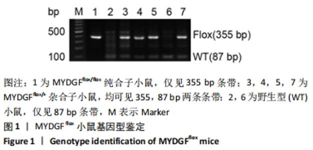
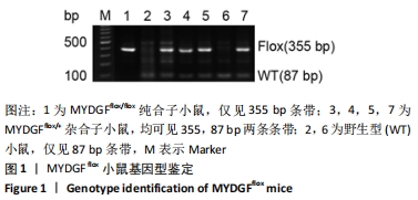
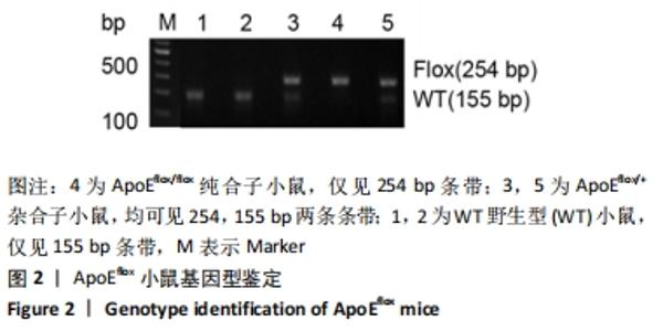
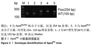
[15] 桑璐,郭英,任立明,等. 利用Cre/lox重组系统在转基因动物中筛选高效表达外源基因的“友好”基因座[J]. 高技术通讯,2004, 14(12):43-49.
[16] ZHU B, LI Y, XIANG L, et al. Alogliptin improves survival and health of mice on a high-fat diet. Aging Cell. 2019;18(2):e12883.
[17] 赖舒畅,邱鸿,王肖,等.条件性胰岛β细胞DEPTOR基因敲除小鼠构建及鉴定[J].实用医学杂志,2018,34(4):552-555.
[18] ZHU B, MEI W, JIAO T, et al. Neuregulin 4 alleviates hepatic steatosis via activating AMPK/mTOR-mediated autophagy in aged mice fed a high fat diet. Eur J Pharmacol. 2020;884:173350.
[19] 丁燕,傅友,单兰兰,等. 佛手柑内酯对双氧水诱导人脐静脉内皮细胞衰老的影响[J].中国组织工程研究,2016,20(46):6885-6892.
[20] 丁燕,赖丽芬,奈文青,等. 血管内皮细胞敲除Rheb基因杂合子小鼠模型建立及意义[J].山东医药,2017,57(36):34-36.
[21] HE M, LI Y, WANG L, et al. MYDGF attenuates podocyte injury and proteinuria by activating Akt/BAD signal pathway in mice with diabetic kidney disease. Diabetologia. 2020;63(9):1916-1931.
[22] DING Y, JIANG H, MENG B, et al. Sweroside-mediated mTORC1 hyperactivation in bone marrow mesenchymal stem cells promotes osteogenic differentiation. J Cell Biochem. 2019;120(9):16025-16036.
[23] BORTNOV V, ANNIS DS, FOGERTY FJ, et al. Myeloid-derived growth factor is a resident endoplasmic reticulum protein. J Biol Chem. 2018; 293(34):13166-13175.
[24] DWIVEDI RC, KROKHIN OV, EL-GABALAWY HS, et al. Development of an SRM method for absolute quantitation of MYDGF/C19orf10 protein. Proteomics Clin Appl. 2016;10(6):663-670.
[25] WEILER T, DU Q, KROKHIN O, et al. The identification and characterization of a novel protein, c19orf10, in the synovium. Arthritis Res Ther. 2007; 9(2):R30.
[26] KORF-KLINGEBIEL M, REBOLL MR, KLEDE S, et al. Myeloid-derived growth factor (C19orf10) mediates cardiac repair following myocardial infarction. Nat Med. 2015;21(2):140-149.
[27] Cully M. MYDGF promotes heart repair after myocardial infarction. Nat Rev Drug Discov. 2015;14(3):164-165.
[28] ZHAO L, FENG S, WANG S, et al. Production of bioactive recombinant human myeloid-derived growth factor inEscherichia coli and its mechanism on vascular endothelial cell proliferation. J Cell Biochem. 2019;24(2):1189-1199.
[29] DING Y, SHAN L, NAI W, et al. DEPTOR Deficiency-Mediated mTORc1 Hyperactivation in Vascular Endothelial Cells Promotes Angiogenesis. Cell Physiol Biochem. 2018;46:520-531.
[30] 丁燕,孟碧莹,向光大. 建立血管内皮细胞敲除DEPTOR基因小鼠模型及鉴定[J].中国组织工程研究,2019,23(23):3698-3704.
[31] LIN Z, PAN X, WU F, et al. Fibroblast growth factor 21 prevents atherosclerosis by suppression of hepatic sterol regulatory element-binding protein-2 and induction of adiponectin in mice. Circulation. 2015;131(21):1861-1871.
[32] SUNAGOZAKA H, HONDA M, YAMASHITA T, et al. Identification of a secretory protein c19orf10 activated in hepatocellular carcinoma. Int J Cancer.2011;129(7):1576-1585.
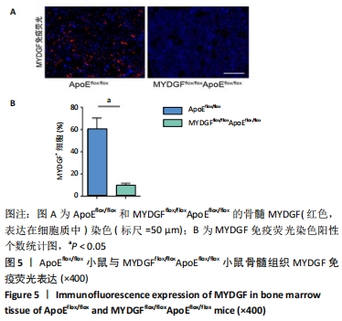
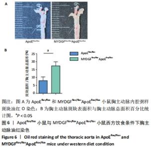
|