Chinese Journal of Tissue Engineering Research ›› 2022, Vol. 26 ›› Issue (19): 3090-3094.doi: 10.12307/2022.391
Previous Articles Next Articles
Mechanism of mesenchymal stem cell transplantation in the treatment of osteoporosis
Tang Zhihong, Duan Hao, Zhong Zongyu, Wang Weizhou, Chen Yongcheng, Li Xiaozhuang, Wang Yanghao, Liu Zhui, He Fei
- First Affiliated Hospital of Kunming Medical University, Kunming 650032, Yunnan Province, China
-
Received:2021-03-08Revised:2021-04-28Accepted:2021-08-04Online:2022-07-08Published:2021-12-29 -
Contact:He Fei, MD, Chief physician, First Affiliated Hospital of Kunming Medical University, Kunming 650032, Yunnan Province, China -
About author:Tang Zhihong, Master candidate, First Affiliated Hospital of Kunming Medical University, Kunming 650032, Yunnan Province, China -
Supported by:Major Science and Technology Project of Yunnan Province (Biomedicine), No. 2019ZF002 (to HF); Yunling Industrial Technology Leading Talent Project (to HF); Kunming Young and Middle-Aged Academic and Technical Leaders Project (to HF); Yunnan Provincial Bone and Joint Disease Clinical Medicine Center Project, No. ZX2019-03-04 (to HF)
CLC Number:
Cite this article
Tang Zhihong, Duan Hao, Zhong Zongyu, Wang Weizhou, Chen Yongcheng, Li Xiaozhuang, Wang Yanghao, Liu Zhui, He Fei. Mechanism of mesenchymal stem cell transplantation in the treatment of osteoporosis[J]. Chinese Journal of Tissue Engineering Research, 2022, 26(19): 3090-3094.
share this article
Add to citation manager EndNote|Reference Manager|ProCite|BibTeX|RefWorks
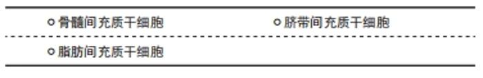
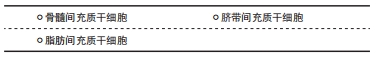
2.1 治疗骨质疏松症的种子细胞选择 目前用于骨质疏松症治疗研究的间充质干细胞来源丰富,相对于传统的治疗药物而言,不论是原发性还是继发性骨质疏松症,间充质干细胞移植都具有更好的治疗效果,但目前间充质干细胞治疗骨质疏松症的研究仍处于基础阶段,随着研究日益增多,相信在未来间充质干细胞移植能非常好地应用于临床治疗骨质疏松症。"
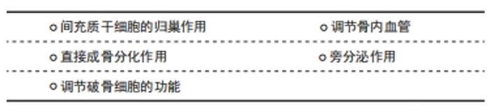
2.1.1 骨髓间充质干细胞 骨髓间充质干细胞主要存在于长骨骨髓中,在健康人群体内,骨髓中骨髓间充质干细胞占0.01%-0.001%[10],随着年龄的增加,人体内的骨髓间充质干细胞含量降低,并且成骨成脂分化失衡,使得骨髓脂肪化,这是导致发生骨质疏松症的其中一个因素[11-12]。骨髓间充质干细胞移植被认为是治疗骨质疏松症的一种新方法,同时多项研究也表明同种异体和自体来源骨髓间充质干细胞均具有治疗骨质疏松症的潜力[13]。 在原发性骨质疏松症的治疗研究中,李云龙等[14]将骨髓间充质干细胞和抗骨质疏松药物雷洛昔芬分别用于大鼠骨质疏松动物模型,结果发现骨髓间充质干细胞干预组大鼠股骨骨密度及各项力学性能明显高于雷洛昔芬组,表明了骨髓间充质干细胞对骨质疏松症的治疗作用优于传统骨质疏松药物;OCARINO等[15]将健康大鼠同种异体骨髓间充质干细胞髓内注射到骨质疏松大鼠股骨中,结果发现大鼠股骨的小梁骨百分比与对照健康大鼠的股骨几乎相似;同时,健康大鼠骨髓间充质干细胞比骨质疏松大鼠骨髓间充质干细胞的成骨作用更强。在与年龄相关的骨质疏松症的治疗研究中,骨髓间充质干细胞同样具有较好的治疗效果。ICHIOKA等[16]构建了加速衰老型小鼠(SAMP6)骨质疏松模型,通过局部注射同种异体小鼠骨髓间充质干细胞,发现成骨细胞生成和破骨细胞生成随年龄的增加而被抑制,同时脂肪生成增强,证明了同种异体骨髓间充质干细胞局部给药可增加骨小梁数量并抑制骨密度的下降;KIERNAN等[17]也利用年龄相关性Sca-1骨质疏松症小鼠模型证明了静脉给予同种异体骨髓间充质干细胞可以改善骨形成并维持骨微结构。SUI等[18]通过腹腔注射糖皮质激素构建继发性骨质疏松小鼠模型,并将骨髓间充质干细胞经尾静脉移植到小鼠体内,结果显示骨质量和强度得到了提高,这意味着骨髓间充质干细胞在继发性骨质疏松的治疗中也起到了比较明显的效果。 此外,非鼠动物模型也被用于研究骨髓间充质干细胞移植治疗骨质疏松症的研究中。将自体骨髓间充质干细胞移植到双侧卵巢切除术诱导的兔骨质疏松模型中,显示出更多的骨附着、更高的骨强度,同时也增加了骨小梁厚度和新形成的骨微结构。将骨髓间充质干细胞注射到绝经后骨质疏松山羊体内,也获得了类似的效果[19]。 2.1.2 脂肪间充质干细胞 脂肪间充质干细胞大量存在于人体脂肪组织中,在2001年由ZUK等[20]首次分离并鉴定,相对于骨髓间充质干细胞而言,除了具有良好的分化能力以外,它们还具备分离容易、在人体内含量更加丰富等优点[21]。这些特点使得脂肪间充质干细胞在临床学科中被广泛研究,也使得其成为治疗骨质疏松症的种子细胞之一。 脂肪间充质干细胞目前主要集中于激素相关性骨质疏松症的治疗研究中,张伟等[22]构建了卵巢切除大鼠骨质疏松模型,并通过尾静脉注射移植脂肪间充质干细胞,结果发现细胞治疗后大鼠股骨骨小梁明显增多,碱性磷酸酶活性增高,这提示脂肪间充质干细胞治疗骨质疏松可能是通过增加成骨来实现的。SHEU等[23]通过尾静脉将脂肪间充质干细胞和富含血小板的纤维蛋白释放物注入卵巢切除小鼠体内,结果发现骨密度、骨体积分数和骨小梁数量在胫骨近端区域显著高于卵巢切除组。URI等[24]采用局部注射的方法将脂肪间充质干细胞移植到卵巢切除大鼠的股骨干骺端,结果发现脂肪间充质干细胞可促进骨再生、增加骨密度。YE等[25]分离了卵巢切除术后的兔脂肪间充质干细胞,并将其用于治疗卵巢切除兔骨质疏松模型,结果发现脂肪间充质干细胞治疗后卵巢切除兔骨密度显著增加。 还有少数研究指出了脂肪间充质干细胞可用于年龄相关性骨质疏松的治疗,LIU等[26]分离了年轻和衰老雌性SAMP8小鼠的脂肪间充质干细胞,并将其用于治疗去卵巢快速老化SAMP8小鼠骨质疏松模型,结果发现年轻SAMP8小鼠脂肪间充质干细胞活性高于衰老小鼠脂肪间充质干细胞,接受年轻脂肪间充质干细胞治疗的小鼠骨密度明显高于接受老年脂肪间充质干细胞治疗的小鼠(平均24.3%的骨密度改善),其结果表明衰老通过减少成骨信号阻碍了脂肪间充质干细胞改善骨质疏松的能力。此外,ZHENG等[27]发现脂肪间充质干细胞移植后保留了原有的抗炎能力,并且能改变骨质疏松动物模型的骨骼微环境,同时还能影响骨重建平衡。 2.1.3 脐带间充质干细胞 脐带间充质干细胞主要是存于新生儿的脐带组织中,其活性与年龄无关,且相较骨髓间充质干细胞而言,其含量、分化能力等方面均存在明显的优势:例如极易进行取材、不会产生伦理方面的问题。脐带间充质干细胞也被相关研究学者当作是治疗骨质疏松症的种子细胞[28]。 目前针对脐带间充质干细胞治疗骨质疏松症的研究尚不多见,AN等[29]构建了卵巢切除裸鼠动物模型,通过尾静脉移植脐带间充质干细胞,结果发现小鼠胫骨骨密度显著提高,并且胫骨的Micro-CT显示骨小梁数量和厚度以及骨体积分数显著增加;HENDRIJANTINI等[30]构建了骨质疏松性下颌骨坏死大鼠模型,通过局部注射方式将脐带间充质干细胞注入下颌骨中,结果发现骨质疏松性下颌骨坏死大鼠骨再生明显增强,并且伴有Runx2等成骨相关蛋白表达与成骨细胞数量增高。虽然脐带间充质干细胞在治疗骨质疏松症方面的优势明显,但这些研究目前尚在基础阶段,尚未见临床研究报道。 2.2 间充质干细胞移植治疗骨质疏松症的机制 引起骨质疏松症的原因多且复杂,各种原因共同作用使得骨小梁结构被破坏,引起骨质量下降,导致了骨质疏松症的发生。应用间充质干细胞治疗骨质疏松症时,需要定向迁移至骨组织才能发挥其最佳治疗效果。间充质干细胞的多向分化能力及各种细胞调节作用使得间充质干细胞在治疗骨质疏松症过程中可以通过不同机制发挥共同的作用,相对于传统药物只针对增加成骨或抑制破骨的单一效果而言,具有更大优势。"
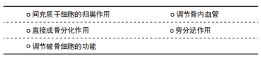

2.2.1 间充质干细胞的归巢作用 间充质干细胞的归巢是指循环干细胞或外源性干细胞定位并进入相应损伤部位的能力,其归巢能力对其治疗效果存在巨大的影响[31]。但间充质干细胞的自发性归巢效率相当低,这也是间充质干细胞治疗骨质疏松症存在的难题之一。研究间充质干细胞在各种疾病中的治疗效果时,无论是通过静脉注射全身给药还是局部原位给药,间充质干细胞均需经过归巢到达病变部位,才能有效发挥其治疗效果[32]。 由于间充质干细胞自发性归巢效率低,因此在研究中常通过增强间充质干细胞归巢效率,从而提高其治疗效果。LIU等[33]将脂肪间充质干细胞经尾静脉注入卵巢切除大鼠体内,同时予以阿司匹林灌胃处理,结果发现经阿司匹林灌胃后,脂肪间充质干细胞向大鼠股骨的归巢数量相对于其对照组明显增多,并且联合应用阿司匹林和脂肪间充质干细胞可以逆转由卵巢切除引起大鼠骨丢失。余翔团队[34]则在使用骨髓间充质干细胞治疗去势大鼠骨质疏松症模型时,给予龟板水灌胃,结果显示经过龟板干预后,骨髓间充质干细胞大量聚集于去势大鼠的椎体等处,而且椎体的骨密度、骨结构及力学性能均得到了明显改善。 在间充质干细胞的归巢过程中,SDF-1/CXCR4轴作为最重要的影响因素被广泛研究[35-36]。虽然基质细胞衍生因子1通过促进间充质干细胞归巢来治疗骨质疏松症的研究少有报道,但YANG等[37]招募了91例绝经后骨质疏松症女性患者,并将88例绝经后非骨质疏松症女性和90例健康女性作为对照,通过检测骨密度和血浆基质细胞衍生因子1水平得出结论:血浆基质细胞衍生因子1水平与腰椎、髋部等处的骨密度呈反比,这提示体内基质细胞衍生因子1水平与骨质疏松症的严重程度呈正相关。同时,PANG等[38]通过检测正常患者和骨质疏松症患者的股骨头中基质细胞衍生因子1基因的表达,发现骨质疏松症患者股骨头内基质细胞衍生因子1水平明显增高,而且基质细胞衍生因子1增多也抑制了成骨细胞的增殖。这些研究虽然没有直接证明基质细胞衍生因子1通过促进间充质干细胞归巢来治疗骨质疏松症,但却在间充质干细胞治疗其他骨疾病的研究中得到了证实。梁至洁等[39]利用基质细胞衍生因子1α修饰大鼠脂肪干细胞后发现其增殖、趋化迁移能力得到明显提高;王鑫等[40]在骨髓间充质干细胞治疗小鼠骨折模型时,通过尾静脉注射基质细胞衍生因子1α特异性阻断剂后发现骨髓间充质干细胞在骨折处聚集的数量明显低于对照组,表明了基质细胞衍生因子1α在体内也具有促进干细胞归巢的作用;XIANG等[41]利用基质细胞衍生因子1α转染的骨髓间充质干细胞治疗骨关节炎时,发现其具有更好的向受损软骨组织迁移的能力。这些研究表明基质细胞衍生因子1理论上可通过在骨质疏松症患者骨组织内高表达使间充质干细胞定向归巢于受损的骨组织,从而达到对骨质疏松症的治疗效果,但仍需进一步实验来证实。CXCR4作为基质细胞衍生因子1的特异性受体,其高表达同样可以促进间充质干细胞的归巢,加强间充质干细胞的治疗效果。SANGHANI等[42]将CXCR4转染的骨髓间充质干细胞通过静脉注射用于卵巢切除大鼠的治疗,结果发现大鼠椎体骨密度和生物力学性能明显提高。这些研究均提示运用间充质干细胞治疗骨质疏松症时,间充质干细胞的归巢在治疗过程中起到决定性作用,可以使得间充质干细胞发挥出更大的治疗效果。 2.2.2 直接成骨分化作用 大量实验证明,人体内的骨髓间充质干细胞受年龄影响,年龄越大,其活性和数量也随之降低,并且其分化方向更加偏向于成脂分化,使得骨重塑过程中成骨与破骨作用发生变化,不再处于平衡状态。WU等[43]敲除了骨髓间充质干细胞的Metll3基因,引起了成骨分化能力减低和骨髓脂肪化增加,导致骨量减少,而骨髓间充质干细胞过表达Mettl3后,预防了卵巢切除小鼠骨质疏松症的发生,这表明骨髓间充质干细胞可通过成骨分化来保持骨量,预防骨质疏松症的发生。LEE等[44]将骨髓间充质干细胞植入大鼠颅骨缺损动物模型中,在缺损处观察到新骨形成,同时碱性磷酸酶、RUNX2和骨钙素表达增高。DEBNATH等[45]在骨膜内发现了骨膜干细胞,其存在于小鼠的长骨和颅骨中,可以直接膜内成骨。DE SANTANA SANTOS等[46]将骨髓间充质干细胞和成骨细胞联合植入颅骨缺损动物模型中,结果表明联合植入较单独植入骨髓间充质干细胞而言,具有更好的成骨作用和骨修复能力。 2.2.3 调节破骨细胞的功能 破骨细胞是从单核细胞/巨噬细胞系产生的,其主要功能是骨吸收作用。破骨细胞的激活与RANKL信号通路相关,其主要调节因子是NFATc1。RANKL信号通路激活诱导破骨细胞生成基因表达,造成破骨细胞数量增加引起了骨质的破坏[47]。体内破骨和成骨作用的相对平衡保证了骨量的平衡,当破骨作用增加,骨生成和骨吸收不再保持原有的平衡状态,导致了骨质疏松症的发生[48]。ABE等[49]将间充质干细胞与破骨前体细胞体外共培养发现,间充质干细胞通过分泌骨保护素抑制破骨前体细胞向破骨细胞分化;WANG等[50]敲除小鼠WNT-1基因,结果发现小鼠骨髓间充质干细胞的WNT-1基因缺失使其成骨分化能力减弱,导致WNT-1基因缺失小鼠出现自发性骨折,同时检测到小鼠骨组织内破骨细胞数量增多,这表明骨髓间充质干细胞可以通过WNT-1的表达来抑制破骨细胞分化,从而维持骨量的平衡 2.2.4 调节骨内血管 以往人们认为骨质疏松症主要是由骨量减少、骨微结构破坏及局部炎症反应引起,但随着研究的深入,发现骨量也会受到骨内血管的影响。骨组织含有丰富的血管网,在静息时,骨内血液含量可占心输出量的10%-15%[51]。骨内的血管网络结构不仅仅起着提供营养和运输代谢废物作用,而且对于骨组织的稳定、发育和修复也必不可少[52]。 目前关于间充质干细胞直接促进骨内血管修复或生成的文献报道较少,ZHANG等[53]研究发现,骨髓间充质干细胞可以通过促进血管生成从而促进成骨,修复大鼠骨不连;JIA等[54]构建大鼠胫骨骨缺损模型,局部注射外泌体,结果发现外泌体对于内皮细胞的迁移能力起到了增强的作用,加快了血管生成,这对于骨修复来说十分有利;QI等[55]提取人诱导多能干细胞的外泌体,并将其用于治疗大鼠颅骨缺损,结果发现外泌体不但具有促成骨作用,而且还可以通过刺激血管生成来加强其成骨效应,且成骨与成血管作用与外泌体的浓度呈正相关。以上研究虽然没有直接证明间充质干细胞在体内可以通过成血管作用来修复受损的骨组织,但证明了其外泌体可以通过调节血管生成来间接促进成骨,这提示了成血管与外泌体的分泌均可能是治疗骨质疏松的机制之一,但仍需进一步研究来加以证明。 2.2.5 旁分泌作用 间充质干细胞的旁分泌作用主要是通过分泌外泌体来实现。间充质干细胞来源外泌体是由间充质干细胞分泌的大小为50-100 nm的微小囊泡,包含多种蛋白质、DNA以及微小RNA[56]。大量研究结果表明,间充质干细胞的治疗作用主要是通过其外泌体来实现的[57]。 研究表明,骨髓间充质干细胞来源外泌体对于成骨细胞的增殖具有促进作用。ZUO等[58]构建了大鼠卵巢切除骨质疏松模型,静脉注射外泌体治疗,结果发现与未治疗组相比,外泌体治疗组大鼠股骨的钙盐沉积明显增多,并且Runx2基因表达明显增高。WEILNE等[59]利用脂肪间充质干细胞来源外泌体治疗骨质疏松也得到了相似的结果。除此之外,脐血间充质干细胞来源外泌体同样对骨质疏松症具有治疗作用。HU等[60]使用脐血间充质干细胞来源外泌体治疗老年性骨质疏松小鼠时发现,治疗组小鼠骨小梁体积分数、厚度、数量和皮质骨厚度增加,同时伴有成骨相关蛋白如碱性磷酸酶、Runx2等增高,表明外泌体可以通过促进成骨达到治疗骨质疏松症的目的。诱导多能干细胞来源外泌体增强了卵巢切除大鼠骨髓间充质干细胞成骨相关蛋白的表达,同时在颅骨缺损动物模型中也取得了良好的治疗效果[55]。这些研究都表明不同间充质干细胞来源外泌体对于骨质疏松症都具有良好的治疗效果,并且与其促进成骨相关。 除外泌体的成骨作用以外,其对破骨细胞和血管也具有一定的调节作用,以此达到治疗骨质疏松症的目的。研究表明,外泌体可以通过RANKL信号通路及微小RNA如miR-218和miR-148a调节骨重塑过程中的破骨细胞功能,以达到维持骨稳态的目的[61]。WANG等[62]发现外泌体改善了卵巢切除小鼠的微血管体积,增加了H型血管的数量、骨矿物质含量和骨密度,这个过程与miR-214-3p的下调有关,LU等[63]提取了小鼠骨髓间充质干细胞来源外泌体,并将其用于治疗年龄相关性骨质疏松症,结果发现骨髓间充质干细胞来源外泌体治疗组小鼠骨组织出现较多内皮细胞,在骨表面出现大量成骨细胞,这个过程与miR-29a的上调有关。由此可见,间充质干细胞分泌的外泌体在骨质疏松症治疗中起着重要作用,多与其包含的微小RNA相关。"

| [1] 夏维波,章振林,林华,等.原发性骨质疏松症诊疗指南(2017)[J].中国骨质疏松杂志,2019,25(3):281-309. [2] 乔林,熊英,徐克惠.绝经后骨质疏松概述[J].实用妇产科杂志, 2020,36(7): 481-484. [3] ARMAS LA, RECKER RR. Pathophysiology of osteoporosis: new mechanistic insights. Endocrinol Metab Clin North Am. 2012;41(3):475-486. [4] BLACK DM, ROSEN CJ. Postmenopausal Osteoporosis. N Engl J Med. 2016;374 (21):2096-2097. [5] 卢春燕,陈德才.绝经后骨质疏松症的药物治疗[J].实用妇产科杂志,2020, 36(7):489-492. [6] BROWN C, MCKEE C, BAKSHI S, et al. Mesenchymal stem cells: Cell therapy and regeneration potential. J Tissue Eng Regen Med. 2019;13(9):1738-1755. [7] KINGERY MT, MANJUNATH AK, ANIL U, et al. Bone Marrow Mesenchymal Stem Cell Therapy and Related Bone Marrow-Derived Orthobiologic Therapeutics. Curr Rev Musculoskelet Med. 2019;12(4): 451-459. [8] GARZA JR, CAMPBELL RE, TJOUMAKARIS FP, et al. Clinical Efficacy of Intra-articular Mesenchymal Stromal Cells for the Treatment of Knee Osteoarthritis: A Double-Blinded Prospective Randomized Controlled Clinical Trial. Am J Sports Med. 2020;48(3):588-598. [9] JIANG Y, ZHANG P, ZHANG X, et al. Advances in mesenchymal stem cell transplantation for the treatment of osteoporosis. Cell Prolif. 2021; 54(1):e12956. [10] GIAMPIETRO PF, MCCARTY C, MUKESH B, et al. The role of cigarette smoking and statins in the development of postmenopausal osteoporosis: a pilot study utilizing the Marshfield Clinic Personalized Medicine Cohort. Osteoporos Int. 2010;21(3):467-477. [11] INFANTE A, RODRÍGUEZ CI. Osteogenesis and aging: lessons from mesenchymal stem cells. Stem Cell Res Ther. 2018;9(1):244. [12] DUQUE G. Bone and fat connection in aging bone. Curr Opin Rheumatol. 2008; 20(4):429-434. [13] MACÍAS I, ALCORTA-SEVILLANO N, RODRÍGUEZ CI, et al. Osteoporosis and the Potential of Cell-Based Therapeutic Strategies. Int J Mol Sci. 2020;21(5):1653. [14] 李云龙,黄炳哲,李正伟,等.骨髓间充质干细胞干预治疗骨质疏松症中的骨组织生物力学性能[J].北京生物医学工程,2021,40(1): 74-78,84. [15] OCARINO NDE M, BOELONI JN, JORGETTI V, et al. Intra-bone marrow injection of mesenchymal stem cells improves the femur bone mass of osteoporotic female rats. Connect Tissue Res. 2010;51(6):426-433. [16] ICHIOKA N, INABA M, KUSHIDA T, et al. Prevention of senile osteoporosis in SAMP6 mice by intrabone marrow injection of allogeneic bone marrow cells. Stem Cells. 2002;20(6):542-551. [17] KIERNAN J, HU S, GRYNPAS MD, et al. Systemic Mesenchymal Stromal Cell Transplantation Prevents Functional Bone Loss in a Mouse Model of Age-Related Osteoporosis. Stem Cells Transl Med. 2016;5(5):683-693. [18] SUI B, HU C, ZHANG X, et al. Allogeneic Mesenchymal Stem Cell Therapy Promotes Osteoblastogenesis and Prevents Glucocorticoid-Induced Osteoporosis. Stem Cells Transl Med. 2016;5(9):1238-1246. [19] CAO L, LIU G, GAN Y, et al. The use of autologous enriched bone marrow MSCs to enhance osteoporotic bone defect repair in long-term estrogen deficient goats. Biomaterials. 2012;33(20):5076-5084. [20] ZUK PA, ZHU M, MIZUNO H, et al. Multilineage cells from human adipose tissue: implications for cell-based therapies. Tissue Eng. 2001; 7(2):211-228. [21] KERN S, EICHLER H, STOEVE J, et al. Comparative analysis of mesenchymal stem cells from bone marrow, umbilical cord blood, or adipose tissue. Stem Cells. 2006;24(5):1294-1301. [22] 张伟,刘媛,白力,等.脂肪间充质干细胞对去卵巢骨质疏松大鼠治疗作用的实验研究[J].中华风湿病学杂志,2013,17(4):236-239,289. [23] SHEU SY, HSU YK, CHUANG MH, et al. Enhanced Bone Formation in Osteoporotic Mice by a Novel Transplant Combined with Adipose-derived Stem Cells and Platelet-rich Fibrin Releasates. Cell Transplant. 2020;29:963689720927398. [24] URI O, BEHRBALK E, FOLMAN Y. Local implantation of autologous adipose-derived stem cells increases femoral strength and bone density in osteoporotic rats: A randomized controlled animal study. J Orthop Surg (Hong Kong). 2018; 26(3):2309499018799534. [25] YE X, ZHANG P, XUE S, et al. Adipose-derived stem cells alleviate osteoporosis by enhancing osteogenesis and inhibiting adipogenesis in a rabbit model. Cytotherapy. 2014;16(12):1643-1655. [26] LIU HY, CHIOU JF, WU AT, et al. The effect of diminished osteogenic signals on reduced osteoporosis recovery in aged mice and the potential therapeutic use of adipose-derived stem cells. Biomaterials. 2012;33(26):6105-6112. [27] ZHENG CX, SUI BD, LIU N, et al. Adipose mesenchymal stem cells from osteoporotic donors preserve functionality and modulate systemic inflammatory microenvironment in osteoporotic cytotherapy. Sci Rep. 2018;8(1):5215. [28] ILIC D, MIERE C, LAZIC E. Umbilical cord blood stem cells: clinical trials in non-hematological disorders. Br Med Bull. 2012;102:43-57. [29] AN JH, PARK H, SONG JA, et al. Transplantation of human umbilical cord blood-derived mesenchymal stem cells or their conditioned medium prevents bone loss in ovariectomized nude mice. Tissue Eng Part A. 2013;19(5-6):685-696. [30] HENDRIJANTINI N, KUSUMANINGSIH T, ROSTINY R, et al. A potential therapy of human umbilical cord mesenchymal stem cells for bone regeneration on osteoporotic mandibular bone. Eur J Dent. 2018; 12(3):358-362. [31] LIESVELD JL, SHARMA N, ALJITAWI OS. Stem cell homing: From physiology to therapeutics. Stem Cells. 2020;38(10):1241-1253. [32] ZHOU Q, YANG C, YANG P. The Promotional Effect of Mesenchymal Stem Cell Homing on Bone Tissue Regeneration. Curr Stem Cell Res Ther. 2017;12(5):365-376. [33] LIU H, LI W, LIU Y, et al. Co-administration of aspirin and allogeneic adipose-derived stromal cells attenuates bone loss in ovariectomized rats through the anti-inflammatory and chemotactic abilities of aspirin. Stem Cell Res Ther. 2015;6:200. [34] 余翔,任辉,尚奇,等.龟板诱导干细胞归巢预防绝经后骨质疏松症的机制探讨[J].中华中医药杂志,2019,34(5):2071-2075. [35] MARIE PJ. Targeting integrins to promote bone formation and repair. Nat Rev Endocrinol. 2013;9(5):288-295. [36] DANIEL SK, SEO YD, PILLARISETTY VG. The CXCL12-CXCR4/CXCR7 axis as a mechanism of immune resistance in gastrointestinal malignancies. Semin Cancer Biol. 2020;65:176-188. [37] YANG XW, HUANG HX, WANG F, et al. Elevated plasma CXCL12/SDF-1 levels are linked with disease severity of postmenopausal osteoporosis. Innate Immun. 2020;26(3):222-230. [38] PANG T, GONG M, HAN J, et al. Relationship between osteoporosis and expression of Bcl-2 and CXCL12. Exp Ther Med. 2018;15(2):1293-1297. [39] 梁至洁,黄东琳,张木子,等.基质细胞衍生因子1α修饰促进大鼠脂肪干细胞迁移能力的体外实验研究[J].中国修复重建外科杂志, 2020,34(10):1305-1312. [40] 王鑫,苟文隆,徐小龙,等.骨髓间充质干细胞归巢促小鼠骨折愈合的实验研究[J].国际骨科学杂志,2015,36(3):218-223. [41] XIANG X, LIU H, WANG L, et al. Ultrasound combined with SDF-1α chemotactic microbubbles promotes stem cell homing in an osteoarthritis model. J Cell Mol Med. 2020;24(18):10816-10829. [42] SANGHANI A, OSAGIE-CLOUARD L, SAMIZADEH S, et al. CXCR4 Has the Potential to Enhance Bone Formation in Osteopenic Rats. Tissue Eng Part A. 2018;24(23-24):1775-1783. [43] WU Y, XIE L, WANG M, et al. Mettl3-mediated m6A RNA methylation regulates the fate of bone marrow mesenchymal stem cells and osteoporosis. Nat Commun. 2018;9(1):4772. [44] LEE YC, CHAN YH, HSIEH SC, et al. Comparing the Osteogenic Potentials and Bone Regeneration Capacities of Bone Marrow and Dental Pulp Mesenchymal Stem Cells in a Rabbit Calvarial Bone Defect Model. Int J Mol Sci. 2019;20(20):5015. [45] DEBNATH S, YALLOWITZ AR, MCCORMICK J, et al. Discovery of a periosteal stem cell mediating intramembranous bone formation. Nature. 2018;562(7725):133-139. [46] DE SANTANA SANTOS T, FLORES ABUNA RP, BACHA LOPES H, et al. Association of mesenchymal stem cells and osteoblasts for bone repair. Regen Med. 2015; 10(2):127-133. [47] ONO T, NAKASHIMA T. Recent advances in osteoclast biology. Histochem Cell Biol. 2018;149(4):325-341. [48] CHEN X, WANG Z, DUAN N, et al. Osteoblast-osteoclast interactions. Connect Tissue Res. 2018;59(2):99-107. [49] ABE T, SUMI K, KUNIMATSU R, et al. The effect of mesenchymal stem cells on osteoclast precursor cell differentiation. J Oral Sci. 2019;61(1): 30-35. [50] WANG F, TARKKONEN K, NIEMINEN-PIHALA V, et al. Mesenchymal Cell-Derived Juxtacrine Wnt1 Signaling Regulates Osteoblast Activity and Osteoclast Differentiation. J Bone Miner Res. 2019;34(6):1129-1142. [51] TOMLINSON RE, SILVA MJ. Skeletal Blood Flow in Bone Repair and Maintenance. Bone Res. 2013;1(4):311-322. [52] HU K, OLSEN BR. The roles of vascular endothelial growth factor in bone repair and regeneration. Bone. 2016;91:30-38. [53] ZHANG L, JIAO G, REN S, et al. Exosomes from bone marrow mesenchymal stem cells enhance fracture healing through the promotion of osteogenesis and angiogenesis in a rat model of nonunion. Stem Cell Res Ther. 2020;11(1):38. [54] JIA Y, ZHU Y, QIU S, et al. Exosomes secreted by endothelial progenitor cells accelerate bone regeneration during distraction osteogenesis by stimulating angiogenesis. Stem Cell Res Ther. 2019;10(1):12. [55] QI X, ZHANG J, YUAN H, et al. Exosomes Secreted by Human-Induced Pluripotent Stem Cell-Derived Mesenchymal Stem Cells Repair Critical-Sized Bone Defects through Enhanced Angiogenesis and Osteogenesis in Osteoporotic Rats. Int J Biol Sci. 2016;12(7):836-849. [56] PEGTEL DM, GOULD SJ. Exosomes. Annu Rev Biochem. 2019;88:487-514. [57] LI Y, JIN D, XIE W, et al. Mesenchymal Stem Cells-Derived Exosomes: A Possible Therapeutic Strategy for Osteoporosis. Curr Stem Cell Res Ther. 2018;13(5):362-368. [58] ZUO R, LIU M, WANG Y, et al. BM-MSC-derived exosomes alleviate radiation-induced bone loss by restoring the function of recipient BM-MSCs and activating Wnt/β-catenin signaling. Stem Cell Res Ther. 2019;10(1):30. [59] WEILNER S, SCHRAML E, WIESER M, et al. Secreted microvesicular miR-31 inhibits osteogenic differentiation of mesenchymal stem cells. Aging Cell. 2016;15(4):744-754. [60] HU Y, XU R, CHEN CY, et al. Extracellular vesicles from human umbilical cord blood ameliorate bone loss in senile osteoporotic mice. Metabolism. 2019;95:93-101. [61] LIU M, SUN Y, ZHANG Q. Emerging Role of Extracellular Vesicles in Bone Remodeling. J Dent Res. 2018;97(8):859-868. [62] WANG X, LI X, LI J, et al. Mechanical loading stimulates bone angiogenesis through enhancing type H vessel formation and downregulating exosomal miR-214-3p from bone marrow-derived mesenchymal stem cells. FASEB J. 2021;35(1):e21150. [63] LU GD, CHENG P, LIU T, et al. BMSC-Derived Exosomal miR-29a Promotes Angiogenesis and Osteogenesis. Front Cell Dev Biol. 2020;8: 608521. |
| [1] | Jiang Huanchang, Zhang Zhaofei, Liang De, Jiang Xiaobing, Yang Xiaodong, Liu Zhixiang. Comparison of advantages between unilateral multidirectional curved and straight vertebroplasty in the treatment of thoracolumbar osteoporotic vertebral compression fracture [J]. Chinese Journal of Tissue Engineering Research, 2022, 26(9): 1407-1411. |
| [2] | Zhu Chan, Han Xuke, Yao Chengjiao, Zhou Qian, Zhang Qiang, Chen Qiu. Human salivary components and osteoporosis/osteopenia [J]. Chinese Journal of Tissue Engineering Research, 2022, 26(9): 1439-1444. |
| [3] | Jin Tao, Liu Lin, Zhu Xiaoyan, Shi Yucong, Niu Jianxiong, Zhang Tongtong, Wu Shujin, Yang Qingshan. Osteoarthritis and mitochondrial abnormalities [J]. Chinese Journal of Tissue Engineering Research, 2022, 26(9): 1452-1458. |
| [4] | Zhang Lichuang, Xu Hao, Ma Yinghui, Xiong Mengting, Han Haihui, Bao Jiamin, Zhai Weitao, Liang Qianqian. Mechanism and prospects of regulating lymphatic reflux function in the treatment of rheumatoid arthritis [J]. Chinese Journal of Tissue Engineering Research, 2022, 26(9): 1459-1466. |
| [5] | Li Wei, Zhu Hanmin, Wang Xin, Gao Xue, Cui Jing, Liu Yuxin, Huang Shuming. Effect of Zuogui Wan on bone morphogenetic protein 2 signaling pathway in ovariectomized osteoporosis mice [J]. Chinese Journal of Tissue Engineering Research, 2022, 26(8): 1173-1179. |
| [6] | Wang Jing, Xiong Shan, Cao Jin, Feng Linwei, Wang Xin. Role and mechanism of interleukin-3 in bone metabolism [J]. Chinese Journal of Tissue Engineering Research, 2022, 26(8): 1260-1265. |
| [7] | Xiao Hao, Liu Jing, Zhou Jun. Research progress of pulsed electromagnetic field in the treatment of postmenopausal osteoporosis [J]. Chinese Journal of Tissue Engineering Research, 2022, 26(8): 1266-1271. |
| [8] | Zhu Chan, Han Xuke, Yao Chengjiao, Zhang Qiang, Liu Jing, Shao Ming. Acupuncture for Parkinson’s disease: an insight into the action mechanism in animal experiments [J]. Chinese Journal of Tissue Engineering Research, 2022, 26(8): 1272-1277. |
| [9] | Tian Chuan, Zhu Xiangqing, Yang Zailing, Yan Donghai, Li Ye, Wang Yanying, Yang Yukun, He Jie, Lü Guanke, Cai Xuemin, Shu Liping, He Zhixu, Pan Xinghua. Bone marrow mesenchymal stem cells regulate ovarian aging in macaques [J]. Chinese Journal of Tissue Engineering Research, 2022, 26(7): 985-991. |
| [10] | Gao Yujin, Peng Shuanglin, Ma Zhichao, Lu Shi, Cao Huayue, Wang Lang, Xiao Jingang. Osteogenic ability of adipose stem cells in diabetic osteoporosis mice [J]. Chinese Journal of Tissue Engineering Research, 2022, 26(7): 999-1004. |
| [11] | Hou Jingying, Guo Tianzhu, Yu Menglei, Long Huibao, Wu Hao. Hypoxia preconditioning targets and downregulates miR-195 and promotes bone marrow mesenchymal stem cell survival and pro-angiogenic potential by activating MALAT1 [J]. Chinese Journal of Tissue Engineering Research, 2022, 26(7): 1005-1011. |
| [12] | Zhou Ying, Zhang Huan, Liao Song, Hu Fanqi, Yi Jing, Liu Yubin, Jin Jide. Immunomodulatory effects of deferoxamine and interferon gamma on human dental pulp stem cells [J]. Chinese Journal of Tissue Engineering Research, 2022, 26(7): 1012-1019. |
| [13] | Liang Xuezhen, Yang Xi, Li Jiacheng, Luo Di, Xu Bo, Li Gang. Bushen Huoxue capsule regulates osteogenic and adipogenic differentiation of rat bone marrow mesenchymal stem cells via Hedgehog signaling pathway [J]. Chinese Journal of Tissue Engineering Research, 2022, 26(7): 1020-1026. |
| [14] | Wang Jifang, Bao Zhen, Qiao Yahong. miR-206 regulates EVI1 gene expression and cell biological behavior in stem cells of small cell lung cancer [J]. Chinese Journal of Tissue Engineering Research, 2022, 26(7): 1027-1031. |
| [15] | Liu Feng, Peng Yuhuan, Luo Liangping, Wu Benqing. Plant-derived basic fibroblast growth factor maintains the growth and differentiation of human embryonic stem cells [J]. Chinese Journal of Tissue Engineering Research, 2022, 26(7): 1032-1037. |
| Viewed | ||||||
|
Full text |
|
|||||
|
Abstract |
|
|||||