中国组织工程研究 ›› 2022, Vol. 26 ›› Issue (30): 4873-4878.doi: 10.12307/2022.768

• 干细胞基础实验 basic experiments of stem cells • 上一篇    下一篇

川楝子醇提物通过线粒体途径诱导白血病CEM细胞的凋亡

吴慧婷,朱大诚,徐笑明,常  娜   

  1. 江西中医药大学,江西省南昌市   330004
  • 收稿日期:2021-08-24 接受日期:2021-10-15 出版日期:2022-10-28 发布日期:2022-03-29
  • 通讯作者: 朱大诚,教授,博士生导师,江西中医药大学,江西省南昌市 330004
  • 作者简介:吴慧婷,女,1993年生,江西省南昌市人,汉族,江西中医药大学在读博士,主要从事中药抗肿瘤方面研究。
  • 基金资助:
    江西省研究生创新专项资金项目(YC2020-B146),项目负责人:吴慧婷

Ethanol extract of toosendan induces apoptosis of leukemia CEM cells through mitochondrial pathway

Wu Huiting, Zhu Dacheng, Xu Xiaoming, Chang Na   

  1. Jiangxi University of Chinese Medicine, Nanchang 330004, Jiangxi Province, China
  • Received:2021-08-24 Accepted:2021-10-15 Online:2022-10-28 Published:2022-03-29
  • Contact: Zhu Dacheng, Professor, Doctoral supervisor, Jiangxi University of Chinese Medicine, Nanchang 330004, Jiangxi Province, China
  • About author:Wu Huiting, Doctoral candidate, Jiangxi University of Chinese Medicine, Nanchang 330004, Jiangxi Province, China
  • Supported by:
    Jiangxi Postgraduate Innovation Special Fund Project, No. YC2020-B146 (to WHT)

摘要:

文题释义:
细胞凋亡的线粒体途径:细胞凋亡主要包括3种途径,其中线粒体途径为最重要的途径之一,当细胞受到如癌基因激活、缺氧等刺激时,可导致线粒体膜电位下降,有序折叠的嵴变得宽大,线粒体通透性转换孔开放诱导线粒体膜通透性增加,进而促进相关凋亡因子的释放,最终导致细胞死亡。
川楝子醇提物:川楝子为楝科植物的干燥成熟果实,经体积分数为80%乙醇提取,乙酸乙酯分离可得到主要药用成分川楝素,其具有神经递质抑制、驱虫及抗癌等作用,能有效抑制肿瘤细胞增殖及促进细胞凋亡,是一个极具潜力的候选抗癌药物。
CME细胞:是一种来源于人急性T淋巴细胞白血病患者外周血中的白血病细胞株,在体外悬浮生长,呈圆球状。

背景:苦寒中药川楝子具有小毒,对生长环境要求不高,在实体瘤中能显著抑制肿瘤细胞的增殖,但在血液肿瘤中报道较少,基于前期研究结果,并结合中医“以毒攻毒”理论探讨其主要成分川楝素抗急性淋巴细胞白血病的作用机制。
目的:探究川楝子醇提物抑制白血病CEM细胞增殖的作用机制。
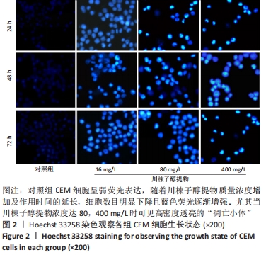
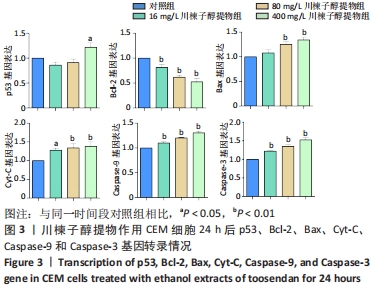
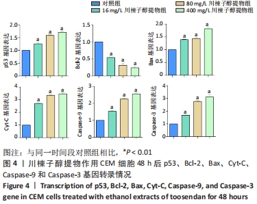
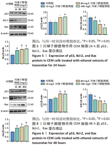
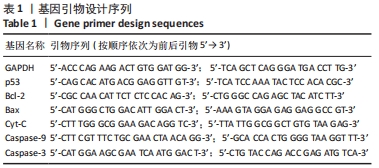
方法:人急性T淋巴细胞白血病CEM细胞培养至对数生长期,经5种浓度梯度川楝子醇提物作用24,48,72 h后,通过MTT实验求得各组细胞抑制率。考虑到受渗透压影响并保证后续实验的开展,筛选出川楝子醇提物低、中、高质量浓度(16,80,400 mg/L),作用大鼠外周血淋巴细胞24,48,72 h,采用MTT实验和吉姆萨-瑞氏染色探究其毒性作用,通过Hoechst 33258染色观察CEM细胞凋亡小体,RT-qPCR实验检测川楝子醇提物作用CEM细胞24,48 h后p53、Bcl-2、Bax、Cyt-C、Caspase-9和Caspase-3 基因转录情况,Western blot实验检测川楝子醇提物作用CEM细胞24,48 h后p53、Bcl-2、Bax蛋白的表达。
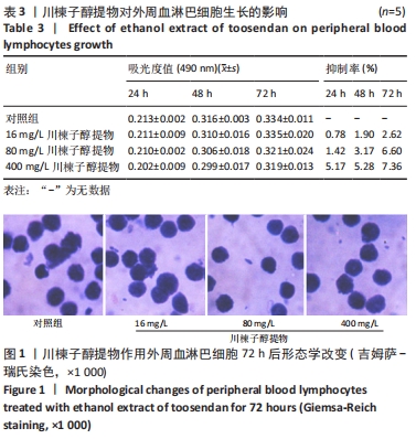
结果与结论:①16,80,400 mg/L川楝子醇提物对正常淋巴细胞无明显抑制作用;②16,80,400 mg/L川楝子醇提物作用CEM细胞经Hoechst 33258染色能看到蓝色亮光的凋亡小体;③与对照组比较,80,400 mg/L川楝子醇提物作用CEM细胞后能提高p53、Bax、Cyt-C、Caspase-9和Caspase-3基因转录水平(P < 0.05或P < 0.01),降低Bcl-2基因转录水平(P < 0.05或P < 0.01),尤其以48 h较为突出;④给药24 h后,相较于对照组,16,80,400 mg/L川楝子醇提物组Bcl-2蛋白表达下调,Bax蛋白表达升高(P < 0.05或P < 0.01),400 mg/L川楝子醇提物组p53蛋白表达升高(P < 0.05),给药48 h后,相较于对照组,80,400 mg/L川楝子醇提物组p53蛋白表达明显上调(P < 0.01),Bcl-2蛋白表达下调(P < 0.05或P < 0.01),Bax蛋白表达则显著升高(P < 0.05或P < 0.01);⑤结果表明,川楝子醇提物能显著抑制白血病CEM细胞的增殖,在一定范围内呈现浓度和时间依赖性,其机制可能是通过诱导线粒体介导的细胞凋亡途径来实现的。

https://orcid.org/0000-0002-1232-2061 (吴慧婷) 

中国组织工程研究杂志出版内容重点:干细胞;骨髓干细胞;造血干细胞;脂肪干细胞;肿瘤干细胞;胚胎干细胞;脐带脐血干细胞;干细胞诱导;干细胞分化;组织工程

关键词: 川楝子醇提物, 中药, 以毒攻毒, 白血病, CEM细胞, 线粒体途径, 凋亡, 分子机制

Abstract: BACKGROUND: Toosendan, a bitter and cold traditional Chinese medicine, has little toxicity and does not require high growth environment. It can significantly inhibit the proliferation of tumor cells in solid tumors, but it is rarely reported in blood tumors. Based on the previous research results, combined with the theory of “combating poison with poison” of traditional Chinese medicine, this paper discusses the mechanism of its main component toosendanin against acute lymphocyte leukemia.  
OBJECTIVE: To explore the mechanism of ethanol extract of toosendan inhibiting proliferation of leukemia CEM cells.
METHODS:  Human acute T lymphoblastic leukemia cell line (CEM cells) was cultured to logarithmic growth stage. After being treated with ethanol extracts of toosendan at five concentration gradients for 24, 48, and 72 hours, their inhibition rates were obtained by MTT assay. Considering the influence of osmotic pressure of cells and ensuring the development of subsequent experiments, the suitable ethanol extracts of toosendan with low, medium, and high concentrations (16, 80, and 400 mg/L) were screened out. The ethanol extract of toosendan had been exposed to peripheral blood lymphocytes of rats for 24, 48, and 72 hours. MTT assay and Giemsa-Reich staining were used to explore its toxic effects. Hoechst 33258 staining was used to observe the apoptotic bodies of CEM cells. RT-qPCR assay was used to detect the transcription of p53, Bcl-2, Bax, Cyt-C, Caspase-9, and Caspase-3 genes in CEM cells after treatment for 24 and 48 hours. Western blot assay was used to detect the expression of p53, Bcl-2, and Bax protein in CEM cells after treatment for 24 and 48 hours.  
RESULTS AND CONCLUSION: (1) Low, medium and high doses (16, 80, and 400 mg/L) of ethanol extract of toosendan had no significant inhibitory effect on normal lymphocytes. (2) After treatment with 16, 80, and 400 mg/L of ethanol extract of toosendan in CEM cells, the apoptotic bodies with blue light could be seen by Hoechst 33258 staining. (3) Compared with the control group, the 80 and 400 mg/L ethanol extract of toosendan increased the transcription of p53, Bax, Cyt-C, Caspase-9, and Caspase-3 genes (P < 0.05 or P < 0.01), and decreased the transcription level of Bcl-2 gene (P < 0.05 or P < 0.01), especially at 48 hours. (4) After administration for 24 hours, compared with the control group, the expression of Bcl-2 protein decreased, the expression of Bax protein increased in the 16, 80, and 400 mg/L extract groups (P < 0.05 or P < 0.01), and the expression of p53 increased in the 400 mg/L extract group (P < 0.05). After treatment for 48 hours, compared with the control group, the p53 protein expression of CEM cells treated with 80 and 400 mg/L ethanol extract of toosendan was significantly increased (P < 0.01), but Bcl-2 protein expression was decreased (P < 0.05 or P < 0.01), and Bax protein expression was significantly increased (P < 0.05 or P < 0.01). (5) The ethanol extract of toosendan can significantly inhibit the proliferation of leukemia CEM cells in a concentration and time dependence and its mechanism may be realized by inducing apoptosis mediated by mitochondria.

Key words: ethanol extract of toosendan, traditional Chinese medicine, combat poison with poison, leukemia, CEM cells, mitochondrial pathway, apoptosis, molecular mechanism

中图分类号: