[1] YANG G, ROTHRAUFF BB, TUAN RS. Tendon and ligament regeneration and repair: clinical relevance and developmental paradigm. Birth Defects Res C Embryo Today. 2013;99(3): 203-222.
[2] SHERMAN SL, CHALMERS PN, YANKE AB, et al. Graft tensioning during knee ligament reconstruction: principles and practice. J Am Acad Orthop Surg. 2012;20(10):633-645.
[3] LU HH, THOMOPOULOS S. Functional attachment of soft tissues to bone: development, healing, and tissue engineering. Annu Rev Biomed Eng. 2013;15:201-226.
[4] LIU YX, THOMOPOULOS S, BIRMAN V, et al. Bi-material attachment through a compliant interfacial system at the tendon-to-bone insertion site. Mech Mater. 2012;49(1): 83-92.
[5] KATZEL EB, WOLENSKI M, LOISELLE AE, et al. Impact of Smad3 loss of function on scarring and adhesion formation during tendon healing. J Orthop Res. 2011;29(5):684-693.
[6] ATESOK K, FU FH, WOLF MR, et al. Augmentation of tendon-to-bone healing. J Bone Joint Surg Am. 2014;96(6): 513-521.
[7] VAN KAMPEN C, ARNOCZKY S, PARKS P, et al. Tissue-engineered augmentation of a rotator cuff tendon using a reconstituted collagen scaffold: a histological evaluation in sheep. Muscles Ligaments Tendons J. 2013; 3(3):229-235.
[8] KAWAKAMI Y, TAKAYAMA K, MATSUMOTO T, et al. Anterior Cruciate Ligament-Derived Stem Cells Transduced With BMP2 Accelerate Graft-Bone Integration After ACL Reconstruction. Am J Sports Med. 2017;45(3):584-597.
[9] SUN L, QU L, ZHU R, et al. Effects of Mechanical Stretch on Cell Proliferation and Matrix Formation of Mesenchymal Stem Cell and Anterior Cruciate Ligament Fibroblast. Stem Cells Int. 2016;2016:9842075.
[10] LU J, CHAMBERLAIN CS, JI ML, et al. Tendon-to-Bone Healing in a Rat Extra-articular Bone Tunnel Model: A Comparison of Fresh Autologous Bone Marrow and Bone Marrow-Derived Mesenchymal Stem Cells. Am J Sports Med. 2019;47(11):2729-2736.
[11] RBIA N, BULSTRA LF, LEWALLEN EA, et al. Seeding decellularized nerve allografts with adipose-derived mesenchymal stromal cells: An in vitro analysis of the gene expression and growth factors produced. J Plast Reconstr Aesthet Surg. 2019;72(8):1316-1325.
[12] KUSUMA GD, MENICANIN D, GRONTHOS S, et al. Ectopic Bone Formation by Mesenchymal Stem Cells Derived from Human Term Placenta and the Decidua. PLoS One. 2015; 10(10):e0141246.
[13] YANG M, LIU H, WANG Y, et al. Hypoxia reduces the osteogenic differentiation of peripheral blood mesenchymal stem cells by upregulating Notch-1 expression. Connect Tissue Res. 2019;60(6):583-596.
[14] HASHIMOTO R, KATOH Y, MIYAMOTO Y, et al. Increased extracellular and intracellular Ca²⁺ lead to adipocyte accumulation in bone marrow stromal cells by different mechanisms. Biochem Biophys Res Commun. 2015;457(4): 647-652.
[15] CHEN X, LIU Y, DING W, et al. Mechanical stretch-induced osteogenic differentiation of human jaw bone marrow mesenchymal stem cells (hJBMMSCs) via inhibition of the NF-κB pathway. Cell Death Dis. 2018;9(2):207.
[16] BORNES TD, JOMHA NM, MULET-SIERRA A, et al. Porous Scaffold Seeding and Chondrogenic Differentiation of BMSC-seeded Scaffolds. Bio Protoc. 2015;5(24): e1693.
[17] QIANG Y, LIANG G, YU L. Human amniotic mesenchymal stem cells alleviate lung injury induced by ischemia and reperfusion after cardiopulmonary bypass in dogs. Lab Invest. 2016;96(5):537-546.
[18] EVANS MA, BROUGHTON BRS, DRUMMOND GR, et al. Amnion epithelial cells - a novel therapy for ischemic stroke?Neural Regen Res. 2018;13(8):1346-1349.
[19] CARLBERG AL, TUAN RS, HALL DJ. Regulation of scleraxis function by interaction with the bHLH protein E47. Mol Cell Biol Res Commun. 2000;3(2):82-86.
[20] HU JS, OLSON EN, KINGSTON RE. HEB, a helix-loop-helix protein related to E2A and ITF2 that can modulate the DNA-binding ability of myogenic regulatory factors. Mol Cell Biol. 1992;12(3):1031-1042.
[21] LIU Y, WATANABE H, NIFUJI A, et al. Overexpression of a single helix-loop-helix-type transcription factor, scleraxis, enhances aggrecan gene expression in osteoblastic osteosarcoma ROS17/2.8 cells. J Biol Chem. 1997;272(47): 29880-29885.
[22] ALBERTON P, POPOV C, PRÄGERT M, et al. Conversion of human bone marrow-derived mesenchymal stem cells into tendon progenitor cells by ectopic expression of scleraxis. Stem Cells Dev. 2012;21(6):846-858.
[23] MADRY H, KOHN D, CUCCHIARINI M. Direct FGF-2 gene transfer via recombinant adeno-associated virus vectors stimulates cell proliferation, collagen production, and the repair of experimental lesions in the human ACL. Am J Sports Med. 2013;41(1):194-202.
[24] WANG CJ, WENG LH, HSU SL, et al. pCMV-BMP-2- transfected cell-mediated gene therapy in anterior cruciate ligament reconstruction in rabbits. Arthroscopy. 2010;26(7): 968-976.
[25] CHEN B, LI B, QI YJ, et al. Enhancement of tendon-to-bone healing after anterior cruciate ligament reconstruction using bone marrow-derived mesenchymal stem cells genetically modified with bFGF/BMP2. Sci Rep. 2016;6:25940.
[26] KAWAKAMI Y, TAKAYAMA K, MATSUMOTO T, et al. Anterior Cruciate Ligament-Derived Stem Cells Transduced With BMP2 Accelerate Graft-Bone Integration After ACL Reconstruction. Am J Sports Med. 2017;45(3):584-597.
[27] PENG W, ZHANG J, ZHANG H, et al. Effects of lentiviral transfection containing bFGF gene on the biological characteristics of rabbit BMSCs. J Cell Biochem. 2018; 119(10): 8389-8397.
[28] ILHAN A, YOLCU U, GUNDOGAN FC. Amniotic membrane: new concepts for an old dressing. Wound Repair Regen. 2015;23(1):145.
[29] KARALASHVILI L, KAKABADZE A, VYSHNEVSKA G, et al. Acellular human amniotic membrane as a three-dimensional scaffold for the treatment of mucogingival defects. Georgian Med News. 2015;(244-245):84-89.
[30] ZELEN CM, SNYDER RJ, SERENA TE, et al. The use of human amnion/chorion membrane in the clinical setting for lower extremity repair: a review. Clin Podiatr Med Surg. 2015; 32(1):135-146.
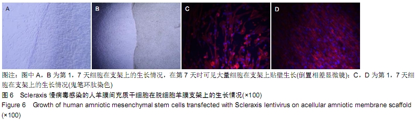
[31] 朱喜忠,刘子铭,吴术红,等.Scleraxis慢病毒基因感染人羊膜间充质干细胞向肌腱细胞的定向分化[J].中国组织工程研究, 2017, 21(33):5382-5387.
[32] LIU PF, GUO L, ZHAO DW, et al. Study of human acellular amniotic membrane loading bone marrow mesenchymal stem cells in repair of articular cartilage defect in rabbits. Genet Mol Res. 2014;13(3):7992-8001.
[33] ZHANG Y, YU J, ZHANG J, et al. Simvastatin With PRP Promotes Chondrogenesis of Bone Marrow Stem Cells In Vitro and Wounded Rat Achilles Tendon-Bone Interface Healing In Vivo. Am J Sports Med. 2019;47(3):729-739.
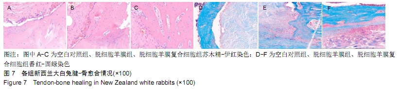
[34] ZHANG J, YUAN T, ZHENG N, et al. The combined use of kartogenin and platelet-rich plasma promotes fibrocartilage formation in the wounded rat Achilles tendon entheses. Bone Joint Res. 2017;6(4):231-244.
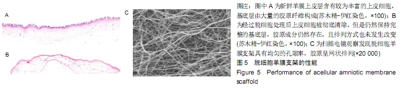
|