中国组织工程研究 ›› 2019, Vol. 23 ›› Issue (24): 3857-3861.doi: 10.3969/j.issn.2095-4344.1296

• 骨与关节图像与影像 bone and joint imaging • 上一篇    下一篇

MRI评价拇外翻畸形:拇趾跖趾关节结构及位置的改变

郭  娟1,钱丽霞1,王晓东2 
  

  1. 1山西大医院磁共振室,山西省太原市  030032;2太原市中心医院骨科,山西省太原市  030009
  • 出版日期:2019-08-28 发布日期:2019-08-28
  • 通讯作者: 钱丽霞,博士,主任医师,硕士生导师,山西大医院磁共振室,山西省太原市 030032
  • 作者简介:郭娟,女,1983年生,山西省忻州市人,汉族,2010年山西医科大学毕业,硕士,主治医师,主要从事磁共振影像诊断方面的研究。
  • 基金资助:
    国家重点研发计划课题(2016YFC0101605),项目参与人:郭娟,钱丽霞

Magnetic resonance imaging in the assessment of hallux valgus deformity: changes in the structure and position of the metatarsophalangeal joints

Guo Juan1, Qian Lixia1, Wang Xiaodong2
  

  1. 1山西大医院磁共振室,山西省太原市  030032;2太原市中心医院骨科,山西省太原市  030009
  • Online:2019-08-28 Published:2019-08-28
  • Contact: Qian Lixia, MD, Chief physician, Master’s supervisor, Magnetic Resonance Room of Shanxi Dayi Hospital, Taiyuan 030032, Shanxi Province, China
  • About author:Guan Juan, Master, Attending physician, Magnetic Resonance Room of Shanxi Dayi Hospital, Taiyuan 030032, Shanxi Province, China
  • Supported by:
    the National Key Research and Development Plan, No. 2016YFC0101605 (to GJ and QLX)

摘要:

文章快速阅读:


文题释义:
拇外翻:是一种复杂的足部畸形,英文名称为“Hallux valgus”,由1871年Carl Heuter介绍。是指拇趾向外倾斜、第一跖骨向内侧倾斜,常伴拇囊炎和第一跖骨头内侧疼痛。
拇外翻角:指第1跖骨中轴线与近节趾骨中轴线之夹角,正常<15°。
第1,2跖骨间角:指第1,2跖骨中轴线之夹角,正常<9°。
 
摘要
背景:目前诊断拇外翻主要依靠临床查体及通过足部X射线正位片,评价第1跖骨与趾骨及第1,2跖骨的位置结构改变,而对于评价并发的骨质内部、跖趾关节囊及周围软组织病变存在局限性,MRI不仅可以显示结构改变,还可以显示其他并发改变。
目的:分析MRI在拇外翻畸形诊断中的应用价值。
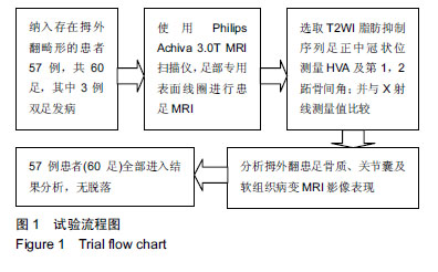
方法:纳入行足部MRI检查的患者187例,通过X射线正位片测量拇外翻角及第1,2跖骨间角,诊断为拇外翻畸形的患者共57例60足,分析足趾位置结构改变和骨质、关节囊及软组织并发改变MRI表现。所有患者对检查方案均知情同意,且得到医院伦理委员会批准。
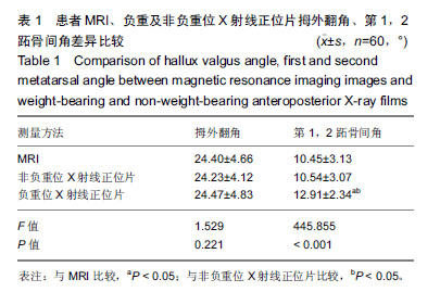
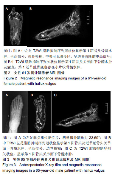
结果与结论:①拇外翻MRI与负重位、非负重位正位片拇外翻角测量值比较差异无显著性意义(P > 0.05);而MRI较负重位X射线正位片第1,2跖骨间角测量值小,差异有显著性意义(P < 0.05);②MRI不仅可以显示拇外翻畸形第1跖趾关节结构及位置的改变,其骨质、关节囊及邻近软组织也存在不同程度的病变;③40%(24/60)的患者存在第一跖骨或趾骨关节面下骨髓水肿及囊变,28%(17/60)的患者存在拇囊炎,30%(18/60)的患者存在第1跖趾关节腔积液,12足同时存在2种或3种骨及软组织并发改变;④提示MRI不仅可以显示拇外翻畸形拇趾跖趾关节结构及位置的改变,还可较好评价拇外翻骨质、关节腔、软组织病变及程度,能够更加全面的评价拇外翻严重程度。


中国组织工程研究
杂志出版内容重点:人工关节;骨植入物;脊柱骨折;内固定;数字化骨科;组织工程
ORCID: 0000-0003-4767-2042(郭娟)

关键词: 足, 拇外翻, 畸形, MRI, X射线, 拇外翻角, 第1 2跖骨间角, 关节囊

Abstract:

BACKGROUND: At present, the diagnosis of hallux valgus mainly depends on clinical examination and weight-bearing and non-weight-bearing X-ray films of the foot to evaluate the structural changes of the first metatarsal bone and phalanges and the first and second metatarsal bones. There are limitations in the changes of the internal bone, metatarsophalangeal joint capsule and surrounding soft tissues. Magnetic resonance imaging can not only show structural changes, but also display other concurrent changes.
OBJECTIVE: To study the value of magnetic resonance imaging in the diagnosis of hallux valgus.
METHODS: Totally 187 patients, who underwent foot magnetic resonance imaging, were enrolled in this study. The hallux valgus angle and the first and second metatarsal angles were measured by foot X-ray. Thus, 57 patients with hallux valgus (60 feet) were diagnosed as hallux valgus deformity. The changes of toe position and structure, bone, articular capsule and soft tissue, and magnetic resonance imaging performance were analyzed. All patients signed informed consent. The study was approved by the Hospital Ethics Committee.
RESULTS AND CONCLUSION: (1) There was no statistical difference between hallux valgus magnetic resonance imaging and weight-bearing and non-weight-bearing anteroposterior X-ray hallux valgus measurements (P > 0.05). The intermetatarsal angle measurements of magnetic resonance imaging were smaller than those of weight-bearing anteroposterior X-ray films (P < 0.05). (2) Magnetic resonance imaging showed that hallux valgus deformity not only had changes in the structure and position of the first metatarsophalangeal joint, but also had different degrees of pathological changes in bone, joint capsule and adjacent soft tissues. (3) 40% (24/60) patients had subarticular bone marrow edema and cystic degeneration of the first metatarsal or phalangeal bone; 28% (17/60) patients had pollicidal bursitis; 30% (18/60) patients had hydrocele of the first metatarsophalangeal joint; and 12 feet had two or three kinds of bone and soft tissue changes simultaneously. (4) Our results indicated that magnetic resonance imaging can reveal change of structure and position of metatarsophalangeal joint in hallux valgus deformity, evaluate the pathological changes and degree of hallux valgus bone, articular capsule and soft tissue, and comprehensively assess the severity of hallux valgus.

Key words: foot, hallux valgus, deformity, magnetic resonance imaging, X-ray, hallux valgus angle, the first and second metatarsal angle, articular capsule

中图分类号: 

','1');return false;" target="_blank">
R459.9