中国组织工程研究 ›› 2026, Vol. 30 ›› Issue (30): 7989-7999.doi: 10.12307/2026.960
• 组织构建综述 tissue construction review • 上一篇
骨软骨界面结构与功能及在骨关节炎中的病理改变
秦可一1,2,陆加霖1,3,徐嘉艺1,2,高 雨1,常 非1
- 1吉林大学第二医院,吉林省长春市 130041;2吉林大学白求恩第二临床医学院,吉林省长春市 130021;3中国科学院长春应用化学研究所,高分子科学与技术全国重点实验室,吉林省长春市 130022
-
收稿日期:2026-05-22修回日期:2026-06-08出版日期:2026-10-28发布日期:2026-09-15 -
通讯作者:常非,博士,主任医师,教授,吉林大学第二医院足踝外科,吉林省长春市 130041 -
作者简介:秦可一,女,河南省郑州市人,汉族,吉林大学白求恩第二临床医学院本科在读,主要从事软骨损伤与修复方面的研究。 -
基金资助:国家重点研发计划项目(2022YFE0107700),项目负责人:常非;国家自然科学基金项目(8257093758),项目负责人:常非
Structural and functional features of the osteochondral interface and its pathological alterations in osteoarthritis
Qin Keyi1, 2, Lu Jialin1, 3, Xu Jiayi1, 2, Gao Yu1, Chang Fei1
- 1The Second Hospital of Jilin University, Changchun 130041, Jilin Province, China; 2Norman Bethune Second Clinical Medical College, Jilin University, Changchun 130021, Jilin Province, China; 3State Key Laboratory of Polymer Science and Technology, Changchun Institute of Applied Chemistry, Chinese Academy of Sciences, Changchun 130022, Jilin Province, China
-
Received:2026-05-22Revised:2026-06-08Online:2026-10-28Published:2026-09-15 -
Contact:Chang Fei, MD, Chief physician, Professor, The Second Hospital of Jilin University, Changchun 130041, Jilin Province, China -
About author:Qin Keyi, The Second Hospital of Jilin University, Changchun 130041, Jilin Province, China; Norman Bethune Second Clinical Medical College, Jilin University, Changchun 130021, Jilin Province, China -
Supported by:The National Key Research & Development Program of China, No. 2022YFE0107700 (to CF); the National Natural Science Foundation of China, No. 8257093758 (to CF)

摘要:

文题释义:
骨软骨界面:指关节软骨与软骨下骨之间由潮线、钙化软骨及骨水泥线构成的过渡连接区域,具有应力传递与结构整合等功能。
骨关节炎:是一种常见的慢性退行性关节疾病,主要特征为关节软骨进行性破坏、丢失,伴随软骨下骨重塑、骨赘形成以及骨软骨界面结构改变。它由年龄、创伤、异常机械应力等多种因素引起,最终导致关节疼痛、僵硬、活动受限和功能障碍,是中老年人残疾的主要原因之一。
背景:关节软骨修复是临床难题,传统修复策略主要关注软骨组织,往往忽视连接软骨与软骨下骨的骨软骨界面,导致出现整合不良等并发症。
目的:综述骨软骨界面的结构特征、生物与生物力学功能及在骨关节炎中的病理改变,并探讨跨物种模型的特征差异与临床修复策略的演进,以期为组织工程修复和仿生支架设计提供理论依据。
方法:基于系统文献检索策略,收集PubMed、Web of Science数据库及中国知网中的相关研究,按照预设纳入标准进行筛选,并对骨软骨界面的结构特征、功能作用及在骨关节炎中的病理改变以及临床转化研究进行归纳与综合分析。
结果与结论:骨软骨界面通过独特的成分与结构实现软硬组织的功能整合。骨软骨界面具有典型微纳米分层结构特征,由潮线、钙化软骨和骨水泥线构成,并通过肌联蛋白和蛋白聚糖梯度、胶原纤维转变、矿化异质性和拓扑互锁结构实现营养交换、分子筛选和应力传递等关键生理功能,从而维持关节稳态。在骨关节炎进展过程中,骨软骨界面结构完整性受损,矿化模式紊乱,同时出现炎症与血管异常侵袭,这些改变破坏骨软骨界面生理功能,最终导致关节微环境失衡并加速退行性变。此外,不同物种及实验模型在界面结构与力学环境方面存在差异,研究结果在向临床外推时需谨慎解读。目前的临床修复策略多未能有效重建界面结构与功能梯度,是影响长期疗效的重要因素之一。未来应发展具备多级梯度及界面仿生特征的修复策略,以实现骨软骨单元的一体化再生。
https://orcid.org/0009-0002-8897-2666 (秦可一);https://orcid.org/0000-0002-7141-5794 (常非)
中国组织工程研究杂志出版内容重点:干细胞;骨髓干细胞;造血干细胞;脂肪干细胞;肿瘤干细胞;胚胎干细胞;脐带脐血干细胞;干细胞诱导;干细胞分化;组织工程
中图分类号:
引用本文
秦可一, 陆加霖, 徐嘉艺, 高 雨, 常 非. 骨软骨界面结构与功能及在骨关节炎中的病理改变[J]. 中国组织工程研究, 2026, 30(30): 7989-7999.
Qin Keyi, Lu Jialin, Xu Jiayi, Gao Yu, Chang Fei. Structural and functional features of the osteochondral interface and its pathological alterations in osteoarthritis[J]. Chinese Journal of Tissue Engineering Research, 2026, 30(30): 7989-7999.
2.1.1 潮线 潮线处于关节软骨非钙化区与钙化区边界,是一薄层嗜碱性波浪状结构,成因可能与原始生长板的残余相关[30]。组织学结果表明,该区域含有磷脂、腺苷三磷酸酶、碱性磷酸酶等生物活性分子,以及凝集素和高迁移率族箱染色体蛋白1等特殊成分。在胶原组成方面,潮线以胶原Ⅱ和胶原Ⅰ为主导;而在细胞组成方面,潮线本身不含细胞,仅有关节软骨细胞与潮线表面接触[31]。值得注意的是,铅元素在潮线区域的特异性富集现象已获证实,但分子机制仍有待阐明[32]。
潮线是一种具有空间梯度特征的复杂三维结构,它的胶原框架呈现3种不同模式:随机排列的致密原纤维,与非钙化区原纤维连续;平行于钙化软骨表面的扁平原纤维;垂直排列的粗大原纤维;同时,钙化物质沿原纤维轴向呈梯度递增分布,这种结构特征可能通过栓系机制维持非钙化区垂直排列胶原纤维的力学稳定,所谓栓系机制是指胶原纤维末端穿透潮线并深埋于矿化程度更高的钙化基质中,形成强力机械锚定效应,从而防止它在锚定位点发生剪切位移[17-18]。
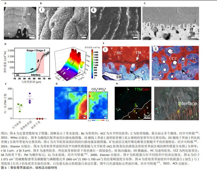
基于钙盐含量差异,WANG等[33]利用冯·科萨染色区分透明软骨与钙化软骨,在二者界面处,波浪状潮线于纵切面清晰可见。鉴于透明软骨与钙化软骨钙盐含量差异,可通过沿潮线的剪切应力实现两组织的分离。扫描电镜观察显示,分离后的透明软骨与钙化软骨界面呈现互补性结构。透明软骨界面表现为规则排列的脊-沟交替构型,其脊部宽大而沟部狭窄,表面分布着大量片状微凸起;与之对应的钙化软骨界面则显示窄脊宽沟的镜像结构,并可见颗粒状表面突起[33],这表明二者连接区域存在精确的形态学匹配。在更高分辨率下,潮线浅层与深层具有不同的组织化学特性,中央过渡层呈现双重染色特征,提示可能存在部分相互渗透现象,但因缺乏特异性标记成分而难以明确分层界限[34]。
在组织学切片中,潮线通常被称为清晰平滑的矿化前沿。然而,潮线与矿化前沿并非同一概念。同一组织可能且常存在多层潮线,但仅存在单一矿化前沿[35]。研究表明,矿化前沿具有清晰边界但形态高度不规则,这与先前文献报道的组织学切片中呈现的波浪状界面不同。同时,三维重建图像显示,矿化前沿呈现粗糙连续的平面特征,表面密布由位于矿化前沿中部软骨细胞退化遗留的近圆形空腔[36]。
生物力学分析显示,透明软骨与钙化软骨间的模量、硬度及钙含量差异显著高于钙化软骨与软骨下骨板间的差异。鉴于骨软骨界面的模量在30 μm内呈指数级增长(从4.15 MPa到1.87 GPa),潮线在其中具有关键生物力学传导作用[17]。此外,潮线是骨骼成熟的标志,会随年龄增长或骨关节炎的发生而复制[32]。因此,潮线不仅是透明软骨与钙化软骨的结合结构,还可作为指示骨关节炎的潜在信号。
2.1.2 钙化软骨 作为骨软骨界面的主体结构,钙化软骨是软骨内成骨的结果,在生长板闭合后依然存在。钙化软骨在组成和结构上展现出独特的梯度过渡特性,是实现软骨与软骨下骨之间力学传导的生物学基础。钙化软骨厚度占关节软骨的3%-8%,此比例在人和犬类等哺乳动物中相对恒定[23]。马属动物的钙化软骨比例较高,在马掌关节中,钙化软骨可占关节软骨厚度的20%[37]。此外,最近研究表明C57/BL6小鼠关节某些区域的钙化软骨厚度甚至可达全层关节软骨厚度的60%。这种厚度差异可能归因于该品系小鼠的特异性和测量方法的差异[38]。钙化软骨的深度及与邻近组织的体积比被认为由骨重塑与潮线侵袭的动态平衡所调控[35,39]。
钙化软骨的基质成分兼具软骨与骨组织特性,呈现出明显的成分梯度,无机成分以羟基磷灰石晶体为主,占比可达(65.09±2.31)%,显著高于上层透明软骨,但低于深层的软骨下骨(85.78±3.42)%,形成连续矿化梯度[40]。此外,钙磷比率从潮线到骨水泥线由1.2升至1.6,碳酸根与磷酸根比率则递减,表明矿化程度和晶体成熟度存在空间异质性[17]。有机成分中Ⅱ型胶原含量占(20.16±0.96)%,虽低于透明软骨(61.39±0.38)%,但沿深度进展梯度递增[39]。基质中还包含蛋白多糖,糖胺聚糖的浓度从软骨深层向钙化软骨方向递减,导致组织的压缩刚度降低。组织学分析表明,钙化软骨内分布肥大软骨细胞,密度约为每平方毫米50个,它们呈柱状排列于钙化基质中,起到调控基质最终矿化作用[39]。另一种在钙化软骨富集的重要功能蛋白是肌联蛋白,可介导机械应力,有助于降低界面处的应力集中[17]。
在微观和超微结构层面,钙化软骨呈现精密的构造和特殊的形态。其中胶原纤维走向具有明确的方向性,沿长骨长轴平行排列,与关节软骨表面垂直。许多纤维与关节软骨深层区的纤维连续,提示它可能促进层间黏附[41]。此外,钙化软骨矿化呈现显著的空间异质性与梯度特性。邻近软骨细胞的基质区域矿化活跃,可直接利用细胞分泌的无机离子参与矿化,而大部分基质的矿化依赖扩散的矿盐,速率较慢。矿化物质在沉积初期表现为直径仅1.0-2.0 μm的球状聚集体,随着时间推移,球状体数量增多、体积增大、密度增高,逐渐融合形成致密矿化区域,但仍可通过较低密度和纤维结构与邻近骨组织区分,这些球状体形在胶原纤维表面形成团簇结构,并与纤维保持明确界限,这种渐进矿化模式通过扩散机制实现,并与肥大软骨细胞分泌的X型胶原密切相关[36]。钙化软骨的无机成分含量虽低于软骨下骨,但矿化程度介于未钙化软骨与软骨下骨之间。该结构特征被认为可能在维持界面力学连续性及调节间质液交换方面发挥一定作用,但具体机制仍有待进一步研究[42]。
此外,钙化软骨通过高度矿化的基质分隔了软骨与骨的微环境,建立了独特的氧分压与营养梯度。三维重构显示,该区域存在源自软骨下骨的封闭式囊泡与开放式通道,但无功能性血管网络,其营养主要依靠扩散和对流运输[43]。
2.1.3 骨水泥线 骨水泥线亦称黏合线,位于钙化软骨远端9-277 μm处,是软骨下骨板与钙化软骨层的中介结构,对维持骨软骨界面的力学完整性具有重要作用[41],该区域整体矿化程度低,基质成分与钙化软骨和软骨下骨不同,可能含有硫酸化黏蛋白等特殊基质分子。骨水泥线可为周围骨基质提供相对延展的界面,它通过局部刚度差异、纤维-基质结合控制性弱化及特殊能量传递特性,在密质骨中发挥着诱导微裂纹萌生并阻滞灾难性扩展的双重效应。该机制的核心在于水泥线与周围基质间的界面结合强度相对较低,在受载过程中允许发生局部微观剥离或弱化,从而将应力集中转化为受控的界面裂纹偏转,这种现象不仅能通过界面滑动摩擦及黏弹性变形实现高效的能量耗散,亦可有效预防大面积应力贯穿导致的结构失效,从而在生物力学层面增强了骨组织的断裂韧性与抗疲劳性能。需要指出的是,该研究基于人源骨骼标本的显微结构观察及计算模型分析,属于结构与力学行为的间接证据,相关结论主要反映骨组织在特定条件下的微观力学特性,其在体内复杂生理环境中的实际作用仍有待进一步验证[44]。此外,骨水泥线还富集了肌联蛋白等功能性大分子,通过可逆形变耗散能量,协同纳米羟基磷灰石抑制裂纹扩展[17],这种成分特异性使骨水泥线在微观尺度上作为一个具有独特力学性能的界面。
基于胶原蛋白类型的差异,钙化软骨可通过番红-固绿染色与富含Ⅰ型胶原的软骨下骨区分,两者之间的黏合结构在纵切面中呈规则的梳齿状,在横切面中则表现为不规则的弧线,表面粗糙度为1.99±0.06[33]。由于钙化软骨与软骨下骨均高度矿化且结合紧密,黏合线具有很强的抵抗机械分离的能力。连续组织切片与计算机建模重建发现,钙化软骨呈现类似倒置甲床的均质钙化基质,而软骨下骨形成与之精确匹配的拓扑凹陷构型,这种类似榫卯式的精密互锁结构使钙化软骨紧密锚定于软骨下骨[30]。
骨水泥线与周围软骨下骨在扫描电镜下边界不清,但在低倍光学显微镜下可见Ⅰ型和Ⅱ型胶原蛋白的明确分界,该现象与机械联锁理论相印证[45]。胶原纤维的连续性中断证实骨软骨连接主要依赖钙化软骨与骨骼的微结构交错实现力学锚定[30]。值得注意的是,有研究者通过荧光显微镜发现马骨样本中存在跨钙化软骨于软骨下骨的胶原纤维穿透现象;二次谐波成像进一步显示胶原纤维呈非均匀分布,在50 μm尺度范围内可见局部非纤维区,提示界面处可能存在微区异质性组装机制[31,35]。
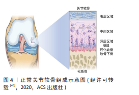
正常关节软骨组成示意图,见图4[46]。
2.2 骨软骨界面的功能特性 骨软骨界面因独特的成分和结构特征,具有选择性物质转运、屏障和高效力学传导三大功能,参与维持关节稳态与力学完整性。具体而言,骨软骨界面通过多孔通道结构与微米级间隙,发挥分子筛效应,并借助机械载荷实现营养物质与代谢废物的选择性转运。同时,骨软骨界面通过高度矿化基质与成分梯度构建独特的微环境,形成生理屏障,严格限制血管与神经侵入软骨组织。此外,界面通过微米至纳米尺度的成分与结构梯度,例如矿化梯度、模量跃升和波形互锁拓扑,实现高效能量耗散、抗剪切以及优化应力分布和应力传递性能,保障软硬组织在长期循环载荷下的稳定。这些功能相互关联、协同作用,是维持关节健康与发挥运动功能的生物学基础。
2.2.1 选择性物质转运 骨软骨界面的选择性物质转运功能主要依赖于其独特的结构和生理活动。首先,骨软骨界面在溶质转运中具有分子筛效应。钙化软骨的非矿化区域(约占体积22%)具有多孔通道结构,支持分子质量低于500 Da的物质从软骨下骨向软骨组织扩散,但严格限制蛋白质、生长因子等大分子物质转运[47-48]。例如,在自发性骨关节炎豚鼠模型中观察到70 kD示踪剂仅富集于骨髓腔,无法到达健康关节软骨,而较小分子质量的示踪剂则可以到达,提示该界面具有尺寸选择性通透能力[49]。结合该区域的致密微孔结构,可推测骨软骨界面存在选择性通透的分子筛效应。此外,小分子物质虽能扩散跨越界面,但其效率受年龄和疾病状态的影响。老年动物组织中的10 kD示踪剂分布体积显著低于年轻个体,提示随年龄增长分子转运效率降低。而健康界面中难以穿透的大分子物质,可能因疾病导致的结构改变获得通透性[49]。
此外,界面的微观结构为运输功能提供了物质基础。在生理状态下,胶原纤维密度与排列方向影响大分子扩散效率,高密度或逆向排列的胶原纤维会降低溶质迁移率。而病理状态(如软骨机械性损伤)破坏纤维结构可增强多种大分子溶质(例如异硫氰酸荧光素、葡聚糖和胰岛素)的扩散能力[19,50]。同时,潮线处存在的微小间隙,也被认为可能是营养物质从软骨下骨向无血管的关节软骨深层输送的潜在途径[47]。
2.2.2 生理屏障 骨软骨界面在生理状态下是骨与软骨间的生理屏障,对维持透明软骨内部微环境稳态至关重要。该界面将关节软骨与软骨下骨分为两个功能区:上层为无血管、无神经的软骨组织,下层高度血管化和神经化的骨组织[51]。这种分区保证了软骨的低氧微环境和营养依赖扩散机制,同时防止血管和神经从软骨下骨侵入,维持关节的机械稳定性和组织稳态。潮线作为薄膜状结构,在一定程度上限制流体流动和分子扩散,进一步强化这一屏障作用[12]。
钙化软骨较高的无机成分能够形成化学隔离效应,并通过波形起伏特征和高度矿化的基质分隔骨与软骨微环境,极大地限制了骨髓腔至关节软骨的营养扩散,从而参与建立了独特的营养梯度与氧分压。在血管化骨组织中,物质交换通过灌注实现。而软骨营养主要依赖滑液扩散及循环载荷驱动的对流运输[25]。在此过程中,蛋白多糖通过调控水合作用实现动态物质交换。具体而言,非载荷期含营养物质的滑液渗入,载荷期则加速代谢产物排出。因此,营养物质输入速率与细胞代谢速率之间的平衡形成了代谢物质梯度[52]。葡萄糖作为关键能量底物及糖胺聚糖合成前体,其浓度从滑膜侧向钙化软骨方向呈梯度递减,直接影响糖胺聚糖合成速率。氧分压分布呈现相似梯度特征,滑液氧分压为50-70 mmHg (6.5%-9.0%),关节表层为53 mmHg (7%),向深层径向区递减至
7.6 mmHg (< 1%),形成渐进性低氧微环境[53-54]。
在抵抗血管侵入方面,钙化软骨同样扮演着关键角色,其高矿化密度和低孔隙率形成物理和化学屏障,显著有效抑制血管内皮细胞的迁移和血管生成,并有效阻挡血管从软骨下骨延伸进入软骨区,从而有助于维持软骨的无血管特性[44]。这种机制依赖于细胞外基质的组成梯度,包括Ⅱ型胶原蛋白和蛋白聚糖的分布,这些成分调控血管生成因子的局部表达,在一定程度上抑制血管内皮生长因子的活性。在生理条件下,这种屏障确保营养主要通过扩散途径供给软骨,而非血管灌注,极大降低了潜在的异常矿化和组织重塑风险[55]。
此外,骨软骨界面的矿化梯度在维持软骨的无神经状态中发挥了重要作用[31]。软骨下骨含有丰富的无髓鞘神经末梢,用于感知机械载荷和营养信号,但界面处的高密度矿化基质和潮线形成连续屏障,限制了神经纤维向上生长[56],矿化梯度不仅阻止了神经生长因子的扩散,还通过局部微环境调控(如低氧张力)抑制轴突延伸[47]。这种精细的分区机制有效避免了使软骨区免受不必要的神经信号干扰,保障了正常的力学承载功能。
2.2.3 生物力学功能 骨软骨界面通过多级梯度实现软硬组织的无缝连接,优化力学功能以维持结构整合与稳定。核心生物力学功能主要体现在以下几个方面:首先,骨软骨界面能有效缓解应力集中和限制微裂纹扩展,渐进式的物理化学梯度将关节载重时产生的集中应力均匀化,有助于降低局部应力峰值,从而减少微裂纹的萌生和扩展,这种功能与骨软骨组织渐进式矿化梯度密切相关。羟基磷灰石晶体的形态从浅层直径20-50 nm的球体转变为深层直径大于100 nm的片层状结构,同时结晶度上升[36]。原位力学测试表明,羟基磷灰石晶体的结晶性指数从0.72升至0.89,界面压缩强度可达120 MPa,介于钙化软骨(80 MPa)与软骨下骨(150 MPa)之间,形成了有效的应力缓冲层,显著提升界面整体抗压强度[17]。此外,界面处密度高达150-200 μm-2的垂直定向排列的胶原纤维通过拉伸抵抗剥离力,将裂纹扩展速率抑制至0.5 μm/循环载荷,进一步阻滞了裂纹的扩展[39]。有限元分析证实,这些结构能显著降低界面处的应力集中系数;同时,纳米压痕实验显示,界面的弹性模量在仅约30 μm的空间内呈指数级增长,横跨约3个数量级[17],见图5。
其次,动态能量耗散与延缓疲劳损伤也是骨软骨界面的重要功能之一。此功能被认为通过两种机制协同实现:一方面,碳酸根取代率由界面浅层向深层递减,浅层高含量碳酸根可降低羟基磷灰石晶体脆性,并通过晶界滑移增强能量耗散,延缓疲劳损伤;另一方面,近年研究发现肌联蛋白在界面区域特异性富集,浓度可达软骨层的3倍,该蛋白的分子链通过可逆形变吸收能量,显著提升动态载荷缓冲效率,有助于维持界面弹性并辅助应力传递,尤其在周期性载荷下展现出抑制层间剥离的潜力[17]。
此外,骨软骨界面还在转化应力模式与抗剪切性能中扮演重要角色,将作用在关节软骨上的剪切应力有效地转化为压缩应力或拉伸应力,增强界面抗剪切能力,降低软骨被撕裂或压碎的风险。此功能主要得益于界面的波形互锁结构[12]。三维重建显示,钙化软骨层与软骨下骨形成类似倒置甲床与凹陷构型的精确匹配,这种齿梳状互锁结构通过增大接触面积来增强界面结合强度,使关节软骨经此结构紧密锚定于软骨下骨,有效抑制骨软骨层间滑移[33]。同时,关节运动产生的剪切应力作用于波形斜面时,被拆分为垂直界面的压缩分力和平行界面的拉伸分力,显著降低界面剪切应力峰值。此外,界面区域垂直排列的胶原纤维通过自身拉伸抵抗剥离力,衰减运动冲击能量,进一步降低关节软骨被撕裂或压碎的风险[10]。
最后,骨软骨界面还具有高效力学传导与负荷传递作用。作为关节软骨与软骨下骨之间的力学桥梁,它能将关节软骨承受的压力垂直传递至软骨下骨,极大程度避免了应力屏蔽或异常应力峰值出现,这一功能是梯度结构协同作用的结果。弹性模量的指数级跃迁平衡了软骨与骨两种材料界面的应力突变,确保了应力分布的均匀性。同时,羟基磷灰石晶体的纳米级异质性排列起到应力缓冲作用。界面深层通过低碳酸根取代率提升羟基磷灰石晶体的结晶度以优化力学传导效率。有限元模拟证实,这种结构能显著提升载荷传递效率,降低软骨下骨应力峰值[12,17]。
骨软骨界面的生物力学功能并非由单一元素决定,而是由成分梯度(如羟基磷灰石结晶度、碳酸根取代率、肌联蛋白)和结构梯度(如矿化梯度、模量梯度、拓扑互锁)在微米与纳米尺度上产生的协同效应,这种多级梯度设计共同实现应力的优化分布、能量的高效耗散、高效的抗剪切能力、负荷的有效传导以及界面的稳固整合,从而保障了关节的长期生物力学稳定。
2.3 骨软骨界面的病理改变 骨关节炎的病理演进是一个涉及力学失稳、结构破坏和生物学应答失衡的复杂过程。在疾病早期,异常机械负荷导致微裂纹和骨赘形成,骨软骨界面结构破坏。随着疾病进展,受累骨软骨单元出现病理性钙化、钙化软骨增厚、矿物梯度分布异常等改变。同时,大量炎细胞浸润,释放促炎因子并通过骨细胞-细胞外囊泡轴等方式穿透骨软骨界面运输至软骨区,最终导致软骨细胞代谢紊乱、基质降解加速与修复失败。此外,受累骨软骨单元还可出现异常血管生成和血管分布紊乱等病理改变。见表1。
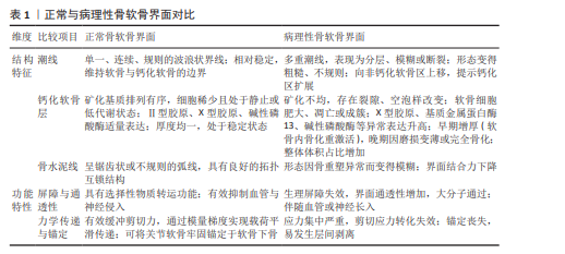
2.3.1 界面结构破坏 骨关节炎可导致潮线重复、界面增厚等结构异常,并重新激活软骨及软骨下骨发育。研究发现,骨关节炎关节的钙化软骨层出现类似骨单位样结构,而软骨下骨板中则存在富含Ⅱ型胶原的软骨样区域,这些结构成分紊乱提示骨软骨界面的病理变化可能是骨关节炎进程中连接软骨退变与软骨下骨重塑的核心环节[57]。骨软骨界面结构破坏时,蛋白多糖流失、胶原纤维网络连续性中断以及波浪状几何结构丧失,导致界面完整性破坏与应力传导功能失效。此类改变引发关节载荷分布异常,加速软骨退变进程。研究显示,当透明软骨损伤侵及钙化软骨但未突破黏合线时,常伴有不同程度的潮线缺失[58]。此外,成人骨折多发生于潮线区域,而在钙化软骨尚未完全分化的青少年群体中,骨折易沿黏合线及软骨下骨的不规则表面发生。这一现象表明,钙化软
骨对抑制界面裂纹扩展具有关键作用[31]。钙化软骨损伤后软骨下骨暴露,为微裂纹形成与扩展提供病理基础。
值得注意的是,微裂纹可能作为破骨细胞-软骨细胞通讯通道,软骨下骨中破骨细胞对软骨细胞的调控加剧了软骨完整性丧失与缺损进展[1]。抗酒石酸酸性磷酸酶染色显示,破骨细胞吸收区域从软骨下骨延伸至钙化软骨,表明破骨细胞存在趋化性迁移,这可能加速骨软骨界面结构破坏。同时,破骨细胞在骨吸收过程中释放多种因子如转化生长因子β1、磷酸钙复合物等,这些分子可跨骨软骨界面扩至上层软骨,促进软骨细胞肥大化并加速软骨降解[59]。微裂纹亦被证实有助于缺损软骨物质传输,基于水介质模型的水力传导率研究进一步支持该结论[58]。
骨赘形成是软骨缺损进展中的另一重要病理事件。肥大软骨细胞介导的软骨内成骨是骨赘形成的关键环节。骨赘形成过程中,肥大软骨细胞首先参与软骨形成并积累蛋白聚糖等基质成分,表层成纤维样细胞增殖分化为软骨细胞,随后中央区肥大软骨细胞进一步分化为骨组织并通过软骨内成骨形成骨髓腔[60]。成熟骨
赘与原有软骨下骨融合,界面正常结构破坏,纤维表层持续存在并被软骨覆盖,从而扩大关节范围。然而,由于缺乏正常应力刺激,新生骨赘的力学性能显著低于正常关节组织[61]。
2.3.2 病理性钙化 在骨关节炎进程中,病理性钙化同样是标志性病理特征之一。研究证实,骨关节炎软骨中多种矿化相关标志物表达异常,例如X型胶原、基质金属蛋白酶、核心结合因子α1、成骨细胞特异性转录因子及低氧诱导因子2α[31,62]。其中,X型胶原作为肥大软骨细胞的经典标志物,通常在骨骺板肥大区特异表达。同时,在病理环境中,X型胶原蛋白相关基因所表达的mRNA和蛋白质水平显著上调。此外,基质金属蛋白酶作为一个包含20余种锌依赖性内肽酶的家族,在软骨缺损进程中发挥关键作用,尤其是基质金属蛋白酶13已被确认为是肥大软骨细胞的分子标志物。部分研究通过局部应用基质金属蛋白酶13抑制剂,增强骨关节炎修复效果并减少不良反应,然而它的临床应用价值尚需进一步讨论验证[63]。此外,低氧可通过降低碱性磷酸酶相关基因表达水平抑制软骨细胞肥大分化,提示软骨细胞肥大与碱性磷酸酶含量升高密切相关[64-65]。
WANG等[3]针对骨关节炎患者关节软骨的研究表明,软骨缺损进展通过关节表面的自上而下钙化和骨软骨界面自下而上钙化两种途径共同实现。早期阶段,自上而下钙化始于关节表面球形矿物颗粒的形成,随疾病进展进一步形成纤维状结构,并向软骨深层转化为致密堆积的钙化物质。软骨缺损早期阶段样本中可观察到厚100-200 μm的钙化层自下而上侵入原始钙化软骨,形成骨软骨界面处的夹层结构,见图6。随时间推移,原钙化层与上层逐渐融合,导致钙化软骨增厚和结构破坏。组织学染色与密度依赖性彩色扫描电镜显示,位于部分钙化骨陷窝内的软骨细胞定位于软骨缺损早期与进展期样本的矿化前沿,提示这些软骨细胞可能通过肥大化或凋亡机制参与钙化过程。
组织学染色与密度依赖性彩色扫描电镜揭示了正常软骨与软骨缺损病理状态的微观结构特征。正常钙化软骨的矿化物呈梯度分布,近软骨侧为球形矿物分布区,深层为高矿化区。能量色散X射线扫描分析显示,缺损骨中球形矿物分布区厚度较正常样本显著增加,支持钙化过程由深层向浅层扩展的观点。此外,所有样本中球形矿物分布区与高矿化区的矿物组成存在差异,且钙磷比值呈逐渐升高趋势。软骨缺损早期钙化软骨呈过度矿化,含有刚度较高的羟基磷灰石,而在进展期矿化程度降低,含有较软的羟基磷灰石,这种差异可能与早期基质小泡成核作用及进展期碳酸盐核心形成有关。相关研究发现,与正常软骨和软骨缺损早期组样本相比,进展期样本中肥大软骨细胞高表达碳酸酐酶Ⅱ和碳酸酐酶IX,这种异常表达可能导致碳酸钙的异常沉积[3,66]。另有研究发现,机械过载可通过诱发骨软骨界面来源的纳米晶体碎裂与深层软骨基质异位沉积启动骨关节炎进程。晶体沉积导致基质刚度升高,重塑肥大软骨细胞所处的力学微环境,并伴随纤连蛋白和玻连蛋白上调及黏着斑相关机械转导激活并促进肥大软骨细胞释放钙化基质囊泡,进一步驱动基质自下而上的矿化,从而加速骨关节炎退变[67]。这些发现提示骨软骨界面这一矿化前沿结构破坏可引起细胞表型转变与病理性钙化扩增进而诱导骨关节炎持续进展。
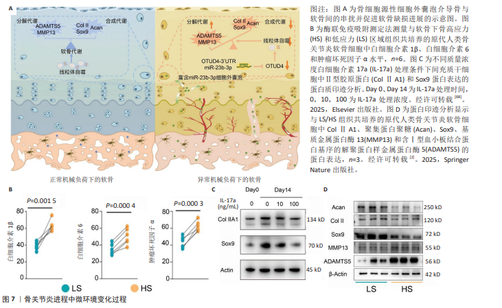
2.3.3 炎症与基质降解 骨关节炎早期阶段,异常机械负荷首先作用于软骨下骨,诱导骨重塑和微结构损伤,软骨细胞的形态与排列仍可维持相对完整。软骨下骨中的骨细胞作为主要力学感受细胞,通过形态改变和表型转化响应机械应力,并启动炎症因子的合成与释放。研究表明,高应力区域(如膝关节内侧平台)骨细胞中,多种白细胞介素如白细胞介素1β和

白细胞介素6,以及肿瘤坏死因子α等表达水平显著上调[2],见图7。研究表明,在骨关节炎早期微环境中,间充质干细胞在启动软骨修复程序时分泌高水平的白细胞介素6,与转化生长因子β1协同驱动CD4+辅助性T细胞向辅助性T细胞17分化,并通过产生大量白细胞介素破坏骨软骨界面的稳态修复。在软骨下骨层面,白细胞介素17通过增强破骨细胞活性,加剧骨吸收与异常骨重塑,破坏骨软骨界面完整性,最终导致软骨下骨支撑功能衰退。在软骨层面,白细胞介素17抑制间充质干细胞的成软骨分化能力,下调关键转录因子Sox9和Ⅱ型胶原蛋白等基质成分的表达,并减少蛋白聚糖的合成与沉积,阻碍软骨再生。同时,白细胞介素17刺激软骨细胞高表达基质金属蛋白酶3和基质金属蛋白酶13,加速软骨基质降解,进一步削弱界面结构的完整性[68]。同时,病变软骨中促炎因子水平升高和滑膜炎症会共同抑制细胞外基质合成并降低软骨细胞合成代谢活性[69]。其中,白细胞介素1β作为骨关节炎软骨退变的关键递质,可显著促进软骨基质降解,最终导致基质修复与降解间的稳态失衡,加速关节退行性变[70-71]。骨关节炎晚期常因细胞凋亡、自噬及慢性低度炎症反应导致浅层区域软骨细胞密度降低,软骨细胞失去线性排列特征并形成簇状聚集,深层区域软骨细胞密度反而增高,该现象可能与软骨祖细胞的激活与募集有关[31]。
此外,在骨关节炎早期,细胞外囊泡生物合成通路高度激活,成为炎症因子向软骨传输的起点。软骨下骨骨细胞在力学刺激下分泌的细胞外囊泡主要通过骨软骨界面的微裂纹和血管通道,迁移至软骨深层区域。同时,细胞外囊泡携带多种生物活性分子,其中miRNA是调控软骨细胞代谢的核心因子。测序分析显示高应力区域骨细胞的细胞外囊泡中miR-23b-3p表达显著升高,并通过抑制靶基因OTUD4破坏线粒体自噬,进而引发基质金属蛋白酶13、含Ⅰ型血小板结合蛋白基序的解聚蛋白样金属蛋白酶5等促分解代谢因子表达上调,加速软骨基质降解,同时Ⅱ型胶原蛋白、Sox9、聚集蛋白聚糖等软骨标志物表达下调,抑制基质修复。此外,功能实验证实,抑制miR-23b-3p可显著缓解软骨退变,恢复代谢平衡[2]。
2.3.4 异常血管生成 在健康关节中,毛细血管分支仅延伸至骨软骨界面,极少穿越潮线侵入透明软骨区域。然而,骨关节炎患者的软骨下骨板与骨赘中存在细微神经血管结构,随疾病进展,这些结构可向上延伸穿过潮线。有研究发现,在潮线邻近通道中可见纤维血管组织分布于软骨与纤维组织交界以及关节面裂隙中[31]。其中,α-肌动蛋白阳性细胞的发现提示软骨下骨存在微血管通道。此外,软骨下间隙内同样发现阳性表达的血管,证明这些血管通道可能穿越潮线。对构成血管的细胞进行检测发现存在CD34阳性内皮细胞、增殖细胞核抗原阳性细胞核以及DAPI染色的非增殖细胞核。软骨下骨血管通道中的细胞中还存在多种生长因子如血管内皮生长因子、血小板衍生生长因子及神经生长因子[43]。
软骨抗血管生成能力减弱是目前骨软骨界面中血管生成的公认机制。免疫组织化学分析表明,在健康软骨中,多种蛋白酶抑制剂与血管内皮生长因子的表达呈现梯度分布,主要富集于软骨表层,在中间层表达减弱,深层几乎不表达。然而,在软骨缺损模型中,上述因子表达普遍上调,且表达强度与软骨退变程度呈显著正相关。在退变早期,蛋白酶抑制剂上调可在一定程度上拮抗血管内皮生长因子的促血管生成作用。但在深层软骨区域,由于缺乏足够的蛋白酶抑制剂表达,最终可能导致血管生成并由钙化软骨区向非钙化软骨区侵入,破坏界面完整性[72-73]。
2.4 跨物种与解剖部位的界面特征差异及外推局限 评估骨软骨界面的生物学特征需充分考虑不同物种在解剖尺度、组织结构及力学环境方面的差异。与人类相比,小型动物(如小鼠、大鼠及兔)骨软骨界面整体较薄,钙化软骨层不发达,潮线结构相对简单,且矿化梯度范围较窄,这类结构特征与其较低的关节负荷水平相一致,但也导致界面在力学缓冲及应力分散能力方面与人类差异较大[12,17]。此外,小型动物软骨层较薄、细胞密度较高,营养供给与代谢方式更依赖扩散过程,界面在物质转运中的调控作用相对有限[22]。因此,基于小型动物模型所得关于界面屏障功能及物质交换机制的结论,在外推至人类时需保持审慎。同时,小型动物模型在模拟骨软骨界面病理改变方面亦存在局限。已有研究表明,小鼠及大鼠模型软骨退变进程较快,且缺乏人类骨关节炎典型的软骨下骨重塑及血管侵入过程,界面结构的长期重塑特征难以充分体现[41]。此外,潮线重复形成及推进过程与人类慢性疾病进展并不完全一致。因此,小型动物更适用于机制探索及早期干预研究,而对于界面稳定性、血管屏障功能及慢性退变过程的评估,代表性仍有限[11]。
相较而言,大型动物(如羊、猪及犬)的骨软骨界面结构与人类更为接近,在软骨、钙化软骨层厚度及潮线结构上均表现出更高的一致性[43]。研究显示,大型动物关节在承受较高生理载荷的条件下,界面区域形成更加明显的矿化梯度及复杂的三维结构,以实现软骨与骨之间的力学过渡与能量耗散[74]。此外,大型动物模型中可观察到与人类相似的软骨下骨重塑、界面微裂隙形成及血管侵入等病理特征,因此在评价骨软骨界面相关修复策略方面具有更高的临床相关性[23]。然而,在大型动物模型中,仍需注意与人类之间的差异。例如,不同物种在关节负重模式、步态特征及运动范围方面存在差异,这些因素均可能影响界面区域的应力分布及长期适应性重塑[11,75]。此外,大多数动物研究基于急性缺损模型,而人类骨关节炎则为长期渐进性疾病,其界面改变涉及复杂的时间依赖过程[11]。因此,尽管大型动物模型在结构与功能层面更接近人类,研究结果在临床转化时仍需结合疾病类型及时间尺度加以谨慎解读。
综上,不同物种及不同模型在骨软骨界面结构与功能方面均存在系统性差异,这些差异不仅限制了实验研究结果向临床应用的直接外推,也对组织工程支架的个性化设计提出了更高要求。未来研究在评价新型仿生材料的修复效能时,应建立在充分模拟特定物种及特定部位力学环境的基础上,并谨慎解读跨物种数据在临床转化中的适用边界。
2.5 骨软骨界面的修复转化 临床现有的骨软骨修复策略普遍侧重于恢复关节面的表面解剖形态,但在重建骨软骨界面生理结构及功能完整性方面仍面临严峻挑战。微骨折术通过穿透软骨下骨板募集内源性间充质干细胞,虽能促进缺损填充,但诱导生成的组织多为缺乏天然矿化梯度的纤维软骨。这种替代组织与宿主骨床之间难以形成稳固的钙化软骨过渡层,导致界面处的生物力学传导紊乱,临床远期观察常伴随软骨下骨硬化及修复组织的继发性剥离[13,76]。与之类似,自体软骨细胞移植虽然在实现软骨层透明化修复方面具有潜力,但在临床应用中仍存在移植物脱落风险,核心原因在于未能有效重建界面层的拓扑互锁结构,导致新生软骨与深层骨组织间的生物整合强度不足,难以抵御关节活动中的剪切应力载荷[11,13]。
骨软骨自体或异体移植虽然保留了天然的骨软骨界面,但在实践中常受限于供受体部位界面厚度的不匹配以及边缘整合不良等问题。这些技术局限引发了一系列界面相关的临床并发症,包括软骨下骨囊肿形成、移植物边缘坏死以及因力学失配导致的异位骨化[11-12]。上述临床局限性进一步提示,忽视界面重建的单相修复策略难以实现骨软骨单元的长效稳态,因此,深入探讨界面在病理状态下的演变机制,并开发具备多级梯度与生理屏障功能的仿生修复材料,已成为突破现有临床瓶颈、阻断骨关节炎进展的关键科研方向[77]。
目前,组织工程修复技术已成为软骨再生的极具潜力的治疗方案之一,其在骨软骨一体化修复中的设计理念已由单一组织替代逐步转向界面导向的分层与梯度重建。根据结构特征,常见支架可分为单相、双相、三相及连续梯度支架,在模拟骨软骨界面方面各具优势与局限[24]。
单相支架通常由单一生物材料制成,具有均一的成分、孔隙结构和力学性能,设计初衷是提供简单的三维结构以支持细胞迁移、增殖及分化为软骨或骨细胞,具有制备简便及力学性能可控等优点。但天然骨软骨单元呈连续梯度结构,生物化学环境和力学性能存在数量级的差异。传统的单相均质支架无法模拟这种梯度变化,使它与宿主组织之间存在严重的力学失配,易导致修复组织在界面处发生应力集中及整合不良。此外,由于未能提供软骨和骨的差异微环境,在促进特异性细胞分化与基质沉积方面存在局限,单一的生化环境无法同时为软骨形成和骨形成提供最优的诱导信号,常导致修复组织为机械性能较差的纤维软骨或发生异位钙化[78]。
双相支架在软骨和骨层间应用不同类型材料或不同结构设计,此类支架有助于形成两个具有特殊生化信号、结构和力学特性的区域。支架通常采用低模量聚合物或水凝胶模拟软骨层,而选用高刚度矿物复合材料构建软骨下骨层[79]。研究已证实这类支架可分别诱导软骨样组织与骨样组织形成,在一定程度上实现了组织分区修复[80]。但由于界面处通常为突变式过渡,缺乏类似天然界面的连续矿化梯度,仍难以有效缓解界面剪切应力,易出现层间剥离和应力集中,长期稳定性有限[81]。
在双相支架的基础上,三相支架通过在软骨层与骨层之间增加中间过渡层,以模拟天然软骨的钙化软骨层的功能。该过渡层通常通过调控矿化程度、材料组成或孔隙结构实现,例如引入含钙无机成分或调节孔径与连通性,从而在不同层之间建立相对连续的力学与生物学过渡,并在一定程度上减小界面剪切应力、改善力学传递[82]。已有研究表明,三相支架能够在一定程度上诱导软骨样组织与骨样组织的分区形成,同时改善界面区域的组织整合[83]。然而,由于多数三相支架仍采用离散分层构建方式,层间界面可能成为力学薄弱区,结构连续性及长期稳定性仍存在一定局限[84]。
相比之下,梯度支架通过在空间上连续调控材料组成、孔隙率及力学性能,被认为更接近天然骨软骨界面的渐变特征。部分研究进一步引入多种生物活性因子梯度,通过调控局部微环境以增强界面区域的再生潜力[79]。在结构层面,通过调控孔径及孔隙连通性形成由致密到疏松的空间变化,在多尺度上影响细胞行为与组织形成。此外,在力学层面,梯度支架设计有助于缓解界面应力集中,并通过调控力学转导信号诱导细胞区域性分化[11,24]。融合结构、成分及力学信号的多维梯度设计被认为是当前骨软骨界面仿生的重要发展方向。尽管梯度支架具有多种优势,但制备复杂性较高,对制造精度与可重复性要求较高,且当前多数证据仍来源于体外或动物模型研究,在长期负荷环境下的稳定性及临床转化效果仍需进一步验证[51]。总体而言,支架设计从单相向梯度结构的演进,反映了研究重心由单纯组织修复向界面重建的转变,但临床应用价值仍需更多长期与高质量研究加以验证。
在生物力学转导层面,动态机械应力与静态力学微环境可通过不同机制影响骨软骨组织的细胞行为和组织再生[85-86]。动态机械应力(如周期性压缩、拉伸、剪切和静水压)能够模拟关节生理运动环境,对骨软骨界面区域的细胞代谢和分化产生重要调控作用[85]。研究显示,适宜强度的动态压缩和静水压可激活瞬时感受器电位香草酸受体4和压电样机械敏感离子通道蛋白1/2等机械敏感离子通道,引发细胞内钙离子内流,进而上调Sox9、Ⅱ型胶原α1链等软骨特异性基因和聚集蛋白聚糖等基质成分的表达,同时抑制部分促炎因子的释放,从而有利于软骨样组织的形成[26,87-88]。此外,动态剪切与压缩的组合加载还可能通过整合素及下游的RhoA/ROCK通路,影响界面区域的力学信号传递和细胞排列,在一定程度上促进修复组织的力学性能改善和界面整合[27]。
静态力学微环境主要指支架材料的刚度、黏弹性和空间几何结构等固有物理特性,这些特性可对细胞施加持续的被动力学线索[26,89]。现有研究表明,基质刚度是调控间充质干细胞分化方向的重要因素之一:较低刚度环境倾向于促进成软骨分化,而较高刚度则可能更有利于成骨相关表型的出现[28,86]。同时,基质的黏弹性(特别是应力松弛特性)也发挥着不可忽视的作用:具有较快应力松弛速率的基质允许细胞更有效地重塑周围微环境,通常能显著促进干细胞的细胞铺展与成骨分化;而相对较慢的松弛特性则更有助于干细胞维持圆形形态,从而促进成软骨分化[90]。上述这些对刚度和黏弹性的力学感知过程,常涉及整合素介导的黏着斑形成以及Yes相关蛋白/PDZ结合域转录共激活因子信号通路的核转位[29,89]。在骨软骨界面仿生设计中,梯度刚度与梯度黏弹性设计可能有助于为不同区域的细胞提供适宜的静态力学微环境,从而在空间上影响细胞黏附、迁移和表型维持,但具体效果仍受材料组成、降解速率等多种因素影响[91]。

| [1] HU W, CHEN Y, DOU C, et al. Microenvironment in subchondral bone: predominant regulator for the treatment of osteoarthritis. Ann Rheum Dis. 2021;80(4):413-422. [2] LIU N, MA Y, GONG W, et al. Osteocyte-derived extracellular vesicles mediate the bone-to-cartilage crosstalk and promote osteoarthritis progression. Nat Commun. 2025;16(1):4746. [3] WANG X, WU Q, ZHANG R, et al. Stage-specific and location-specific cartilage calcification in osteoarthritis development. Ann Rheum Dis. 2023;82(3):393-402. [4] DAS GUPTA S, FINNILÄ MAJ, KARHULA SS, et al. Raman microspectroscopic analysis of the tissue-specific composition of the human osteochondral junction in osteoarthritis: A pilot study. Acta Biomater. 2020;106:145-155. [5] FAWNS HT, LANDELLS JW. Histochemical studies of rheumatic conditions. I. Observations on the fine structures of the matrix of normal bone and cartilage. Ann Rheum Dis. 1953;12(2):105-113. [6] LORIES RJ, LUYTEN FP. The bone-cartilage unit in osteoarthritis. Nat Rev Rheumatol. 2011;7(1): 43-49. [7] PRITZKER KP, GAY S, JIMENEZ SA, et al. Osteoarthritis cartilage histopathology: grading and staging. Osteoarthritis Cartilage. 2006;14(1): 13-29. [8] GBD 2021 Osteoarthritis Collaborators. Global, regional, and national burden of osteoarthritis, 1990-2020 and projections to 2050: a systematic analysis for the Global Burden of Disease Study 2021. Lancet Rheumatol. 2023;5(9):e508-e522. [9] GBD 2019 Diseases and Injuries Collaborators. Global burden of 369 diseases and injuries in 204 countries and territories, 1990-2019: a systematic analysis for the Global Burden of Disease Study 2019. Lancet. 2020;396(10258):1204-1222. [10] SILVA B, DOMINGOS M, AMADO S, et al. Toward Integrative Biomechanical Models of Osteochondral Tissues: A Multilayered Perspective. Bioengineering (Basel). 2025;12(6):649. [11] CHEN R, PYE JS, LI J, et al. Multiphasic scaffolds for the repair of osteochondral defects: Outcomes of preclinical studies. Bioact Mater. 2023;27: 505-545. [12] NORDBERG RC, WEN DH, WANG D, et al. Challenges and recent advances in engineering the osteochondral interface. Curr Opin Biomed Eng. 2024;31:100546. [13] KUTAISH H, KLOPFENSTEIN A, OBEID ADORISIO SN, et al. Current trends in the treatment of focal cartilage lesions: a comprehensive review. EFORT Open Rev. 2025;10(4):203-212. [14] GOPINATTH V, JACKSON GR, TOUHEY DC, et al. Microfracture for medium size to large knee chondral defects has limited long-term efficacy: A systematic review. J Exp Orthop. 2024;11(4):e70060. [15] NUELLE CW, GELBER PE, WATERMAN BR. Osteochondral Allograft Transplantation in the Knee. Arthroscopy. 2024;40(3):663-665. [16] ENTESSARI M, BARGHI A, DEES A, et al. Current evidence on medial unicompartmental knee arthroplasty in patients 60 and younger: a narrative review. Ann Jt. 2025;10:42. [17] WANG X, LIN J, LI Z, et al. Identification of an Ultrathin Osteochondral Interface Tissue with Specific Nanostructure at the Human Knee Joint. Nano Lett. 2022;22(6):2309-2319. [18] WEI W, DAI H. Articular cartilage and osteochondral tissue engineering techniques: Recent advances and challenges. Bioact Mater. 2021;6(12):4830-4855. [19] HUANG Y, CHEN C, WANG F, et al. Observation of Solute Transport between Articular Cartilage and Subchondral Bone in Live Mice. Cartilage. 2021;13(2_suppl):398S-407S. [20] MIGLIORINI F, SIMEONE F, BARDAZZI T, et al. Regenerative Cartilage Treatment for Focal Chondral Defects in the Knee: Focus on Marrow-Stimulating and Cell-Based Scaffold Approaches. Cells. 2025;14(15):1217. [21] ZHAO Z, SUN X, TU P, et al. Mechanisms of vascular invasion after cartilage injury and potential engineering cartilage treatment strategies. FASEB J. 2024;38(6):e23559. [22] COPE PJ, OURRADI K, LI Y, et al. Models of osteoarthritis: the good, the bad and the promising. Osteoarthritis Cartilage. 2019;27(2): 230-239. [23] LEMIRRE T, SANTSCHI E, GIRARD C, et al. Maturation of the equine medial femoral condyle osteochondral unit. Osteoarthr Cartil Open. 2020;2(1):100029. [24] XIONG Z, HONG F, WU Z, et al. Gradient scaffolds for osteochondral tissue engineering and regeneration. Chem Eng J. 2024;498:154797. [25] NIU X, LI N, DU Z, et al. Integrated gradient tissue-engineered osteochondral scaffolds: Challenges, current efforts and future perspectives. Bioact Mater. 2022;20:574-597. [26] JIA Y, LE H, WANG X, et al. Double-edged role of mechanical stimuli and underlying mechanisms in cartilage tissue engineering. Front Bioeng Biotechnol. 2023;11:1271762. [27] GILBERT SJ, BONNET CS, BLAIN EJ. Mechanical Cues: Bidirectional Reciprocity in the Extracellular Matrix Drives Mechano-Signalling in Articular Cartilage. Int J Mol Sci. 2021;22(24):13595. [28] EL-RASHIDY AA, EL MOSHY S, RADWAN IA, et al. Effect of Polymeric Matrix Stiffness on Osteogenic Differentiation of Mesenchymal Stem/Progenitor Cells: Concise Review. Polymers (Basel). 2021;13(17):2950. [29] DIETERLE MP, HUSARI A, ROLAUFFS B, et al. Integrins, cadherins and channels in cartilage mechanotransduction: perspectives for future regeneration strategies. Expert Rev Mol Med. 2021;23:e14. [30] KAZEMI M, WILLIAMS JL. Properties of Cartilage-Subchondral Bone Junctions: A Narrative Review with Specific Focus on the Growth Plate. Cartilage. 2021;13(2_suppl):16S-33S. [31] FAN X, WU X, CRAWFORD R, et al. Macro, Micro, and Molecular. Changes of the Osteochondral Interface in Osteoarthritis Development. Front Cell Dev Biol. 2021;9:659654. [32] RAUWOLF M, TURYANSKAYA A, WOBRAUSCHEK P, et al. Thickness determination of the tidemark of human articular cartilage using high-resolution micro-XRF imaging of zinc and lead. Osteoarthr Cartil Open. 2021;3(3):100182. [33] WANG F, YING Z, DUAN X, et al. Histomorphometric analysis of adult articular calcified cartilage zone. J Struct Biol. 2009;168(3):359-365. [34] LYONS TJ, STODDART RW, MCCLURE SF, et al. The tidemark of the chondro-osseous junction of the normal human knee joint. J Mol Histol. 2005;36(3):207-215. [35] BOYDE A. The Bone Cartilage Interface and Osteoarthritis. Calcif Tissue Int. 2021;109(3):303-328. [36] ZECCA PA, REGUZZONI M, PROTASONI M, et al. The chondro-osseous junction of articular cartilage. Tissue Cell. 2023;80:101993. [37] PUHAKKA PH, TE MOLLER NC, TANSKA P, et al. Optical coherence tomography enables accurate measurement of equine cartilage thickness for determination of speed of sound. Acta Orthop. 2016;87(4):418-424. [38] ZHENG W, LI X, LI J, et al. Mechanical loading mitigates osteoarthritis symptoms by regulating the inflammatory microenvironment in a mouse model. Ann N Y Acad Sci. 2022;1512(1):141-153. [39] WANG W, YE R, XIE W, et al. Roles of the calcified cartilage layer and its tissue engineering reconstruction in osteoarthritis treatment. Front Bioeng Biotechnol. 2022;10:911281. [40] FINNILÄ MAJ, DAS GUPTA S, TURUNEN MJ, et al. Mineral Crystal Thickness in Calcified Cartilage and Subchondral Bone in Healthy and Osteoarthritic Human Knees. J Bone Miner Res. 2022;37(9): 1700-1710. [41] EVANS LAE, PITSILLIDES AA. Structural clues to articular calcified cartilage function: A descriptive review of this crucial interface tissue. J Anat. 2022;241(4):875-895. [42] LI G, LUO Y, HU Z, et al. Recent Advances in Peptide-Functionalized Hydrogels for Bone Tissue Engineering. ACS Biomater Sci Eng. 2025; 11(4):1970-1989. [43] YILDIRIM N, AMANZHANOVA A, KULZHANOVA G, et al. Osteochondral Interface: Regenerative Engineering and Challenges. ACS Biomater Sci Eng. 2023;9(3):1205-1223. [44] ALLAHYARI P, SILANI M, YAGHOUBI V, et al. On the fracture behavior of cortical bone microstructure: The effects of morphology and material characteristics of bone structural components. J Mech Behav Biomed Mater. 2023;137:105530. [45] HAYAT K, MARR N, MAK KKL, et al. Type-I and -II collagens from bone and cartilage colocalize at the osteochondral cement line. Bone Joint Res. 2025;14(8):735-744. [46] FRASSICA MT, GRUNLAN MA. Perspectives on Synthetic Materials to Guide Tissue Regeneration for Osteochondral Defect Repair. ACS Biomater Sci Eng. 2020;6(8):4324-4336. [47] OLIVEIRA SILVA M, GREGORY JL, ANSARI N, et al. Molecular Signaling Interactions and Transport at the Osteochondral Interface: A Review. Front Cell Dev Biol. 2020;8:750. [48] PENG Y, ZHUANG Y, LIU Y, et al. Bioinspired gradient scaffolds for osteochondral tissue engineering. Exploration (Beijing). 2023;3(4):20210043. [49] NGO L, KNOTHE LE, KNOTHE TATE ML. Knee Joint Tissues Effectively Separate Mixed Sized Molecules Delivered in a Single Bolus to the Heart. Sci Rep. 2018;8(1):10254. [50] POURAN B, RAOOF A, DE WINTER DAM, et al. Topographic features of nano-pores within the osteochondral interface and their effects on transport properties -a 3D imaging and modeling study. J Biomech. 2021;123:110504. [51] BARUI S, GHOSH D, LAURENCIN CT. Osteochondral regenerative engineering: challenges, state-of-the-art and translational perspectives. Regen Biomater. 2022;10:rbac109. [52] KARPIŃSKI R, PRUS A, BAJ J, et al. Articular Cartilage: Structure, Biomechanics, and the Potential of Conventional and Advanced Diagnostics. Appl Sci-Basel. 2025;15(12):6896. [53] ZHOU S, CUI Z, URBAN JP. Nutrient gradients in engineered cartilage: metabolic kinetics measurement and mass transfer modeling. Biotechnol Bioeng. 2008;101(2):408-421. [54] FERMOR B, CHRISTENSEN SE, YOUN I, et al. Oxygen, nitric oxide and articular cartilage. Eur Cell Mater. 2007;13:56-65. [55] QIAO K, CHEN Q, CAO Y, et al. Diagnostic and Therapeutic Role of Extracellular Vesicles in Articular Cartilage Lesions and Degenerative Joint Diseases. Front Bioeng Biotechnol. 2021;9: 698614. [56] MORGAN M, THAI J, NAZEMIAN V, et al. Changes to the activity and sensitivity of nerves innervating subchondral bone contribute to pain in late-stage osteoarthritis. Pain. 2022;163(2):390-402. [57] MAINARDI A, BÖRSCH A, OCCHETTA P, et al. An Organ-on-Chip Platform for Strain-Controlled, Tissue-Specific Compression of Cartilage and Mineralized Osteochondral Interface to Study Mechanical Overloading in Osteoarthritis. Adv Healthc Mater. 2025;14(23):e2501588. [58] XU J, JI J, JIAO J, et al. 3D Printing for Bone-Cartilage Interface Regeneration. Front Bioeng Biotechnol. 2022;10:828921. [59] 葛梦涛,王威,刘曦明,等.破骨-软骨细胞交互调控在骨关节炎作用的研究现状[J].中国矫形外科杂志,2025,33(6):505-510. [60] CHAWLA S, MAINARDI A, MAJUMDER N, et al. Chondrocyte Hypertrophy in Osteoarthritis: Mechanistic Studies and Models for the Identification of New Therapeutic Strategies. Cells. 2022;11(24):4034. [61] RIM YA, NAM Y, JU JH. The Role of Chondrocyte Hypertrophy and Senescence in Osteoarthritis Initiation and Progression. Int J Mol Sci. 2020; 21(7):2358. [62] HAN T, ZHU T, LU Y, et al. Collagen type X expression and chondrocyte hypertrophic differentiation during OA and OS development. Am J Cancer Res. 2024;14(4):1784-1801. [63] JAHANGIR S, EGLIN D, PÖTTER N, et al. Inhibition of hypertrophy and improving chondrocyte differentiation by MMP-13 inhibitor small molecule encapsulated in alginate-chondroitin sulfate-platelet lysate hydrogel. Stem Cell Res Ther. 2020;11(1):436. [64] MONACO G, LADNER YD, EL HAJ AJ, et al. Mesenchymal Stromal Cell Differentiation for Generating Cartilage and Bone-Like Tissues In Vitro. Cells. 2021;10(8):2165. [65] WANG Y, WU S, KUSS MA, et al. Effects of Hydroxyapatite and Hypoxia on Chondrogenesis and Hypertrophy in 3D Bioprinted ADMSC Laden Constructs. ACS Biomater Sci Eng. 2017;3(5):826-835. [66] YAN M, CAI L, DUAN X, et al. Carbonic anhydrase 2 is important for articular chondrocyte function and metabolic homeostasis. Bone. 2025;190:117313. [67] 任荣,苏展,周家豪,等.骨软骨界面处纳米矿物微晶扰动引发骨关节炎[C].南京:2024年第21次颞下颌关节病学及牙合学研讨会论文集,2024. [68] LIN C, YING C, XU Y, et al. Synergizing adaptive immunity and regenerative signals to enhance osteochondral defects repair. Bioact Mater. 2024;46:242-258. [69] YE T, WANG C, YAN J, et al. Lysosomal destabilization: A missing link between pathological calcification and osteoarthritis. Bioact Mater. 2023;34:37-50. [70] SAMIE M, KHAN AF, RAHMAN SU, et al. Drug/bioactive eluting chitosan composite foams for osteochondral tissue engineering. Int J Biol Macromol. 2023;229:561-574. [71] LONGLEY R, FERREIRA AM, GENTILE P. Recent Approaches to the Manufacturing of Biomimetic Multi-Phasic Scaffolds for Osteochondral Regeneration. Int J Mol Sci. 2018;19(6):1755. [72] HU Y, CHEN X, WANG S, et al. Subchondral bone microenvironment in osteoarthritis and pain. Bone Res. 2021;9(1):20. [73] PEI YA, CHEN S, PEI M. The essential anti-angiogenic strategies in cartilage engineering and osteoarthritic cartilage repair. Cell Mol Life Sci. 2022;79(1):71. [74] HONTOIR F, PIRSON R, SIMON V, et al. Age-related morphometric changes of the tidemark in the ovine stifle. Anat Histol Embryol. 2019;48(4):366-374. [75] KOH RH, KIM J, KIM JU, et al. Bioceramic-mediated chondrocyte hypertrophy promotes calcified cartilage formation for rabbit osteochondral defect repair. Bioact Mater. 2024;40:306-317. [76] 刘语嫣.梯度分布手性羟基磷灰石的仿生支架用于骨软骨缺损修复[D].上海:上海海洋大学, 2025. [77] MOLFETTA L, CASABELLA A, ROSINI S, et al. Role of the Osteochondral Unit in the Pathogenesis of Osteoarthritis: Focus on the Potential Use of Clodronate. Curr Rheumatol Rev. 2022;18(1): 2-11. [78] ZHOU L, GJVM VO, MALDA J, et al. Innovative Tissue-Engineered Strategies for Osteochondral Defect Repair and Regeneration: Current Progress and Challenges. Adv Healthc Mater. 2020;9(23):e2001008. [79] DORCEMUS DL, KIM HS, NUKAVARAPU SP. Gradient scaffold with spatial growth factor profile for osteochondral interface engineering. Biomed Mater. 2021;16(3):035006. [80] PITROLINO KA, FELFEL RM, PELLIZZERI LM, et al. Development and in vitro assessment of a bi-layered chitosan-nano-hydroxyapatite osteochondral scaffold. Carbohydr Polym. 2022; 282:119126. [81] VÉLEZ-SLIMANI H, HERNÁNDEZ-MONTELONGO J, SALAZAR LA. Recent Developments in Osteoarthritis Research: Innovative Therapeutic Approaches and the Role of Polyphenols and Nanotechnology. Int J Mol Sci. 2025;26(18):8925. [82] LIU J, LI L, SUO H, et al. 3D printing of biomimetic multi-layered GelMA/nHA scaffold for osteochondral defect repair. Mater Des. 2019;171:107708. [83] KORPAYEV S, KAYGUSUZ G, ŞEN M, et al. Chitosan/collagen based biomimetic osteochondral tissue constructs: A growth factor-free approach. Int J Biol Macromol. 2020;156:681-690. [84] BITTNER SM, SMITH BT, DIAZ-GOMEZ L, et al. Fabrication and mechanical characterization of 3D printed vertical uniform and gradient scaffolds for bone and osteochondral tissue engineering. Acta Biomater. 2019;90:37-48. [85] ANDERSON DE, JOHNSTONE B. Dynamic Mechanical Compression of Chondrocytes for Tissue Engineering: A Critical Review. Front Bioeng Biotechnol. 2017;5:76. [86] SUN Y, WAN B, WANG R, et al. Mechanical Stimulation on Mesenchymal Stem Cells and Surrounding Microenvironments in Bone Regeneration: Regulations and Applications. Front Cell Dev Biol. 2022;10:808303. [87] LIU Y, JIA F, LI K, et al. Critical signaling molecules in the temporomandibular joint osteoarthritis under different magnitudes of mechanical stimulation. Front Pharmacol. 2024;15:1419494. [88] LÓPEZ-JIMÉNEZ C, CHIU LLY, WALDMAN SD, et al. TRPV4 activation enhances compressive properties and glycosaminoglycan deposition of equine neocartilage sheets. Osteoarthr Cartil Open. 2022;4(2):100263. [89] SHOU Y, TEO XY, WU KZ, et al. Dynamic Stimulations with Bioengineered Extracellular Matrix-Mimicking Hydrogels for Mechano Cell Reprogramming and Therapy. Adv Sci (Weinh). 2023;10(21):e2300670. [90] SEMERARO F, HERRERA MILLAR VR, AIDOS L, et al. Beyond Biomaterials: Engineering Bioactive Hydrogels as Immuno-Mechanobiological Niches for Osteochondral Regeneration. Gels. 2025;11(8):658. [91] NARASIMHAN BN, HORROCKS MS, MALMSTRÖM J. Hydrogels With Tunable Physical Cues and Their Emerging Roles in Studies of Cellular Mechanotransduction. Adv Nanobiomed Res. 2021;1(10):2100059. [92] KRAKOWSKI P, REJNIAK A, SOBCZYK J, et al. Cartilage Integrity: A Review of Mechanical and Frictional Properties and Repair Approaches in Osteoarthritis. Healthcare (Basel). 2024; 12(16):1648. [93] 徐扬.具生物屏障界面的多相支架诱导骨—软骨缺损再生修复的研究[D].成都:四川大学,2022. |
| [1] | 郑汪杨, 费 冀, 杨 砥, 赵 浪, 王伶俐, 刘 鹏, 李海洋. 有限元分析肱骨外展与前屈运动中冈上肌腱及盂肱关节的受力变化[J]. 中国组织工程研究, 2026, 30(9): 2199-2207. |
| [2] | 蔡其锐, 戴小玮, 郑晓斌, 简思莉, 卢绍平, 刘特熹, 刘国科, 林远方. 龙氏牵引下正骨法对颈椎功能单元的力学影响:头颈生物力学模型定量分析[J]. 中国组织工程研究, 2026, 30(9): 2208-2216. |
| [3] | 饶敬澄, 李豫皖, 郑红兵, 徐 志, 朱爱祥, 史 册, 王 冰, 杨 春, 孔祥如, 朱大伟. 新型股骨近端稳定髓内钉与传统髓内钉生物力学的差异[J]. 中国组织工程研究, 2026, 30(9): 2217-2225. |
| [4] | 陈 龙, 王小阵, 席金涛, 鲁齐林. 短节段置钉联合可扩张聚醚醚酮置换体在骨质疏松椎体中的生物力学性能[J]. 中国组织工程研究, 2026, 30(9): 2226-2235. |
| [5] | 闫相宁, 陈 雷, 陈永欢, 王 超, 李小生. 下蹲动作中不同深度和负荷对膝关节力学和周围肌肉力量特征的影响[J]. 中国组织工程研究, 2026, 30(9): 2236-2247. |
| [6] | 张先绪, 马 忠, 刘 欣, 黄 磊, 沈文翔, 罗志强. 腰椎融合联合单侧固定治疗腰椎退行性疾病:生物力学、技术演化及临床应用[J]. 中国组织工程研究, 2026, 30(9): 2334-2342. |
| [7] | 曾 轩, 翁 汭, 叶仕成, 唐佳栋, 莫 凌, 李文超. 两种腰椎旋扳手法治疗腰椎间盘突出症:生物力学差异的有限元分析[J]. 中国组织工程研究, 2026, 30(9): 2153-2161. |
| [8] | 武泓序, 刘轩宇, 王韬宇, 王诗尧, 程靖祎, 张鸣文, 张银霞, 刘治华, 王晓洁. 引入肌肉单元的脊柱侧弯有限元仿真:双向载荷的矫形效果验证[J]. 中国组织工程研究, 2026, 30(9): 2172-2181. |
| [9] | 刘佳富, 任茹霞, 廖州伟, 周夏丽, 吴益宏, 张少群. 颈性眩晕模型大鼠颈椎生物力学特性的三维有限元分析[J]. 中国组织工程研究, 2026, 30(9): 2182-2190. |
| [10] | 周道斌, 王科豪, 谢 洋, 宁仁德. 掌侧锁定钢板与联合背侧钢板固定桡骨远端骨折尺背侧骨折块的生物力学特征[J]. 中国组织工程研究, 2026, 30(9): 2255-2261. |
| [11] | 张 楠, 孟庆华, 鲍春雨. 踝关节有限元模型的特性及临床应用[J]. 中国组织工程研究, 2026, 30(9): 2343-2349. |
| [12] | 张子华. 从坐到站动作老年下肢肌群仿真及用力特征分析[J]. 中国组织工程研究, 2026, 30(6): 1407-1416. |
| [13] | 钟彩红, 肖晓歌, 李 明, 林剑虹, 洪 靖. 运动相关髌腱炎发病的生物力学机制[J]. 中国组织工程研究, 2026, 30(6): 1417-1423. |
| [14] | 韦浩凯, 左匡时, 柳秋丽, 李智斐, 韩 斌, 张展鸣, 周劲衍. MRI、虚拟仿真及动作捕捉技术分析脊髓型颈椎病颈椎旁肌的生物力学特征[J]. 中国组织工程研究, 2026, 30(36): 9497-9504. |
| [15] | 袁 玥, 常国欣, 林定梅, 莫梓轩, 余晶晶, 朱艺霞, 郑兆广, 王 岩. 组织/器官灌流技术的应用与进展[J]. 中国组织工程研究, 2026, 30(35): 9231-9238. |
目前,临床针对关节软骨损伤及骨关节炎的干预手段呈阶梯式,但总体仍难以实现骨软骨单元的协同修复,关键限制之一在于未能有效重建骨软骨界面的结构连续性与功能梯度。在疾病早期,非类固醇抗炎药、物理干预及关节腔内注射(如透明质酸或富血小板血浆)等保守治疗虽可缓解症状,但主要用于调控关节环境,对骨软骨界面结构及软骨下骨重塑缺乏直接干预,难以阻断界面相关的退变进程[13]。随着病情进展,通常实施关节镜下微骨折术,然而该技术虽可招募骨髓间充质干细胞参与修复,但再生组织多为机械性能较差的纤维软骨,且未能形成与软骨下骨之间稳定的界面过渡结构,因而在长期负荷作用下易发生界面剪切失稳及结构退变,并在远期与骨关节炎进展加速及较高的手术失败率相关[14]。对于骨软骨自体或异体移植术,虽然提供成熟的透明软骨,但

治疗瓶颈在于移植物与宿主组织之间的侧向界面整合不良,而且缺乏功能性的梯度过渡区,界面处严重的应力集中常导致移植物周缘剥离或软骨下骨囊肿形成[13,15]。对于终末期患者,全关节置换虽可显著改善功能,但忽略了自然骨软骨界面对载荷的均匀分布作用,假体与宿主骨之间的力学模量失配易引发应力屏蔽及假体松动[16]。因此,现有临床策略在骨软骨界面重建方面的不足,被认为是影响长期疗效与组织稳定性的关键因素之一,提示未来修复策略有必要从界面结构与功能一体化重建的角度进行优化。
骨软骨界面由潮线、钙化软骨和骨水泥线构成,是一厚度仅为20-30 μm却横跨3个数量级模量的超薄过渡结构[17]。该界面并非单纯粘合结构,而是一个兼具多级梯度与拓扑互锁的独立功能单元,它的结构完整性直接决定关节稳态与病理转归[17-18]。作为关节软骨与软骨下骨之间的天然过渡区,它不仅通过指数级模量跃迁实现软硬组织的高效力学整合与应力缓冲,还扮演着分子筛与生物屏障的双重角色,既维持透明软骨的无血管低氧微环境,又精确调控跨层的物质转运[12,19]。骨软骨界面的结构完整性直接决定着关节的稳态与病理转归,一旦该屏障受损,如潮线前移或互锁结构崩塌,常诱发应力传导失衡,进而导致血管异常侵入、炎症因子扩散及软骨基质不可逆降解[20-21]。值得注意的是,在研究骨软骨界面的生理与病理机制时,物种间的解剖与力学差异是不可忽视的因素。小型实验动物虽便于机制探索,但在界面厚度、潮线复杂程度及应力缓冲能力上与人类存在显著差异,而大型动物模型在模拟慢性病理演变及血管屏障功能方面更具临床外推价值[22-23]。
针对界面修复的严峻挑战,组织工程支架的设计理念已从早期的单一组织替代,进化为以界面为导向的分层与梯度重建[24]。相较于易引发应力集中和异位钙化的单相或双相支架,具备多级梯度特征的仿生支架能更精准地模拟天然界面的矿化异质性与拓扑互锁结构,从而在多尺度上调控细胞行为并增强界面整合强度[11,25]。在生物力学层面,骨软骨再生的成效高度依赖于动、静态力学线索的协同调控[26]。动态应力通过瞬时感受器电位香草酸受体4等通道维持软骨稳态,而静态微环境(如刚度与黏弹性)则经Yes相关蛋白/PDZ结合域转录共激活因子通路介导干细胞分化[27-29]。这种多维力学信号与梯度结构融合,已成为当前骨软骨界面仿生设计的核心方向。
尽管近年来骨软骨界面作为一种关键生物结构正受到越来越多的重视,但目前的临床干预与组织工程策略仍普遍侧重于浅层软骨重建或软骨下骨填充,对界面这一中介结构的生理重建缺乏充分考量,这种研究视角的分散导致工程组织在实际应用中频发生物力学失配、组织整合强度不足及异位骨化等问题[12]。此外,既往综述多集中于界面的静态组织学特征描述,对骨关节炎演进过程中界面结构的动态退变模式及功能级联影响尚缺乏系统梳理,导致组织工程支架设计缺乏明确的界面导向依
据[11-12]。针对这些问题,此文章系统综述了骨软骨界面的生理结构和功能特征,并聚焦于界面结构与功能特性在软骨缺损病理进展中发生的改变。通过总结现有研究,文章旨在明确骨软骨界面在维持关节稳态中的地位,并梳理在骨关节炎进展中的动态改变规律,从而为构建具备界面修复效能的一体化仿生支架提供理论支撑。
中国组织工程研究杂志出版内容重点:干细胞;骨髓干细胞;造血干细胞;脂肪干细胞;肿瘤干细胞;胚胎干细胞;脐带脐血干细胞;干细胞诱导;干细胞分化;组织工程
1.1 资料来源
1.1.1 检索人及检索时间 第一作者在2026年4月进行检索。
1.1.2 检索文献时限 2016年1月至2026年4月。
1.1.3 检索数据库 Web of Science、PubMed 数据库和中国知网。
1.1.4 检索词 英文检索词为“Osteochondral interface,Tidemark,Calcified cartilage,Cement line,Osteochondral defect”, 中文检索词为“骨软骨界面,骨关节炎,潮线,钙化软骨层,骨水泥线”。
1.1.5 检索文献类型 研究原著、综述、临床研究和荟萃分析。
1.1.6 手工检索情况 无。
1.1.7 检索策略 以PubMed数据库检索策略为例,见图2。

1.2 资料筛选
1.2.1 纳入标准 按PICOS原则进行筛选。研究对象(P):涉及骨软骨界面(包括潮线、钙化软骨、骨水泥线)及骨关节炎病变的研究;干预/重点(I):探讨界面的成分、微纳米分层结构、功能特性、病理退变机制以及仿生支架的梯度设计策略;对照(C):健康组织与病理组织的对比研究,或新型仿生材料与传统单一材料的对比评价;结局指标(O):包括但不限于界面整合效果、应力传导特征、血管侵入规律、软骨基质代谢改变等;研究设计(S):优先选择近5-10年发表的核心期刊、高水平综述、原创性实验研究及临床转化研究。
1.2.2 排除标准 ①虽涉及骨软骨组织,但未讨论骨软骨界面及其深层结构的研究;②内容陈旧、证据质量等级较低、方法学描述不详的文献;③无法获取全文的会议摘要、简报、撤稿文献及重复发表的文献。
1.3 资料提取与质量评价 由2名研究员独立进行文献筛选与数据提取。首先,阅读文章题目及摘要进行初筛,排除与研究目的明显不符的文献;随后,对剩余文献进行全文审阅,根据纳入与排除标准评估证据水平及研究设计的严谨性。若在筛选过程中产生分歧,则通过通讯作者协助或共同讨论达成一致。最终根据PRISMA 2020标准流程图(图3)确定纳入93篇符合标准的文献进行综述,确保综述内容的代表性与客观性。

3.1 既往研究贡献与存在的问题 既往针对软骨缺损及继发性骨关节炎的研究,主要集中于关节软骨组织本身的修复,重点在于重建透明软骨特有的细胞外基质结构,如Ⅱ型胶原纤维网络及富含蛋白多糖的基质环境,以恢复力学性能及生物学功能[43,58,92]。基于这一认识发展起来的多种软骨修复策略,如自体软骨细胞移植及组织工程支架等,在一定程度上改善了局部软骨缺损的结构与功能,为软骨组织工程领域奠定了重要基础[18,43]。随着研究的深入,越来越多的证据表明,关节软骨与软骨下骨在结构和功能上存在密切联系,骨软骨界面在维持界面力学连续性及物质交换过程中发挥关键作用[47]。在此基础上,部分研究开始关注界面区域在软骨修复中的重要性,并尝试通过多层或梯度结构设计以模拟骨软骨界面的组织特征[24,93]。
然而,现有研究仍存在若干不足。首先,在多数修复策略中,软骨与软骨下骨仍常被作为相对独立的修复对象,界面结构的完整性及其梯度特征未得到充分重视,这可能导致修复组织在生理载荷下出现应力集中、界面结合不良甚至异位骨化等问题[51];其次,当前关于骨软骨界面的研究多集中于形态学观察及成分分析,对于界面微观结构与功能之间的具体对应关系以及在病理状态下的动态演变规律,仍缺乏系统深入的阐释[30-31]。因此,有必要在整合既有结构、力学及生物学研究的基础上,对骨软骨界面的功能特性及在疾病状态下的变化进行系统梳理,以弥补现有研究在整体性与机制层面的不足。
3.2 作者综述区别于他人他篇的特点 与以往侧重于单一组织修复或仅关注界面形态的综述不同,此综述基于现有结构与力学研究,将骨软骨界面视为一个具有多级梯度和拓扑互锁结构的独立功能单元。文章不仅详细阐述了潮线、钙化软骨和骨水泥线的成分与结构特征,还深入剖析了肌联蛋白梯度、矿化异质性等微观特征在物质转运、生理屏障及生物力学功能中的具体作用。更重要的是,此综述聚焦于骨关节炎进展中的动态病理改变,特别关注了自上而下与自下而上的双向病理性钙化机制,以及骨细胞外囊泡和miRNA在骨软骨串扰及炎症传递中的分子机制。这种从微纳米结构到分子生物学机制的跨尺度分析,为理解骨软骨单位的一体化功能提供了更为全面的视角。
3.3 综述的局限性 首先,鉴于骨软骨界面结构的复杂性,部分微观机制(如铅元素在潮线的富集机制)尚未完全阐明,限制了对此类现象生物学意义的深入讨论;其次,文中引用的部分病理机制研究,如特定miRNA通路,主要基于啮齿类动物损伤模型,鉴于不同物种在钙化软骨厚度、代谢速率及力学环境上的显著差异,此类基础研究结论向人类慢性骨关节炎临床实践转化的外推边界仍需谨慎界定;此外,由于生物制造技术迭代迅速,部分前沿的高精度打印策略在临床转化中的长期安全性与生物整合效能亦有待更多循证数据补充;最后,文中涵盖了至2026年的文献,但该领域发展迅速,部分最新的生物制造技术可能未能详尽囊括。
3.4 综述的重要意义 此综述通过系统梳理骨软骨界面的结构、功能与病理演变,为骨软骨组织工程从传统的分层修复向一体化梯度修复转变提供了理论依据。文中总结的界面梯度特征,如模量跃迁和矿化梯度,为仿生支架的设计提供了关键参数参考。同时,对骨关节炎进程中炎症微环境和血管异常侵袭机制的阐述,提示通过阻断界面病理通讯,如靶向抑制白细胞介素17或干预特定细胞外囊泡的定向运输,可能成为延缓骨关节炎进展的新型干预靶点,这对于优化未来临床治疗策略、提升软骨损伤修复的远期疗效具有重要参考价值。
3.5 未来建议 基于现有研究进展与挑战,课题组对未来研究提出以下建议:一是深化界面多细胞交互的分子通讯机制研究,建议利用单细胞测序与时空转录组学技术,进一步探索多种细胞在骨关节炎微环境下的空间分布及其分子串扰规律,特别是通过细胞外囊泡介导的非编码RNA交换,以期发现干预病理性界面重构与矿化异常的潜在分子节点;二是开发仿生梯度支架制造策略,利用多尺度3D生物打印与微流控技术,尝试在微纳米水平精确模拟天然界面的矿化梯度、孔隙梯度及拓扑互锁结构,旨在仿生重构骨软骨界面中胶原纤维取向转变及矿化梯度特征,为实现移植物与宿主组织在解剖学及生物力学上的无缝整合提供理论与技术储备;三是构建智能响应型微环境调节策略,建议研发具备时空受控释放特征的功能化材料,通过原位抑制促炎细胞因子并阻断异常血管内皮生长因子诱导的血管侵袭,探索重建骨软骨界面生理屏障功能的新路径,为从病理源头阻断关节组织退变提供新思路;四是加强大动物长期追踪与临床转化研究,宜建立标准化、长周期的骨关节炎综合评价体系,通过羊或猪等大型动物模型验证一体化梯度修复策略的远期生物安全性与载荷稳定性,并结合高场强MRI等多模态影像技术评估界面修复的完整性,为最终实现临床转化积累严谨的循证依据。
中国组织工程研究杂志出版内容重点:干细胞;骨髓干细胞;造血干细胞;脂肪干细胞;肿瘤干细胞;胚胎干细胞;脐带脐血干细胞;干细胞诱导;干细胞分化;组织工程
骨软骨界面的损伤机制与再生研究是目前骨科与组织工程领域公认的难点。传统的修复手段,如微骨折术和软骨移植术,往往因忽视骨软骨界面的梯度特性而导致移植物纤维化、整合不良或早期塌陷。目前的研究热点已从单一组织修复转向骨-界面-软骨一体化构建。随着空间转录组学和纳米力学,以及材料科学和增材制造打印技术的飞速发展,研究者们正致力于在微纳米尺度上揭示潮线及钙化软骨的稳态维持机制,并通过多相甚至梯度支架精准模拟界面的成分与力学跃迁。本综述通过系统梳理骨软骨界面的结构、功能及缺损病理演变规律,旨在为跨尺度的界面仿生设计及寻找阻断退行性变的分子靶点提供理论支撑。
中国组织工程研究杂志出版内容重点:干细胞;骨髓干细胞;造血干细胞;脂肪干细胞;肿瘤干细胞;胚胎干细胞;脐带脐血干细胞;干细胞诱导;干细胞分化;组织工程
| 阅读次数 | ||||||
|
全文 |
|
|||||
|
摘要 |
|
|||||