[1] KIRKWOOD TB. Understanding the odd science of aging. Cell. 2005; 120(4):437-447.
[2] WOOD JW. Fecundity and natural fertility in humans. Oxf Rev Reprod Biol. 1989;11:61-109.
[3] BROEKMANS FJ, KNAUFF EA, TE VELDE ER, et al. Female reproductive ageing: current knowledge and future trends. Trends Endocrinol Metab. 2007;18(2):58-65.
[4] LIU Y, HAN M, LI X, et al. Age-related changes in the mitochondria of human mural granulosa cells. Hum Reprod. 2017;32(12):2465-2473.
[5] QIAN Y, SHAO L, YUAN C, et al. Implication of Differential Peroxiredoxin 4 Expression with Age in Ovaries of Mouse and Human for Ovarian Aging. Curr Mol Med. 2016;16(3):243-251.
[6] CUKURCAM S, BETZENDAHL I, MICHEL G, et al. Influence of follicular fluid meiosis-activating sterol on aneuploidy rate and precocious chromatid segregation in aged mouse oocytes. Hum Reprod. 2007; 22(3):815-828.
[7] KOBAYASHI H, YOSHIMOTO C, MATSUBARA S, et al. Altered Energy Metabolism, Mitochondrial Dysfunction, and Redox Imbalance Influencing Reproductive Performance in Granulosa Cells and Oocyte During Aging. Reprod Sci. 2024;31(4):906-916.
[8] HOEIJMAKERS JH. DNA damage, aging, and cancer. N Engl J Med. 2009; 361(15):1475-1485.
[9] ASHWOOD-SMITH MJ, EDWARDS RG. DNA repair by oocytes. Mol Hum Reprod. 1996;2(1):46-51.
[10] ZHANG D, ZHANG X, ZENG M, et al. Increased DNA damage and repair deficiency in granulosa cells are associated with ovarian aging in rhesus monkey. J Assist Reprod Genet. 2015;32(7):1069-1078.
[11] JOHNSON J, KEEFE DL. Ovarian aging: breaking up is hard to fix. Sci Transl Med. 2013;5(172):172fs5.
[12] 崔浩亮,高锦春,史佩华,等.衰老对小鼠卵巢颗粒细胞DNA甲基化及氧化应激水平的影响[J].黑龙江畜牧兽医,2023(11): 126-130+139.
[13] LIU S, JIA Y, MENG S, et al. Mechanisms of and Potential Medications for Oxidative Stress in Ovarian Granulosa Cells: A Review. Int J Mol Sci. 2023;24(11):9205.
[14] TATONE C, AMICARELLI F. The aging ovary--the poor granulosa cells. Fertil Steril. 2013;99(1):12-17.
[15] OLSEN KW, CASTILLO-FERNANDEZ J, ZEDELER A, et al. A distinctive epigenetic ageing profile in human granulosa cells. Hum Reprod. 2020;35(6):1332-1345.
[16] SARNIAK A, LIPIŃSKA J, TYTMAN K, et al. Endogenous mechanisms of reactive oxygen species (ROS) generation. Postepy Hig Med Dosw (Online). 2016;70(0):1150-1165.
[17] DEVINE PJ, PERREAULT SD, LUDERER U. Roles of reactive oxygen species and antioxidants in ovarian toxicity. Biol Reprod. 2012; 86(2):27.
[18] WANG S, ZHENG Y, LI J, et al. Single-Cell Transcriptomic Atlas of Primate Ovarian Aging. Cell. 2020;180(3):585-600.e19.
[19] VENUGOPAL R, JAISWAL AK. Nrf1 and Nrf2 positively and c-Fos and Fra1 negatively regulate the human antioxidant response element-mediated expression of NAD(P)H:quinone oxidoreductase1 gene. Proc Natl Acad Sci U S A. 1996;93(25):14960-14965.
[20] ZHAO Y, PAN S, WU X. Human umbilical cord mesenchymal stem cell-derived exosomes inhibit ovarian granulosa cells inflammatory response through inhibition of NF-κB signaling in polycystic ovary syndrome. J Reprod Immunol. 2022;152:103638.
[21] SAMMAD A, LUO H, HU L, et al. Transcriptome Reveals Granulosa Cells Coping through Redox, Inflammatory and Metabolic Mechanisms under Acute Heat Stress. Cells. 2022;11(9):1443.
[22] DU X, LI Q, CAO Q, et al. Integrated Analysis of miRNA-mRNA Interaction Network in Porcine Granulosa Cells Undergoing Oxidative Stress. Oxid Med Cell Longev. 2019;2019:1041583.
[23] SALMINEN A, KAARNIRANTA K. AMP-activated protein kinase (AMPK) controls the aging process via an integrated signaling network. Ageing Res Rev. 2012;11(2):230-241.
[24] SHIDA M, KITAJIMA Y, NAKAMURA J, et al. Impaired mitophagy activates mtROS/HIF-1α interplay and increases cancer aggressiveness in gastric cancer cells under hypoxia. Int J Oncol. 2016;48(4): 1379-1390.
[25] MIWA S, KASHYAP S, CHINI E, et al. Mitochondrial dysfunction in cell senescence and aging. J Clin Invest. 2022;132(13):e158447.
[26] ABATE M, FESTA A, FALCO M, et al. Mitochondria as playmakers of apoptosis, autophagy and senescence. Semin Cell Dev Biol. 2020;98: 139-153.
[27] GUO Y, GUAN T, SHAFIQ K, et al. Mitochondrial dysfunction in aging. Ageing Res Rev. 2023;88:101955.
[28] FRANCESCHI C, BONAFÈ M, VALENSIN S, et al. Inflamm-aging. An evolutionary perspective on immunosenescence. Ann N Y Acad Sci. 2000;908:244-254.
[29] LI X, LI C, ZHANG W, et al. Inflammation and aging: signaling pathways and intervention therapies. Signal Transduct Target Ther. 2023;8(1):239.
[30] FERRUCCI L, FABBRI E. Inflammageing: chronic inflammation in ageing, cardiovascular disease, and frailty. Nat Rev Cardiol. 2018;15(9): 505-522.
[31] TEISSIER T, BOULANGER E, COX LS. Interconnections between Inflammageing and Immunosenescence during Ageing. Cells. 2022; 11(3):359.
[32] CAMAIONI A, UCCI MA, CAMPAGNOLO L, et al. The process of ovarian aging: it is not just about oocytes and granulosa cells. J Assist Reprod Genet. 2022;39(4):783-792.
[33] BAYNE S, LI H, JONES ME, et al. Estrogen deficiency reversibly induces telomere shortening in mouse granulosa cells and ovarian aging in vivo. Protein Cell. 2011;2(4):333-346.
[34] SHEN M, JIANG Y, GUAN Z, et al. FSH protects mouse granulosa cells from oxidative damage by repressing mitophagy. Sci Rep. 2016;6: 38090.
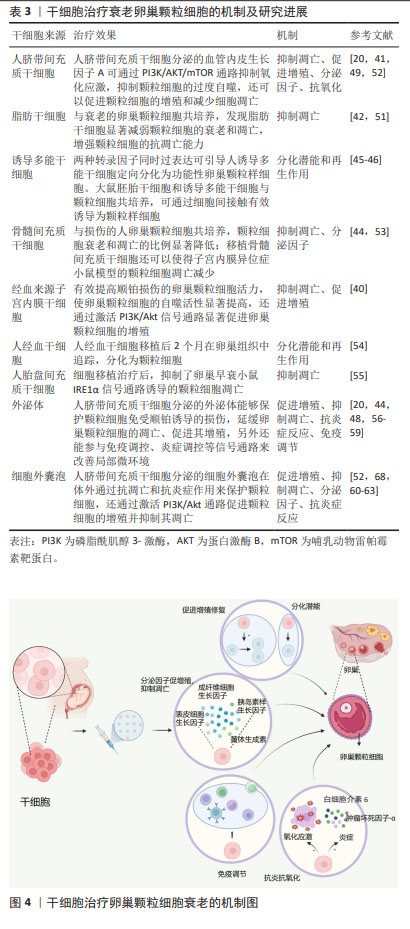
[35] JIN J. Stem Cell Treatments. JAMA. 2017;317(3):330.
[36] ZAKRZEWSKI W, DOBRZYŃSKI M, SZYMONOWICZ M, et al. Stem cells: past, present, and future. Stem Cell Res Ther. 2019;10(1):68.
[37] CAPLAN AI. Mesenchymal stem cells. J Orthop Res. 1991;9(5):641-650.
[38] TAKAHASHI K, YAMANAKA S. Induction of pluripotent stem cells from mouse embryonic and adult fibroblast cultures by defined factors. Cell. 2006;126(4):663-676.
[39] HWANG JJ, RIM YA, NAM Y, et al. Recent Developments in Clinical Applications of Mesenchymal Stem Cells in the Treatment of Rheumatoid Arthritis and Osteoarthritis. Front Immunol. 2021;12: 631291.
[40] FU X, ZHANG S, LI T, et al. Menstrual blood-derived endometrial stem cells ameliorate the viability of ovarian granulosa cells injured by cisplatin through activating autophagy. Reprod Toxicol. 2022;110:39-48.
[41] WANG J, ZHAO Y, ZHENG F, et al. Activated Human Umbilical Cord Blood Platelet-Rich Plasma Enhances the Beneficial Effects of Human Umbilical Cord Mesenchymal Stem Cells in Chemotherapy-Induced POF Rats. Stem Cells Int. 2021;2021:8293699.
[42] AI G, MENG M, GUO J, et al. Adipose-derived stem cells promote the repair of chemotherapy-induced premature ovarian failure by inhibiting granulosa cells apoptosis and senescence. Stem Cell Res Ther. 2023;14(1):75.
[43] SEOK J, PARK HS, CETIN E, et al. The potent paracrine effect of umbilical cord mesenchymal stem cells mediates mitochondrial quality control to restore chemotherapy-induced damage in ovarian granulosa cells. Biomed Pharmacother. 2024;172:116263.
[44] CHEN S, WANG Y, LIAO L, et al. Similar Repair Effects of Human Placenta, Bone Marrow Mesenchymal Stem Cells, and Their Exosomes for Damaged SVOG Ovarian Granulosa Cells. Stem Cells Int. 2020; 2020:8861557.
[45] PIERSON SMELA MD, KRAMME CC, FORTUNA PRJ, et al. Directed differentiation of human iPSCs to functional ovarian granulosa-like cells via transcription factor overexpression. Elife. 2023;12:e83291.
[46] ZHANG J, LI H, WU Z, et al. Differentiation of rat iPS cells and ES cells into granulosa cell-like cells in vitro. Acta Biochim Biophys Sin (Shanghai). 2013;45(4):289-295.
[47] SUN B, MA Y, WANG F, et al. miR-644-5p carried by bone mesenchymal stem cell-derived exosomes targets regulation of p53 to inhibit ovarian granulosa cell apoptosis. Stem Cell Res Ther. 2019;10(1):360.
[48] SUN YT, CAI JH, BAO S. Overexpression of lncRNA HCP5 in human umbilical cord mesenchymal stem cell-derived exosomes promoted the proliferation and inhibited the apoptosis of ovarian granulosa cells via the musashi RNA-binding protein 2/oestrogen receptor alpha 1 axis. Endocr J. 2022;69(9):1117-1129.
[49] STAVELY R, NURGALI K. The emerging antioxidant paradigm of mesenchymal stem cell therapy. Stem Cells Transl Med. 2020;9(9): 985-1006.
[50] DAI W, YANG H, XU B, et al. Human umbilical cord-derived mesenchymal stem cells (hUC-MSCs) alleviate excessive autophagy of ovarian granular cells through VEGFA/PI3K/AKT/mTOR pathway in premature ovarian failure rat model. J Ovarian Res. 2023;16(1):198.
[51] DING C, ZOU Q, WANG F, et al. HGF and BFGF Secretion by Human Adipose-Derived Stem Cells Improves Ovarian Function During Natural Aging via Activation of the SIRT1/FOXO1 Signaling Pathway. Cell Physiol Biochem. 2018;45(4):1316-1332.
[52] DENG T, HE J, YAO Q, et al. Human Umbilical Cord Mesenchymal Stem Cells Improve Ovarian Function in Chemotherapy-Induced Premature Ovarian Failure Mice Through Inhibiting Apoptosis and Inflammation via a Paracrine Mechanism. Reprod Sci. 2021;28(6):1718-1732.
[53] DWININGSIH SR, DARMOSOEKARTO S, HENDARTO H, et al. Effects of bone marrow mesenchymal stem cell transplantation on tumor necrosis factor-alpha receptor 1 expression, granulosa cell apoptosis, and folliculogenesis repair in endometriosis mouse models. Vet World. 2021;14(7):1788-1796.
[54] NOORY P, NAVID S, ZANGANEH BM, et al. Human Menstrual Blood Stem Cell-Derived Granulosa Cells Participate in Ovarian Follicle Formation in a Rat Model of Premature Ovarian Failure In Vivo. Cell Reprogram. 2019;21(5):249-259.
[55] LI H, ZHAO W, WANG L, et al. Human placenta-derived mesenchymal stem cells inhibit apoptosis of granulosa cells induced by IRE1α pathway in autoimmune POF mice. Cell Biol Int. 2019;43(8):899-909.
[56] QU Q, LIU L, CUI Y, et al. miR-126-3p containing exosomes derived from human umbilical cord mesenchymal stem cells promote angiogenesis and attenuate ovarian granulosa cell apoptosis in a preclinical rat model of premature ovarian failure. Stem Cell Res Ther. 2022;13(1):352.
[57] GAO T, CHEN Y, HU M, et al. MicroRNA-22-3p in human umbilical cord mesenchymal stem cell-secreted exosomes inhibits granulosa cell apoptosis by targeting KLF6 and ATF4-ATF3-CHOP pathway in POF mice. Apoptosis. 2023;28(7-8):997-1011.
[58] IZADI M, REZVANI ME, ALIABADI A, et al. Mesenchymal stem cells-derived exosomes as a promising new approach for the treatment of infertility caused by polycystic ovary syndrome. Front Pharmacol. 2022;13:1021581.
[59] SUN L, LI D, SONG K, et al. Exosomes derived from human umbilical cord mesenchymal stem cells protect against cisplatin-induced ovarian granulosa cell stress and apoptosis in vitro. Sci Rep. 2017; 7(1):2552.
[60] ZHANG J, YIN H, JIANG H, et al. The protective effects of human umbilical cord mesenchymal stem cell-derived extracellular vesicles on cisplatin-damaged granulosa cells. Taiwan J Obstet Gynecol. 2020; 59(4):527-533.
[61] LI N, FAN X, LIU L, et al. Therapeutic effects of human umbilical cord mesenchymal stem cell-derived extracellular vesicles on ovarian functions through the PI3K/Akt cascade in mice with premature ovarian failure. Eur J Histochem. 2023;67(3):3506.
[62] GAO T, CAO Y, HU M, et al. Human Umbilical Cord Mesenchymal Stem Cell-Derived Extracellular Vesicles Carrying MicroRNA-29a Improves Ovarian Function of Mice with Primary Ovarian Insufficiency by Targeting HMG-Box Transcription Factor/Wnt/β-Catenin Signaling. Dis Markers. 2022;2022:5045873.
[63] PU X, ZHANG L, ZHANG P, et al. Human UC-MSC-derived exosomes facilitate ovarian renovation in rats with chemotherapy-induced premature ovarian insufficiency. Front Endocrinol (Lausanne). 2023; 14:1205901.
[64] ZHOU Y, ZHOU J, XU X, et al. Matrigel/Umbilical Cord-Derived Mesenchymal Stem Cells Promote Granulosa Cell Proliferation and Ovarian Vascularization in a Mouse Model of Premature Ovarian Failure. Stem Cells Dev. 2021;30(15):782-796.
[65] CHANG L, FAN W, PAN X, Zhu X. Stem cells to reverse aging. Chin Med J (Engl). 2022;135(8):901-910.
[66] LO KC, CHUANG WW, LAMB DJ. Stem cell research: the facts, the myths and the promises. J Urol. 2003;170(6 Pt 1):2453-2458.
[67] 李仲康,郑嘉华,田彦鹏,等.间充质干细胞治疗卵巢早衰的最新进展及机制[J].中国组织工程研究,2022,26(1):141-147. |