中国组织工程研究 ›› 2025, Vol. 29 ›› Issue (18): 3811-3818.doi: 10.12307/2025.653

• 组织构建实验造模 experimental modeling in tissue construction • 上一篇    下一篇

电针干预脑缺血再灌注模型大鼠的神经保护机制

吴海洋1,段  冕2,李成龙3,张君宇1,计海生1,王海涛1,茅  伟4,王  颖1   

  1. 1安徽中医药大学第二附属医院,安徽省合肥市  230001;2合肥市第二人民医院,安徽省合肥市  230001;3安徽中医药大学研究生院,安徽省合肥市  230000;4广州医科大学附属脑科医院中医科,广东省广州市  510370

  • 收稿日期:2024-05-08 接受日期:2024-07-26 出版日期:2025-06-28 发布日期:2024-11-28
  • 通讯作者: 王颖,博士,主任医师,安徽中医药大学第二附属医院,安徽省合肥市 230001
  • 作者简介:吴海洋,男,1986年生,安徽省合肥市人,汉族,2014年安徽中医药大学毕业,硕士,副主任医师,主要从事针灸防治脑病的临床及机理研究。
  • 基金资助:
    安徽省高等学校科学研究项目(2022AH050503),项目负责人:吴海洋;安徽省高等学校科学研究项目(2023AH040111),项目负责人:王颖;安徽省高等学校科学研究项目(2023AH050816),项目负责人:计海生;安徽省高等学校科学研究项目(2023AH050739),项目负责人:张君宇;安徽省卫生健康委科研项目(AHWJ2023A20030),项目负责人:吴海洋;安徽省名中医王颖工作室建设项目(中发展[2022]5号),项目负责人:王颖;安徽省临床医学研究转化专项项目(202304295107020103),项目负责人:王颖;安徽中医药大学临床科研项目(2021efylc11),项目负责人:吴海洋

Neuroprotective mechanism of electroacupuncture in cerebral ischemia-reperfusion model rats

Wu Haiyang1, Duan Mian2, Li Chenglong3, Zhang Junyu1, Ji Haisheng1, Wang Haitao1, Mao Wei4, Wang Ying1   

  1. 1The Second Affiliated Hospital of Anhui University of Chinese Medicine, Hefei 230001, Anhui Province, China; 2Hefei Second People’s Hospital, Hefei 230001, Anhui Province, China; 3School of Graduate, Anhui University of Chinese Medicine, Hefei 230000, Anhui Province, China; 4Department of TCM, Affiliated Brain Hospital of Guangzhou Medical University, Guangzhou 510370, Guangdong Province, China 
  • Received:2024-05-08 Accepted:2024-07-26 Online:2025-06-28 Published:2024-11-28
  • Contact: Wang Ying, MD, Chief physician, The Second Affiliated Hospital of Anhui University of Chinese Medicine, Hefei 230001, Anhui Province, China
  • About author:Wu Haiyang, Master, Associate chief physician, The Second Affiliated Hospital of Anhui University of Chinese Medicine, Hefei 230001, Anhui Province, China
  • Supported by:
    Scientific Research Project of Higher Education Institutions in Anhui Province, Nos. 2022AH050503 (to WHY), 2023AH040111 (to WY), 2023AH050816 (to JHS), and 2023AH050739 (to ZJY); Scientific Research Project of Anhui Provincial Health Care Commission, No. AHWJ2023A20030 (to WHY); Construction Project of Wang Ying Studio of Famous Chinese Medicine Practitioners in Anhui Province, No. [2022]5 (to WY); Special Project of Clinical Medical Research Translation in Anhui Province, No. 202304295107020103 (to WY); Clinical Research Project of Anhui University of Chinese Medicine, No. 2021efylc11 (to WHY) 

摘要:


文题释义:
脑缺血再灌注损伤:作为急性脑卒中溶栓治疗后的重要病理环节不可避免地加重神经功能损伤,以脑损伤和神经功能障碍为主要特征。研究发现,脑组织血流再灌注后会出现局部组织水肿甚至出血的现象,细胞中钙超载、氧化应激、线粒体损伤和兴奋性氨基酸毒性均参与到再灌注损伤中,可激活凋亡、程序性死亡、自噬、焦亡、铁死亡、坏死等多种细胞死亡途径。
细胞焦亡:是一种典型依赖于炎症反应的程序性细胞死亡方式,以质膜快速破裂、随后释放细胞内容物和促炎递质为特征,其中经典焦亡途径依赖于Caspase-1激活后诱导的炎症反应。NLRP3炎症小体作为脑缺血后中枢神经系统无菌性炎症反应的始动因子,在中枢神经系统中诱导细胞焦亡和一系列炎症反应,导致神经细胞损伤,在中枢神经系统细胞焦亡中发挥关键作用。

背景:前期研究表明,针刺督脉治疗缺血性脑卒中的疗效确切,可通过减轻细胞焦亡改善脑缺血再灌注损伤,但上游调控机制尚不完全明确。
目的:分析电针对脑缺血再灌注损伤模型大鼠的脑保护作用机制。
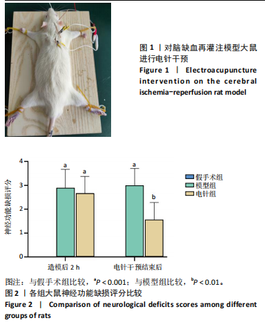
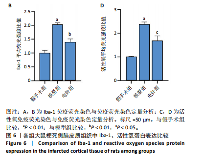
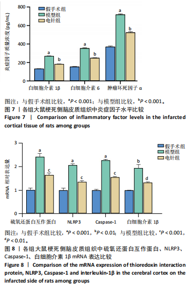
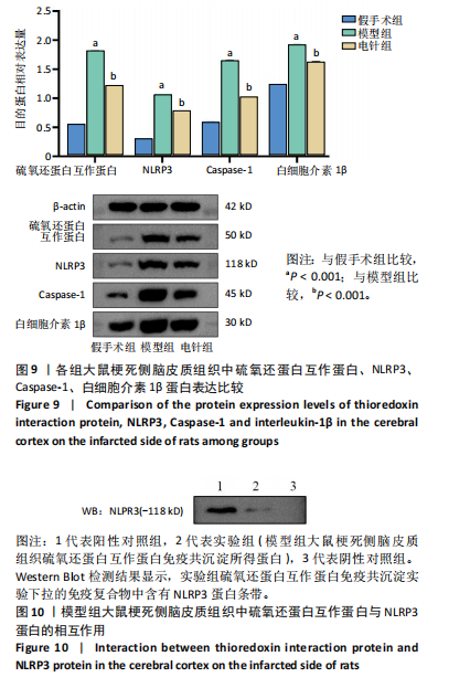
方法:采用随机数字表法将27只SD大鼠随机分为假手术组、模型组和电针组,每组9只。取模型组和电针组大鼠,采用改良线栓法建立脑缺血再灌注损伤模型,验证造模成功后,对电针组大鼠施予电针干预,选取百会、风府、大椎3个穴位,1次/d,20 min/次,连续干预7 d。电针干预结束后,利用神经功能缺损评分、爬杆实验评估大鼠行为学改变,TTC染色评估大鼠脑梗死体积,苏木精-伊红染色观察大鼠梗死侧脑皮质组织形态学变化,免疫荧光染色分析梗死侧脑皮质中Iba-1及活性氧表达,ELISA法检测梗死侧脑皮质组织中白细胞介素1β、白细胞介素6和肿瘤坏死因子α水平,RT-qPCR及Western blot检测大鼠梗死侧脑皮质组织中硫氧还蛋白互作蛋白、NLRP3、Caspase-1、白细胞介素1β的mRNA和蛋白表达,免疫共沉淀法分析大鼠梗死侧脑皮质组织中硫氧还蛋白互作蛋白与NLRP3的相互作用。
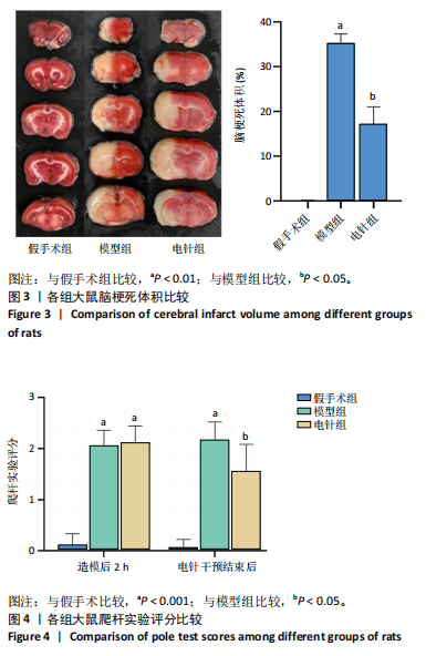
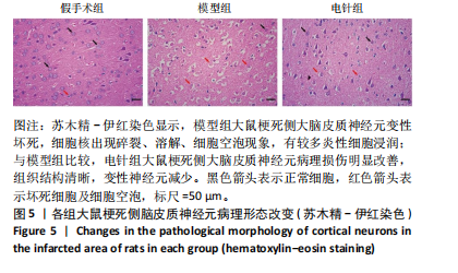
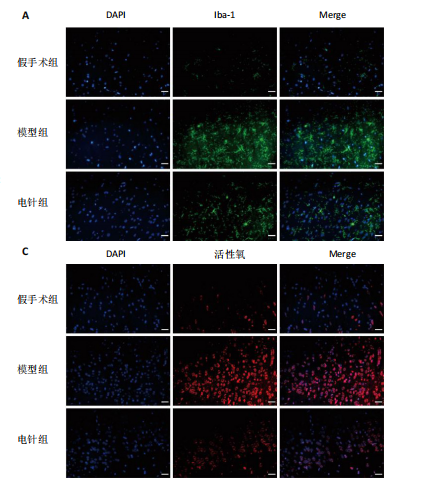
结果与结论:①与假手术组比较,模型组大鼠神经功能缺损评分、爬杆实验评分及脑梗死体积均升高(P < 0.05),Iba-1、活性氧免疫荧光表达增强(P < 0.05),白细胞介素1β、白细胞介素6、肿瘤坏死因子α水平均升高(P < 0.05),硫氧还蛋白互作蛋白、NLRP3、Caspase-1、白细胞介素1β的mRNA和蛋白表达均升高(P < 0.05),苏木精-伊红染色显示模型组大鼠梗死侧脑皮质神经元变性坏死,细胞核出现碎裂溶解及细胞空泡现象;②与模型组比较,电针组大鼠神经功能缺损评分、爬杆实验评分及脑梗死体积均降低(P < 0.05),Iba-1、活性氧免疫荧光表达减弱(P < 0.05),白细胞介素1β、白细胞介素6、肿瘤坏死因子α水平均降低(P < 0.05),硫氧还蛋白互作蛋白、NLRP3、Caspase-1、白细胞介素1β的mRNA和蛋白表达均降低(P < 0.05),苏木精-伊红染色显示电针组大鼠梗死侧脑皮质神经元病理学损伤明显减轻,细胞坏死及空泡现象明显减少;③免疫共沉淀检测显示,模型组大鼠梗死侧脑皮质组织中硫氧还蛋白互作蛋白与NLRP3存在相互作用;④结果表明,电针干预可能通过抑制活性氧/硫氧还蛋白互作蛋白/NLRP3细胞焦亡信号通路及小胶质细胞的活化来减少炎症因子释放,进而发挥抗脑缺血再灌注损伤作用。
https://orcid.org/0009-0003-8986-1578(吴海洋)

中国组织工程研究杂志出版内容重点:组织构建;骨细胞;软骨细胞;细胞培养;成纤维细胞;血管内皮细胞;骨质疏松;组织工程

关键词: 脑缺血再灌注损伤, 电针, NLRP3, 活性氧, 硫氧还蛋白互作蛋白, 小胶质细胞

Abstract: BACKGROUND: Previous studies have demonstrated that acupuncture at the governor meridian has precise efficacy in the treatment of ischemic stroke and can improve cerebral ischemia-reperfusion injury by attenuating pyroptosis, but the upstream regulatory mechanisms are not yet fully clarified.
OBJECTIVE: To observe the neuroprotective effect of electroacupuncture in model rats of cerebral ischemia-reperfusion injury.
METHODS: Twenty-seven Sprague-Dawley rats were randomly divided into sham surgery, model, and electroacupuncture groups, with nine rats in each group. Modified suture method was used to establish cerebral ischemia-reperfusion model rats in the model and electroacupuncture groups. The electroacupuncture group was subjected to electroacupuncture at “Baihui,” “Fengfu,” and “Dazhui” acupoints, 20 minutes each, once a day, for 7 consecutive days. After treatment, neurological deficit scoring and pole test were performed to assess behavioral changes. Tri-phenyl tetrazolium chloride staining was used to assess cerebral infarction size in rats. Hematoxylin-eosin staining was performed to observe morphological changes in cerebral cortex tissue on the infarcted side of rats. Immunofluorescence analysis was used to determine Iba-1 and reactive oxygen species levels in cerebral cortex tissue on the infarcted side of rats, ELISA method was used for measuring interleukin-1β, interleukin-6 and tumor necrosis factor α levels in cerebral cortex tissue on the infarcted side of rats. Real-time fluorescence quantitative PCR and western blot were used to detect mRNA and protein expression levels of thioredoxin interaction protein, nod-like receptor associated protein 3 (NLRP3), Caspase-1 and interleukin-1β in cerebral cortex tissue on the infarcted side of rats respectively, and the interaction between thioredoxin interaction protein and NLRP3 was analyzed by immunoprecipitation. 
RESULTS AND CONCLUSION: (1) Compared with the sham surgery group, rats in the model group showed an increase in neurological deficit score, pole test score, cerebral infarction volume (P < 0.05), the immunofluorescence expression of Iba-1 and reactive oxygen species (P < 0.05), the levels of interleukin-1β, interleukin-6 and tumor necrosis factor α (P < 0.05), and the mRNA and protein expression of thioredoxin interaction protein, NLRP3, Caspase-1 and interleukin-1β in cerebral cortex tissue (P < 0.05). Hematoxylin-eosin staining in the model group showed neuronal degeneration and necrosis, with fragmented and dissolved nuclei and cellular vacuoles. (2) Compared with the model group, rats in the electroacupuncture group showed a reduction in neurological deficit score, pole climbing test score, cerebral infarction volume (P < 0.05), the immunofluorescence expression of Iba-1 and reactive oxygen species (P < 0.05), the levels of interleukin-1β, interleukin-6 and tumor necrosis factor α (P < 0.05), and the mRNA and protein expression of thioredoxin interaction protein, NLRP3, Caspase-1 and interleukin-1β in cerebral cortex tissue (P < 0.05). Hematoxylin-eosin staining showed that the pathological damage of neurons in cerebral cortex tissue on the infarcted side of rats in the electroacupuncture group was significantly attenuated, with significantly reduced cell necrosis and vacuolation. (3) Immunoprecipitation assay showed an interaction between thioredoxin interaction proteins and NLRP3 in the cerebral cortical tissues on the infarcted side of rats in the model group. To conclude, electroacupuncture has a significant therapeutic effect against cerebral ischemia-reperfusion injury, possibly by inhibiting the reactive oxygen species/thioredoxin interaction protein/NLRP3 cell pyroptosis signaling pathway and activation of microglia to reduce the release of inflammatory factors.

Key words: cerebral ischemia-reperfusion injury, electroacupuncture, NLRP3, reactive oxygen species, thioredoxin interaction protein, microglia

中图分类号: