[1] AYERS C, KANSAGARA D, LAZUR B, et al. Effectiveness and Safety of Treatments to Prevent Fractures in People With Low Bone Mass or Primary Osteoporosis: A Living Systematic Review and Network Meta-analysis for the American College of Physicians. Ann Intern Med. 2023; 176(2):182-195.
[2] LORENZO J. From the gut to bone: connecting the gut microbiota with Th17 T lymphocytes and postmenopausal osteoporosis. J Clin Invest. 2021;131(5):e146619.
[3] SONG S, GUO Y, YANG Y, et al. Advances in pathogenesis and therapeutic strategies for osteoporosis, Pharmacol Ther. 2022;237:108168.
[4] LEE S, KIM GJ, KWON H, et al. Estrogenic Effects of Extracts and Isolated Compounds from Belowground and Aerial Parts of Spartina anglica. Mar Drugs. 2021;19(4):210.
[5] RODRÍGUEZ V, RIVOIRA M, PICOTTO G, et al. Analysis of the Molecular Mechanisms by Flavonoids with Potential Use for Osteoporosis Prevention or Therapy. Curr Med Chem. 2022;29(16):2913-2936.
[6] MCDONALD MM, KHOO WH, NG PY, et al. Osteoclasts recycle via osteomorphs during RANKL-stimulated bone resorption. Cell. 2021; 184(5):1330-1347.e13.
[7] MENG J, ZHANG X, GUO X, et al. Briarane-type diterpenoids suppress osteoclastogenisis by regulation of Nrf2 and MAPK/NF-kB signaling pathway. Bioorg Chem. 2021;112:104976.
[8] ALIPRANTIS AO, UEKI Y, SULYANTO R, et al. NFATc1 in mice represses osteoprotegerin during osteoclastogenesis and dissociates systemic osteopenia from inflammation in cherubism. J Clin Invest. 2008; 118(11):3775-3789.
[9] MA JD, JING J, WANG JW, et al. Activation of the Peroxisome Proliferator-Activated Receptor gamma Coactivator 1beta/NFATc1 Pathway in Circulating Osteoclast Precursors Associated With Bone Destruction in Rheumatoid Arthritis. Arthritis Rheumatol. 2019;71(8): 1252-1264.
[10] DENG W, DING Z, WANG Y, et al. Dendrobine attenuates osteoclast differentiation through modulating ROS/NFATc1/ MMP9 pathway and prevents inflammatory bone destruction. Phytomedicine. 2022; 96:153838.
[11] ZHAO Y, GAO J, ZHANG Y, et al. Cyclosporine A Promotes Bone Remodeling in LPS-Related Inflammation via Inhibiting ROS/ERK Signaling: Studies In Vivo and In Vitro. Oxid Med Cell Longev. 2021; 2021:8836599.
[12] BIRTIĆ S, DUSSORT P, PIERRE FX, et al. Carnosic acid. Phytochemistry. 2015;115:9-19.
[13] ZHENG ZG, CHENG HM, ZHOU YP, et al. Dual targeting of SREBP2 and ERRalpha by carnosic acid suppresses RANKL-mediated osteoclastogenesis and prevents ovariectomy-induced bone loss.Cell Death Differ. 2020;27(7):2048-2065.
[14] LI H, DENG W, QIN Q, et al. Isoimperatorin attenuates bone loss by inhibiting the binding of RANKL to RANK. Biochem Pharmacol. 2023;211:115502.
[15] HONG G, CHEN Z, HAN X, et al. A novel RANKL-targeted flavonoid glycoside prevents osteoporosis through inhibiting NFATc1 and reactive oxygen species. Clin Transl Med. 2021;11(5):e392.
[16] LI Y, ZHUANG Q, TAO L, et al. Urolithin B suppressed osteoclast activation and reduced bone loss of osteoporosis via inhibiting ERK/NF-kappaB pathway. Cell Prolif. 2022;55(10):e13291.
[17] WEI L, CHEN W, HUANG L, et al. Alpinetin ameliorates bone loss in LPS-induced inflammation osteolysis via ROS mediated P38/PI3K signaling pathway. Pharmacol Res. 2022;184:106400.
[18] SU X, SHEN Z, YANG Q, et al. Vitamin C kills thyroid cancer cells through ROS-dependent inhibition of MAPK/ERK and PI3K/AKT pathways via distinct mechanisms. Theranostics. 2019;9(15):4461-4473.
[19] SHADEL GS, HORVATH TL. Mitochondrial ROS signaling in organismal homeostasis. Cell. 2015;163(3):560-569.
[20] NI S, QIAN Z, YUAN Y, et al. Schisandrin A restrains osteoclastogenesis by inhibiting reactive oxygen species and activating Nrf2 signalling.Cell Prolif. 2020;53(10):e12882.
[21] LI B, LEE WC, SONG C, et al. Both aerobic glycolysis and mitochondrial respiration are required for osteoclast differentiation. FASEB J. 2020; 34(8):11058-11067.
[22] YANG B, SU Y, HAN S, et al. Aminooxyacetic acid hemihydrochloride inhibits osteoclast differentiation and bone resorption by attenuating oxidative phosphorylation. Front Pharmacol. 2022;13:980678.
[23] SIMS NA, MARTIN TJ. Coupling the activities of bone formation and resorption: a multitude of signals within the basic multicellular unit.Bonekey Rep. 2014;3:481.
[24] YUAN F, PENG W, YANG C, et al. Teriparatide versus bisphosphonates for treatment of postmenopausal osteoporosis: A meta-analysis. Int J Surg. 2019;66:1-11.
[25] ROZENBERG S, AL-DAGHRI N, AUBERTIN-LEHEUDRE M, et al. Is there a role for menopausal hormone therapy in the management of postmenopausal osteoporosis? Osteoporos Int. 2020;31(12): 2271-2286.
[26] KHOSLA S, HOFBAUER LC. Osteoporosis treatment: recent developments and ongoing challenges. Lancet Diabetes Endocrinol. 2017;5(11):898-907.
[27] LIU Y, WANG C, WANG G, et al. Loureirin B suppresses RANKL-induced osteoclastogenesis and ovariectomized osteoporosis via attenuating NFATc1 and ROS activities. Theranostics. 2019;9(16):4648-4662.
[28] LEE DK, JANG HD. Carnosic Acid Attenuates an Early Increase in ROS Levels during Adipocyte Differentiation by Suppressing Translation of Nox4 and Inducing Translation of Antioxidant Enzymes. Int J Mol Sci. 2021;22(11):6096.
[29] HU S, LIU B, YANG M, et al. Carnosic acid protects against doxorubicin-induced cardiotoxicity through enhancing the Nrf2/HO-1 pathway. Food Funct. 2023;14(8):3849-3862.
[30] ZUO RM, JIAO JY, CHEN N, et al. Carnosic acid suppressed the formation of NETs in alcoholic hepatosteatosis based on P2X7R-NLRP3 axis.Phytomedicine. 2023;110:154599.
[31] FISCHER V, HAFFNER-LUNTZER M. Interaction between bone and immune cells: Implications for postmenopausal osteoporosis. Semin Cell Dev Biol. 2022;123:14-21.
[32] LIU M, LIU S, ZHANG Q, et al. Curculigoside attenuates oxidative stress and osteoclastogenesis via modulating Nrf2/NF-kappaB signaling pathway in RAW264.7 cells. J Ethnopharmacol. 2021; 275:114129.
[33] KIM HJ, PARK C, KIM GY, et al. Sargassum serratifolium attenuates RANKL-induced osteoclast differentiation and oxidative stress through inhibition of NF-kappaB and activation of the Nrf2/HO-1 signaling pathway. Biosci Trends. 2018;12(3):257-265.
[34] YAN C, SHI Y, YUAN L, et al. Mitochondrial quality control and its role in osteoporosis. Front Endocrinol (Lausanne). 2023;14:1077058.
[35] GAO J, FENG Z, WANG X, et al. SIRT3/SOD2 maintains osteoblast differentiation and bone formation by regulating mitochondrial stress.Cell Death Differ. 2018;25(2):229-240.
[36] KIM HN, PONTE F, NOOKAEW I, et al. Estrogens decrease osteoclast number by attenuating mitochondria oxidative phosphorylation and ATP production in early osteoclast precursors. Sci Rep. 2020; 10(1):11933. |

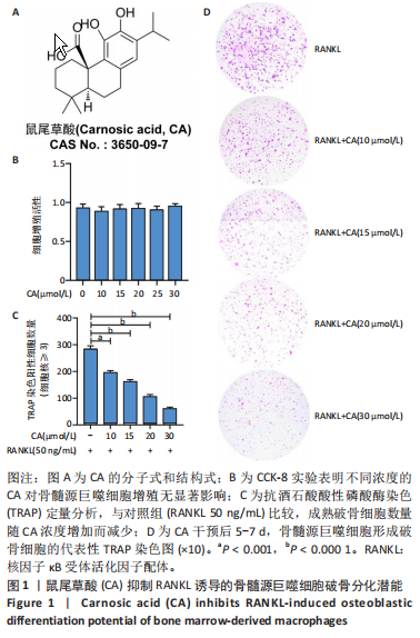
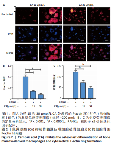
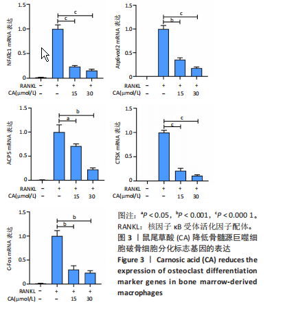
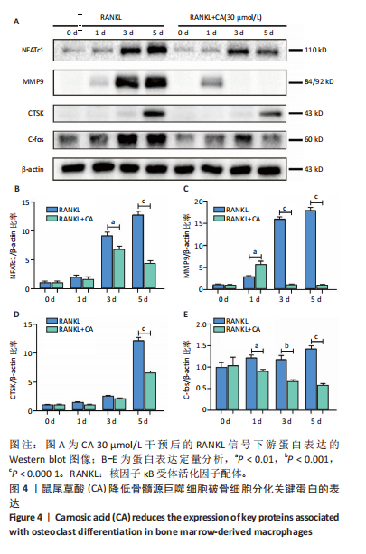

 #br#
#br#