| [1] 范瑞新,吴若彬,肖学钧,等.二尖瓣主动脉瓣三尖瓣同时置换治疗重症风湿性瓣膜病[J].中华胸心血管外科杂志, 2003,19(4): 208-210.
[2] 赵铁夫,王盛宇,周其文,等.心脏瓣膜种类选择对老年患者生活质量影响的临床对照研究[J].中国胸心血管外科临床杂志, 2013,20(4):430-434.
[3] Conrotto F, D'Ascenzo F,Salizzoni S,et al. A gender based analysis of predictors of all cause death after transcatheter aortic valve implantation.Am J Cardiol.2014;114(8): 1269-1274.
[4] Chopard R,Perrotti A,Durst C,et al. Six-year outcomes after non-resective mitral valve repair with artificial chordae using removable clips.J Heart Valve Dis.2014;23(3): 364-369.
[5] Noble S,Frangos E,Samaras N,et al.Transcatheter aortic valve implantation in nonagenarians: effective and safe. Eur J Intern Med.2013;24(8):750-755.
[6] Baraki H,Al Ahmad A,Jeng-Singh S,et al.Pacemaker dependency after isolated aortic valve replacement: do conductance disorders recover over time? Interact Cardiovasc Thorac Surg.2013;16(4):476-481.
[7] Sfeir PM,Abchee AB,Ghazzal Z,et al.Endovascular transcatheter aortic valve implantation: an evolving standard.J Cardiothorac Vasc Anesth.2013;27(4):765-778.
[8] Said SM,Burkhart HM,Dearani JA.Surgical management of congenital (non-Ebstein) tricuspid valve regurgitation. Semin Thorac Cardiovasc Surg Pediatr Card Surg Annu. 2012;15(1): 46-60.
[9] Coats L,Tsang V,Khambadkone S,et al.The potential impact of percutaneous pulmonary valve stent implantation on right ventricular outflow tract re-intervention.Eur J Cardiothorac Surg.2005;27(4):536-543.
[10] Sun W,Martin C,Pham T.Computational modeling of cardiac valve function and intervention. Annu Rev Biomed Eng. 2014;16(1):53-76.
[11] Zahn R,Gerckens U,Linke A,et al.Predictors of one-year mortality after transcatheter aortic valve implantation for severe symptomatic aortic stenosis.Am J Cardiol.2013; 112(2):272-9.
[12] Helder MR,Schaff HV,Dearani JA,et al.Management of mitral regurgitation in Marfan syndrome: Outcomes of valve repair versus replacement and comparison with myxomatous mitral valve disease.J Thorac Cardiovasc Surg.2014;148(3): 1020-1024.
[13] Claiborne TE,Sheriff J,Kuetting M,et al.In vitro evaluation of a novel hemodynamically optimized trileaflet polymeric prosthetic heart valve.J Biomech Eng.2013;135(2):21021.
[14] Jackson MJ,Robinson GM,Ali N,et al.Surface engineering of artificial heart valve disks using nanostructured thin films deposited by chemical vapour deposition and sol-gel methods.J Med Eng Technol.2006;30(5):323-329.
[15] Ma L,Sines G.Fatigue behavior of a pyrolytic carbon. J Biomed Mater Res.2000; 51(1):61-68.
[16] Sneckenberger DS,Stinebring DR,Deutsch S,et al. Mitral heart valve cavitation in an artificial heart environment.J Heart Valve Dis.1996;5(2):216-227.
[17] Fenglan X,Yubao L,Xiaoming Y,et al.Preparation and in vivo investigation of artificial cornea made of nano-hydroxyapatite/ poly(vinyl alcohol) hydrogel composite.J Mater Sci Mater Med. 2007;18(4):635-640.
[18] Ito M,Kotani Y,Hojo Y,et al.Evaluation of hydroxyapatite ceramic vertebral spacers with different porosities and their binding capability to the vertebral body: an experimental study in sheep.J Neurosurg Spine.2007;6(5):431-437.
[19] Siriphannon P,Monvisade P,Jinawath S,et al.Preparation and characterization of hydroxyapatite/poly (ethylene glutarate) biomaterials.J Biomed Mater Res A. 2007;81(2):381-91.
[20] 王晓伟,叶福林,徐志云,等.人脐静脉血管内皮细胞构建组织工程心脏瓣膜及其生理功能[J].江苏医药,2007,33(2):156-158.
[21] 王晓伟,徐志云,张宝仁,等.人脐静脉内皮细胞用于构建组织工程心脏瓣膜的实验研究[J].第二军医大学学报,2003,24(12): 1287-120.
[22] Weymann A,Schmack B,Okada T,et al.Reendothelialization of human heart valve neoscaffolds using umbilical cord-derived endothelial cells.Circ J.2013;77(1): 207-216.
[23] Sha JM,Yan ZY,Cheng GC,et al.In-vitro seeding of human umbilical cord vein endothelial cells on hydroxyapatite for mechanical heart valve applications.J Heart Valve Dis.2010; 19(4):506-512.
[24] Martínez GJ,Seco M,Jaijee SK,et al.Introduction of an interdisciplinary heart team-based transcatheter aortic valve implantation programme: short and mid-term outcomes.Intern Med J.2014;44(9):876-883.
[25] 郝和平.医疗器械生物学评价标准实施指南[M].北京:中国标准出版社,2002: 100-10.
[26] Orme NM,Wright TC,Harmon GE,et al.Imaging Pandora's Box: incidental findings in elderly patients evaluated for transcatheter aortic valve replacement.Mayo Clin Proc.2014; 89(6):747-753.
[27] Sadat-Shojai M,Khorasani MT,Jamshidi A.3-Dimensional cell-laden nano-hydroxyapatite/protein hydrogels for bone regeneration applications.Mater Sci Eng C Mater Biol Appl. 2015;49:835-843.
[28] de Bruyn JR,Goiko M,Mozaffari M,et al.Dynamic light scattering study of inhibition of nucleation and growth of hydroxyapatite crystals by osteopontin. PLoS One.2013; 8(2):e56764.
[29] Moroi A,Ueki K,Okabe K,et al.Comparison between unsintered hydroxyapatite/poly-L-lactic acid mesh and titanium mesh in bone regeneration of rabbit mandible. Implant Dent.2013;22(3):255-262.
[30] Smolen D,Chudoba T,Malka I,et al.Highly biocompatible, nanocrystalline hydroxyapatite synthesized in a solvothermal process driven by high energy density microwave radiation.Int J Nanomedicine.2013;8:653-668. |
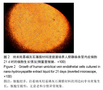
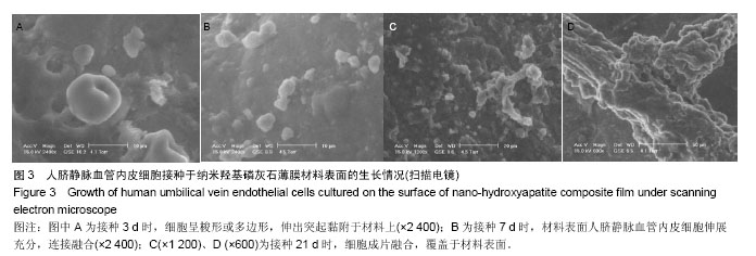
.jpg)
.jpg)