中国组织工程研究 ›› 2016, Vol. 20 ›› Issue (37): 5538-5544.doi: 10.3969/j.issn.2095-4344.2016.37.010

• 肌肉肌腱韧带组织构建 tissue construction of the muscle, tendon and ligament • 上一篇    下一篇

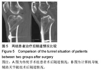
带血管蒂髌韧带修复前交叉韧带损伤:计算机导航辅助关节镜的应用

张  凯1,王伟伟1,王向青2   

  1. 1临沂市沂水中心医院,山东省临沂市   276400;2青岛大学附属医院,山东省青岛市  266000
  • 出版日期:2016-09-09 发布日期:2016-09-09
  • 通讯作者: 王向青,博士,主任医师,博士生导师,青岛大学附属医院,山东省青岛市 266000
  • 作者简介:张凯,男,1978年生,山东省沂水县人,汉族,2004年滨州医学院毕业,主治医师,主要从事关节镜研究。
  • 基金资助:

    国家自然科学基金(81401833)

Patellar ligament with vascular pedicle for anterior cruciate ligament reconstruction: the intraoperative use of computer assisted navigation system combinied with arthroscopy

Zhang Kai1, Wang Wei-wei1, Wang Xiang-qing2   

  1. 1Yishui Central Hospital of Linyi, Linyi 276400, Shandong Province, China; 2Affiliated Hospital of Qingdao University, Qingdao 266000, Shandong Province, China
  • Online:2016-09-09 Published:2016-09-09
  • Contact: Wang Xiang-qing, M.D., Chief physician, Doctoral supervisor, Affiliated Hospital of Qingdao University, Qingdao 266000, Shandong Province, China
  • About author:Zhang Kai, Attending physician, Yishui Central Hospital of Linyi, Linyi 276400, Shandong Province, China
  • Supported by:

    the National Natural Science Foundation of China, No. 81401833

摘要:

文章快速阅读:
 

文题释义:
前交叉韧带:属于稳定膝关节重要的结构之一,由前内侧束和后外侧束两个部分组成。当人体处于不同屈曲角度下时,前交叉韧带不同纤维束处于紧张状态,维持机体稳定。前交叉韧带损伤后如果得不到及时有效的治疗,将会引起膝关节松弛、半月板磨损等,演变为骨性关节炎,因此,加强前交叉韧带诊断治疗成为医学界研究重点。
前交叉韧带重建中股骨隧道的定位:关节镜下重建前交叉韧带保证手术疗效的关键是正确建立股骨隧道。经胫骨隧道入路和经前内侧入路建立股骨隧道是目前临床医师最常用的两种股骨隧道定位的方法,但目前仍然存在争议是哪种方式建立的股骨隧道更符合生理功能。Muneta等研究表明前交叉韧带重建术中经胫骨隧道建立的股骨隧道在冠状面角度与胫骨隧道的角度偏差小,可以减少移植肌腱的磨损,同时术者主要专注于建立胫骨隧道,从理论上可缩短手术的时间。

摘要
背景:
目前,前交叉韧带重建过程中定位胫骨隧道标准为隧道冠状位和关节面水平呈65°-70°,角度过大容易引起撞击,而角度过小则会损伤胫骨内侧平台关节面。
目的:分析计算机联合关节镜利用带血管蒂髌韧带修复交叉韧带方法及效果。
方法:选取40例膝关节前交叉韧带损伤患者资料,随机分为2组,每组20例。两组患者均采用相同定位胫骨隧道标准,传统手术组术中凭借医师经验进行重建,计算机导航辅助关节镜组术中采用计算机联合关节镜利用带血管蒂髌韧带修复,修复后患者行CT连续断层扫描并测量前交叉韧带胫骨隧道,比较两组重建效果。
结果与结论:①计算机导航辅助关节镜组治疗后股骨隧道位置、胫骨隧道位置,显著高于传统手术组   (P < 0.05);②计算机导航辅助关节镜组治疗后3,6个月以及治疗后1年Lysholm评分显著高于传统手术组(P < 0.05);③计算机导航辅助关节镜组治疗后透射次数显著少于传统手术组(P < 0.05);④CT矢状位断层及三维CT显示计算机导航辅助关节镜组治疗后隧道后壁与胫骨近端后方皮质均紧密贴近,贴合距离在2 mm内;传统手术组患者术后隧道近端1/3后壁出口部位出现轻度破裂。⑤结果提示,计算机导航辅助关节镜下重建前交叉韧带效果理想,使得术中股骨隧道定位更加准确,通过导航虚拟探针能准确预测隧道长度、角度,避免了凭借医师肉眼进行主观定位,实用性较强,具有较高的临床应用价值。

中国组织工程研究杂志出版内容重点:组织构建;骨细胞;软骨细胞;细胞培养;成纤维细胞;血管内皮细胞;骨质疏松组织工程
ORCID: 0000-0002-1749-0945(张凯)

关键词: 组织构建, 组织工程, 计算机导航, 关节镜, 前交叉韧带, 重建效果, 定位标准, 传统手术, 断层扫描, 胫骨隧道, 矢状位断层, Lysholm评分, 国家自然科学基金

Abstract:

BACKGROUND: The standard angle between the coronal level of tibial tunnel and the joint surface is 65°-70°. The larger angle is easy to cause impacts, and inversely, the medial joint surface of the tibia plateau will be worn.
OBJECTIVE: To investigate the application and effects of patellar ligament with vascular pedicle for anterior cruciate ligament reconstruction under computer assisted navigation system combined with arthroscopy.
METHODS: Forty patients with anterior cruciate ligament injury were selected, and randomly allotted into two groups (n=20 per group). Patients in traditional surgery group underwent reconstruction by the operator’s experiences, and patients in combination surgery group received the patellar ligament with vascular pedicle for anterior cruciate ligament reconstruction under computer assisted navigation system combinied with arthroscopy, both based on the same location standard. Subsequently, patients underwent CT continuous CT scans, and the tibial tunnel of anterior cruciate ligament was measured to compare the reconstruction effects.
RESULTS AND CONCLUSION: The tibial tunnel and femoral tunnel positions in the combination surgery group were significantly higher than those in the traditional surgery group (P < 0.05). The Lysholm scores in the combination surgery group were significantly higher than those in the traditional surgery group at 3, 6 and 12 months after surgery (P < 0.05). Compared with the traditional surgery group, the number transmission times was significantly decreased in the combination surgery group (P < 0.05). Furthermore, sagittal CT and three-dimensional CT results showed that, in the combination surgery group, the posterior wall of the tibial tunnel closely adhered to the rear cortical bone of the proximal tibia with a distance of < 2 mm; a mild rupture appeared at the posterior wall excit of the 1/3 proximal tunnel in traditional surgery group. These results suggest that anterior cruciate ligament reconstuction under computer assisted navigation system combined with arthroscopy achieves satisfactory effects on location of the femoral tunnel. The use of navigation virtual probe avoids the subjective location by surgeons; therefore, it is feasible for clinical treatment.

中国组织工程研究杂志出版内容重点:组织构建;骨细胞;软骨细胞;细胞培养;成纤维细胞;血管内皮细胞;骨质疏松组织工程

Key words: Anterior Cruciate Ligament, Knee Joint, Osteoarthritis

中图分类号: