中国组织工程研究 ›› 2015, Vol. 19 ›› Issue (8): 1184-1189.doi: 10.3969/j.issn.2095-4344.2015.08.007

• 材料生物相容性 material biocompatibility • 上一篇    下一篇

异种肌腱修复肌腱缺损微观实验:验证可作为临床肌腱修复的生长支架

聂继平,温树正,白志刚   

  1. 内蒙古医科大学第二附属医院手足显微外科,内蒙古自治区呼和浩特市 010030
  • 修回日期:2015-01-14 出版日期:2015-02-19 发布日期:2015-02-19
  • 作者简介:聂继平,男,1980年生,内蒙自治区古凉城县人,汉族,2008年内蒙古医科大学毕业,硕士,主治医师,主要从事手足显微外科创伤、矫形、组织修复等研究。

A micro-experimental study on tendon defect rapair with xenogeneic tendon as a growth scaffold

Nie Ji-ping, Wen Shu-zheng, Bai Zhi-gang   

  1. Department of Hand and Foot Microsurgery, the Second Affiliated Hospital of Inner Mongolia Medical University, Hohhot 010030, Inner Mongolia Autonomous Region, China
  • Revised:2015-01-14 Online:2015-02-19 Published:2015-02-19
  • About author:Nie Ji-ping, Master, Attending physician, Department of Hand and Foot Microsurgery, the Second Affiliated Hospital of Inner Mongolia Medical University, Hohhot 010030, Inner Mongolia Autonomous Region, China

摘要:

背景:自体肌腱修复肌腱缺损因可用肌腱有限且形成供区功能障碍,同种异体肌腱同样来源有限,并且价格昂贵,在临床上很难满足其需要。

目的:观察不同时期异种肌腱修复肌腱缺损的微观变化,为异种肌腱作为临床组织工程化肌腱生长支架提供理论依据。
方法:取6月龄的Leghorn鸡屈趾肌腱经化学去细胞处理后作为异种肌腱移植供体,健康成熟日本大耳白兔36只,建立双后肢跟腱中间束2 cm缺损模型,随机分为异种肌腱移植组和自体肌腱移植组,每组18只。肌腱移植缝合用4-0无创伤肌腱缝合线行双“8”字缝合,移植后伸直位管型石膏固定2周,对供体肌腱行去细胞前后大体观察、生物力学测定、组织学光镜及电镜观察,术后2,4,9周每组取6只兔对标本行组织学光镜及电镜检测。
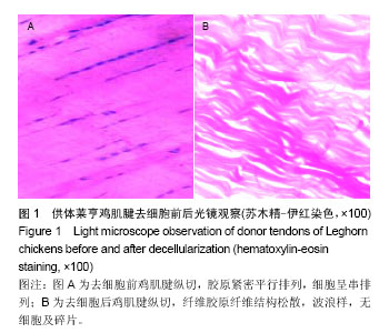
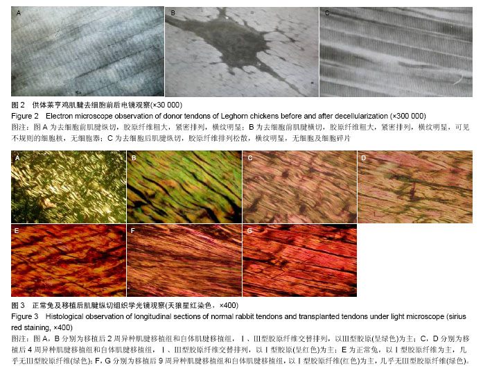
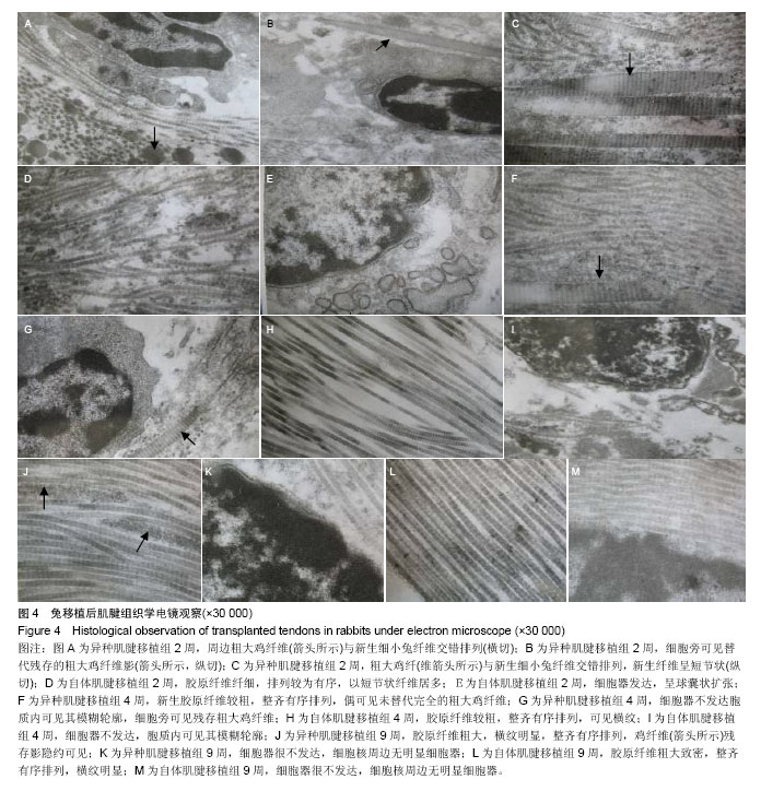
结果与结论:①肌腱经过化学去细胞处理后色泽变白,质地较前柔软,去细胞前可见细胞与胶原纤维交替紧密排列,去细胞后胶原排列相对松散,且无细胞及细胞碎片,去细胞后肌腱的力学强度较术前减弱。②由电镜图片直观看到:随移植时间的延长,移植的粗大鸡肌腱胶原纤维逐渐被再生的兔肌腱胶原纤维最终替代,而新生成的纤细胶原纤维经改造塑形变为粗细相等的较粗大纤维,排列方向逐渐趋于平行,在结构和功能上达到正常肌腱水平。结果表明,去细胞后的最大抗拉力是去细胞前的83.44%,能够满足肌腱移植生物力学的要求。最终肌腱修复是再生胶原纤维形成的结果,异种肌腱经过理化方法处理后可作为临床肌腱修复的生长支架使用。

中国组织工程研究杂志出版内容重点:生物材料;骨生物材料; 口腔生物材料; 纳米材料; 缓释材料; 材料相容性;组织工程


全文链接:

关键词: 生物材料, 材料相容性, 去细胞, 异种肌腱, 组织工程, 肌腱修复, 生物力学

Abstract:

BACKGROUND: Autologous tendons for repair of tendon defects are limited due to the supply of available and can induce tendon dysfunction. Limited sources and high cost of tendon allograft are also difficult to meet the clinical needs.

OBJECTIVE: To provide n theoretical basis for xenogeneic tendon as a clinical tissue-engineered tendon scaffold by observing microscopic changes of xenogeneic tendon in repair of tendon defects at different periods.
METHODS: Flexor tendons from Leghorn chickens (6 months old) were treated chemically as xenogeneic tendon donors. Thirty-six Japanese white rabbits were used to make 2 cm defect models of the intermediate beam in the Achilles tendon of bilateral hind limbs and then divided into two groups randomly, 18 in each group. The experimental group was treated with acellular xenogeneic tendon transplantation and the control group was treated with autologous tendon transplantation. Tendon suture was done using 4-0 non-invasive tendon suture line in double “8” way and the grafts were straightened and immobilized by plaster for 2 weeks. The donor tendons were conducted by general observation, biomechanical test, and engineering observation by optical and electron microscope before and after decellularization. At 2, 4, 9 weeks after transplantation, six rabbits in each group were evaluated by means of optical and electron microscope observation.
RESULTS AND CONCLUSION: (1) The color of tendon cells treated chemically became white and the texture was softer than that before decellularization. The cells and collagen fibers arranged closely and alternately before decellularization, but the arrangement of the collagen fibers was loose relatively and there were no cells and cell debris after decellularization. Moreover, the mechanical strength of the acellular tendon was reduced than that before decellularization. (2) Electron microscope observation: With the increase of transplantation time, the collagen fibers of the transplanted thick chicken tendons were gradually replaced by the ones of the regenerated rabbit tendons. The newly generated collagen fibers were transferred to same-thickness fibers and arrange parallelly, and their structure and function were close to the normal tendons. The maximum tensile resistance after decellularization was 83.44% as before, which could meet the needs of tendon mechanical strength. Ultimately tendon repair is the result of the collagen fiber regeneration, and the xenogeneic tendon treated by chemical methods can be used as the growth scaffold for clinical tendon repair.

中国组织工程研究杂志出版内容重点:生物材料;骨生物材料; 口腔生物材料; 纳米材料; 缓释材料; 材料相容性;组织工程


全文链接:

Key words: Tendons, Transplantation, Heterologous, Biomechanics

中图分类号: