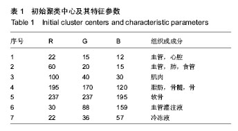
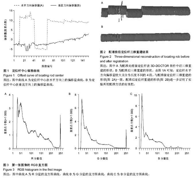
| [1] 姚震,陈林.我国心血管疾病现状与展望[J].海南医学,2013,(13): 1873-1876.
[2] Chen XJ, Nacif MS, Liu ST, et al. A framework of whole heart extracellular volume fraction estimation for low-dose cardiac CT images. IEEE Trans Inf Technol Biomed. 2012;16(5): 842-852.
[3] 周贤惠,翟梅玲,李晋新,等.基于CARTO系统的三维可视化心脏模型在心血管教学中的应用价值[J].新疆医科大学学报,2013, (6):885-887.
[4] 盖长清.基于GPU的心脏体绘制与电生理仿真方法研究[D].哈尔滨:哈尔滨工业大学,2011.
[5] 张雷.心脏电生理的快速仿真和交互式可视化方法研究[D].哈尔滨:哈尔滨工业大学,2013.
[6] 钟春燕,郭燕丽,张绍祥,等.基于中国可视化人体数据集和双源CT数据集的心脏三维可视化模型[J].中国医学影像技术,2011, (10):2127-2130.
[7] 蒋峻,谢叻,方彬吉,等.基于中国数字人数据的心脏三维模型的重建[J].组织工程与重建外科杂志,2014,(1):8-10.
[8] 邱鹏,李桥,刘兵,等.基于虚拟人数据的心脏表面模型三维重建及显示[J].生物医学工程研究,2005,(3):150-152.
[9] 李俊峡.心血管疾病介入治疗发展概述及展望[J].解放军医药杂志,2012,(9):1-6.
[10] 罗渝兰,王景熙,郑昌琼.图像分割在生物医学工程中的应用[J].计算机应用,2002,(8):20-22.
[11] 郭艳蓉,蒋建国,郝世杰,等.统计相似度特征的医学图像分割[J].中国图象图形学报,2013,(2):225-234.
[12] 王弈,李传富.人工智能方法在医学图像处理中的研究新进展[J].中国医学物理学杂志,2013,(3):4138-4143.
[13] 兰红.多阈值优化的交互式医学图像分割方法[J].计算机科学, 2013,(9):296-299.
[14] 许新征,丁世飞,史忠植,等.图像分割的新理论和新方法[J].电子学报,2010,(S1):76-82.
[15] Piecuch P, Wloch M. Renormalized coupled-cluster methods exploiting left eigenstates of the similarity-transformed Hamiltonian. J Chem Phys. 2005;123(22):224105.
[16] Yu QY, Clausi DA. IRGS: image segmentation using edge penalties and region growing. IEEE Trans Pattern Anal Mach Intell. 2008;30(12):2126-2139.
[17] Dehmeshi J, Amin H, Valdivieso M, et al. Segmentation of pulmonary nodules in thoracic CT scans: a region growing approach. IEEE Trans Med Imaging. 2008;27(4):467-480.
[18] 朱蛟英.心内膜电生理标测导航技术中电场的有限元分析[D].郑州:郑州大学,2012.
[19] 隆兴银.基于人体实体切片的图像处理及三维重建技术研究[D].成都:四川大学,2005.
[20] 张贵英,张先杰.医学图像分割技术研究[J].医学信息(上旬刊), 2011,(1):533-535.
[21] Cardenes R, de Luis-Garcia R, Bach-Cuadra M. A multidimensional segmentation evaluation for medical image data. Comput Methods Programs Biomed. 2009;96(2):108-125.
[22] 陈宇飞,吴启迪,赵卫东,等.基于图像熵的快速Chan-Vese模型分割算法[J].同济大学学报(自然科学版),2011,(5):738-744.
[23] 神方舟.多模态虚拟心脏可视化方法研究[D].哈尔滨:哈尔滨工业大学,2012.
[24] 乔海峰.心内导管三维定位导航软件系统设计及关键技术研究[D]. 郑州大学,2011.
[25] 乔海峰,苏智剑.消融导管三维定位导航系统中心脏多腔模型的构建[J].中国组织工程研究与临床康复,2011,(4):643-647.
[26] 钟世镇.数字人-信息与生命科学结合的新领域[J].科技导报, 2005,(2):9-12.
[27] Rastgarpour M, Shanbehzadeh J, Soltanian-Zadeh H. A hybrid method based on fuzzy clustering and local region-based level set for segmentation of inhomogeneous medical images. J Med Syst. 2014;38(8):68-83.
[28] 黄文博,燕杨,王云吉.医学图像分割方法综述[J].长春师范学院学报,2013,(4):22-25.
[29] Sharma N, Aggarwal LM. Automated medical image segmentation techniques. J Med Phys. 2010;35(1):3-15.
[30] Aja-Fernandez S, Vegas-Sanchez-Ferrero G, Martin F, et al. Soft thresholding for medical image segmentation. 2010 Annual International Conference of the IEEE Engineering in Medicine and Biology Society (EMBC). 2010.
[31] Gong X, Zhou Y, Zhou H, et al. Ultrasound Image Edge Detection Based on a Novel Multiplicative Gradient and Canny Operator. Ultrason Imaging. 2014 [Epub ahead of print].
[32] Jia T, Zhang H, Meng H. A novel lung nodules detection scheme based on vessel segmentation on CT images. Biomed Mater Eng. 2014;24(6):3179-3186.
[33] Panetta K, Gao C, Agaian S, et al. Nonreference medical image edge map measure. Int J Biomed Imaging. 2014; 2014: 931375.
[34] Koprowski R, Nowińska A, Wyl?ga?a E, et al. A new algorithm and problems in automatic anterior eye chamber volume determining. Comput Biol Med. 2014;52:144-152.
[35] Vunckx K, Dupont P, Goffin K, et al. Voxel-based comparison of state-of-the-art reconstruction algorithms for 18F-FDG PET brain imaging using simulated and clinical data. Neuroimage. 2014 [Epub ahead of print].
[36] Sárándi I, Claßen DP, Astvatsatourov A, Pfaar O, et al. Quantitative conjunctival provocation test for controlled clinical trials. Methods Inf Med. 2014;53(4):238-244.
[37] Gwo CY, Gwo A, Wei CH, et al. Identification of breast contour for nipple segmentation in breast magnetic resonance images. Med Phys. 2014;41(2):022304.
[38] Ben Chaabane S, Fnaiech F. Color edges extraction using statistical features and automatic threshold technique: application to the breast cancer cells. Biomed Eng Online. 2014;13:4.
[39] Skoura A, Nuzhnaya T, Megalooikonomou V. Integrating edge detection and fuzzy connectedness for automated segmentation of anatomical branching structures. Int J Bioinform Res Appl. 2014;10(1):93-109.
[40] Navlakha S, Ahammad P, Myers EW. Unsupervised segmentation of noisy electron microscopy images using salient watersheds and region merging. BMC Bioinformatics. 2013;14:294.
[41] Gillner M, Eppig T, Langenbucher A. Automatic intraocular lens segmentation and detection in optical coherence tomography images. Z Med Phys. 2014;24(2):104-111.
[42] Toossi MT, Pourreza HR, Zare H, et al. An effective hair removal algorithm for dermoscopy images. Skin Res Technol. 2013;19(3):230-235.
[43] Cuevas E, Oliva D, Díaz M, et al. White blood cell segmentation by circle detection using electromagnetism-like optimization. Comput Math Methods Med. 2013; 2013: 395071.
[44] Qian X, Wang J, Guo S, et al. An active contour model for medical image segmentation with application to brain CT image. Med Phys. 2013;40(2):021911.
[45] Kurugol S, San Jose Estepar R, Ross J, et al. Aorta segmentation with a 3D level set approach and quantification of aortic calcifications in non-contrast chest CT. Conf Proc IEEE Eng Med Biol Soc. 2012;2012:2343-2346.
[46] Tomasi G, Shepherd T, Turkheimer F, et al. Comparative assessment of segmentation algorithms for tumor delineation on a test-retest [(11)C]choline dataset. Med Phys. 2012; 39(12): 7571-7579.
[47] Ortiz CG, Martel AL. Automatic atlas-based segmentation of the breast in MRI for 3D breast volume computation. Med Phys. 2012;39(10):5835-5848.
[48] Xu T, Mandal M, Long R, et al. An edge-region force guided active shape approach for automatic lung field detection in chest radiographs. Comput Med Imaging Graph. 2012;36(6): 452-463.
[49] Bergmeir C, García Silvente M, Benítez JM. Segmentation of cervical cell nuclei in high-resolution microscopic images: A new algorithm and a web-based software framework. Comput Methods Programs Biomed. 2012;107(3):497-512.
[50] Kazerooni AF, Ahmadian A, Serej ND, et al. Segmentation of brain tumors in MRI images using multi-scale gradient vector flow. Conf Proc IEEE Eng Med Biol Soc. 2011;2011: 7973- 7976.
[51] Li K, Tang Z, Liu GJ, et al. Three-dimensional reconstruction of paracentesis approach in transjugular intrahepatic portosystemic shunt. Anat Sci Int. 2012;87(2):71-79.
[52] Tsantis S, Kagadis GC, Katsanos K, et al. Automatic vessel lumen segmentation and stent strut detection in intravascular optical coherence tomography. Med Phys. 2012;39(1): 503-513.
[53] Dizdaro?lu B, Ataer-Cansizoglu E, Kalpathy-Cramer J, et al. Level sets for retinal vasculature segmentation using seeds from ridges and edges from phase maps. IEEE Int Workshop Mach Learn Signal Process. 2012:1-6.
[54] Lecron F, Mahmoudi SA, Benjelloun M, et al. Heterogeneous computing for vertebra detection and segmentation in x-ray images. Int J Biomed Imaging. 2011;2011:640208.
[55] 刘萍.正常女性骨盆数字化三维重建的研究[D].广州:南方医科大学,2012.
[56] 吴景鹏,冯杭,黄臣,等.中国数字人血管的分割和重建方法研究[J].计算机与数字工程,2010,(11):132-135.
[57] 晏义.三维人体模型重建、分割及尺寸提取技术研究[D].株洲:湖南工业大学,2012. |
.jpg)