| [1]Piirsoo M, Kaljas A, Tamm K, et al. Expression of NGF and GDNF family members and their receptors during peripheral nerve development and differentiation of Schwann cells in vitro. J Neurosci Lett.2010;469(1):135-140.
[2]Ma Z, Wang J, Song F, et al. Critical period of axoglial signaling between Neuregulin-1 and BDNF required for early Schwann cell survival and differentiation. J Neurosci. 2011; 31(26): 9630-9640.
[3]Bhatheja K, Field J. Schwann cells: origins and role in axonal main-tenance and regeneration. Int J Biochem Cell Biol. 2006; 38(12):1995-1999.
[4]Tao HY, He B, Liu SQ, et al. Effect of carboxymethylated chitosan on the biosynthesis of NGF and activation of the Wnt/β-catenin signaling pathway in the proliferation of Schwann cells. Eur J Pharmacol, 2013;702(1-3):85-92.
[5]Torigoe K, H ashimoto K, Lundborg G. A role of migratory Schwann cells in a conditioning effect of peripheral nerve regeneration. J Exp Neurol.1999;160(1):99-108.
[6]Yao D, Li M, Shen D, et al. Expression changes and bioinformatic analysis of Wallerian degeneration after sciatic nerve injury in rat. Neurosci Bull,2013,29(3): 321-32.
[7]Frostick SP, Yin Q, Kemp GJ. Schwann cells, neurotrophic factors, and peripheral nerve regeneration. Microsurgery. 1998;18(7): 397-405.
[8]Sangsanoh P, Waleetorncheepsawat S, Suwantong O, et al. In vitro Bio-Compatibility of schwann cells on surfaces of biocompatible polymeric electrospun fibrous and solution-cast film scaffolds. Biomacromolecules.2007;8(5): 1587-1594.
[9]Sinis N, Schaller HE, Becker ST, et al. Long nerve gaps limit the Regenerative potential of bioartificial nerve conduits filled with Schwann cells. Restor Neurol Neurosci.2007;25(2): 131-141.
[10]Waller A. Experiments on the section of the glossopharyngeal and hypoglossal nerves of the frog, and observations of the alterations produced thereby in the structure of their primitive ?bres. Philos. Trans. R. Soc. London B, No. 140. (1850): 423-429.
[11]赵富强,姜保国,张培训,等.鼠坐骨神经损伤后雪旺细胞增殖规律的免疫组织化学观察[J].中华外科杂志,2006,44(4): 268-270.
[12]Li RH, Sliwkowski MX, Lo J, et al. Establishment of Schwann cell lines from normal adult and embryonic rat dorsal root ganglia. J Neurosci Methods.1996;67(1):57-69.
[13]付国强,李学拥.兔坐骨神经电损伤后许旺细胞的增殖变化[J]. 中国临床康复,2005, 9(34):18-19.
[14]Ide C, Tohyama K, Yokota R,et al. Schwann cell processes guide regeneration of peripheral axons. Brain Res.1983; 288(1-2):61-75.
[15]Dong Z, Brennan A, Liu N,et al. Neu differentiation factor is a neuron-glia signal and regulates survival, proliferation, and maturation of rat Schwann cell precursors. Neuron. 1995;1 5(3):585-596.
[16]Wang DY, Huang YC, Chiang H, et al. Microcontact printing of laminin on oxygen plasma activated substrates for the alignment and growth of Schwann cells. J Biomed Mater Res B Appl Biomater.2007;80(2):447-453.
[17]Bunge RP, Bunge MB, Eldridge CF. Linkage between axonal ensheathment and basal lamina production by Schwann cells. Annu Rev Neurosci.1986;9:305-328.
[18]Maisonpierre PC, Belluscio L, Friedman B, et al. NT-3, BDNF, and NGF in the developing rat nervous system: parallel as well as reciprocal patterns of expression. Neuron. 1990;5(4): 501-509.
[19]Lindsay RM. Nerve growth factors (NGF, BDNF) enhance axonal regeneration but are not required for survival of adult sensory neurons. J Neurosci.1988;8(7): 2394-2405.
[20]Shakhbazau A, Kawasoe J, Hoyng SA, et al.Early regenerative effects of NGF-transduced Schwann cells in peripheral nerve repair. Mol Cell Neurosci.2012;50(1): 103-112.
[21]Shin YH, Lee SJ, Jung J.Extracellular ATP inhibits Schwann cell dedifferentiation and proliferation in an ex vivo model of Wallerian degeneration.Biochem Biophys Res Commun. 2013;430(2):852-857.
[22]Somandin C, Gerber D, Pereira JA,et al.LITAF (SIMPLE) regulates Wallerian degeneration after injury but is not essential for peripheral nerve development and maintenance: implications for Charcot-Marie-Tooth disease. Glia. 2012; 60(10):1518-1528.
[23]Gaudet AD, Popovich PG, Ramer MS. Wallerian degeneration: gaining perspective on inflammatory events after peripheral nerve injury.J Neuroinflammation. 2011;8:110.
[24]Jung J, Cai W, Jang SY,et al.Transient lysosomal activation is essential for p75 nerve growth factor receptor expression in myelinated Schwann cells during Wallerian degeneration. Anat Cell Biol. 2011;44(1):41-49.
[25]Jung J, Cai W, Lee HK, et al. Actin polymerization is essential for myelin sheath fragmentation during Wallerian degeneration. J Neurosci.2011;31(6):2009-2015.
[26]Chaballe L, Close P, Sempels M,et al.Involvement of placental growth factor in Wallerian degeneration.Glia. 2011; 59(3):379-396.
[27]Girolami EI, Bouhy D, Haber M, et al.Differential expression and potential role of SOCS1 and SOCS3 in Wallerian degeneration in injured peripheral nerve.Exp Neurol. 2010; 223(1):173-182.
[28]Lee HK, Seo IA, Suh DJ, et al.Interleukin-6 is required for the early induction of glial fibrillary acidic protein in Schwann cells during Wallerian degeneration.J Neurochem. 2009;108(3): 776-786.
[29]Keilhoff G, Schild L, Fansa H.Minocycline protects Schwann cells from ischemia-like injury and promotes axonal outgrowth in bioartificial nerve grafts lacking Wallerian degeneration.Exp Neurol. 2008;212(1):189-200.
[30]刘璋寅.周围神经中许旺细胞特异性标记物[J].组织工程与重建外科杂志,2012,8(5):292-297.
[31]Kubota A, Komiyama A, Matsumoto M, et al.Effect of macrophage suppression with silica on the proliferation of Schwann cells during Wallerian degeneration.Brain Res. 1998;802(1-2):254-258.
[32]Jung J, Jo HW, Kwon H,et al.ATP Release through Lysosomal Exocytosis from Peripheral Nerves: The Effect of Lysosomal Exocytosis on Peripheral Nerve Degeneration and Regeneration after Nerve Injury.Biomed Res Int. 2014;2014: 936891. |
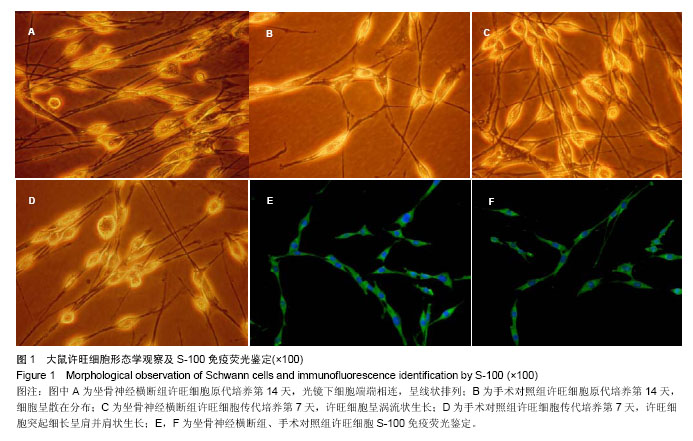
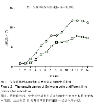
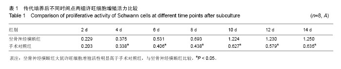
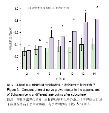
.jpg)