中国组织工程研究 ›› 2013, Vol. 17 ›› Issue (37): 6636-6640.doi: 10.3969/j.issn.2095-4344.2013.37.015

• 组织构建与生物活性因子 tissue construction and bioactive factors • 上一篇    下一篇

眼针治疗急性脑缺血再灌注损伤大鼠海马组织细胞间黏附因子1的变化

潘  茜,王  哲,高  原,于  丹,王  莹,井  欢,王守岩,王  健,关洪全   

  1. 辽宁中医药大学,辽宁省沈阳市  110847
  • 收稿日期:2013-01-18 修回日期:2013-05-22 出版日期:2013-09-10 发布日期:2013-09-10
  • 通讯作者: 关洪全,博士,教授,辽宁中医药大学,辽宁省沈阳市 110847 hongquanguan@sina.com
  • 作者简介:潘茜☆,女,1980年生,博士,讲师,主要从事病理学方面的研究。 panqian_pan@163.com
  • 基金资助:

    国家重点基础研究发展计划项目(2007CB512702)*;辽宁省教育厅课题(L2010351)基金资助*

Eye acupuncture therapy affects intercellular adhesion molecule 1 expression in rat hippocampus of acute cerebral ischemia-reperfusion injury

Pan Qian, Wang Zhe, Gao Yuan, Yu Dan, Wang Ying, Jing Huan, Wang Shou-yan, Wang Jian,  Guan Hong-quan   

  1. Liaoning University of Traditional Chinese Medicine, Shenyang  110847, Liaoning Province, China
  • Received:2013-01-18 Revised:2013-05-22 Online:2013-09-10 Published:2013-09-10
  • Contact: Guan Hong-quan, M.D., Professor, Liaoning University of Traditional Chinese Medicine, Shenyang 110847, Liaoning Province, China hongquanguan@sina.com
  • About author:Pan Qian☆, M.D., Lecturer, Liaoning University of Traditional Chinese Medicine, Shenyang 110847, Liaoning Province, China panqian_pan@163.com
  • Supported by:

    National Key Basic Research Development Project, No. 2007CB512702*; Project of Liaoning Provincial Department of Education, No. L2010351*

摘要:

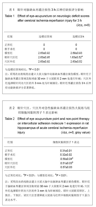
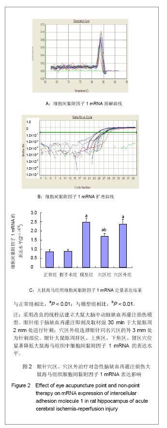
背景:病理情况下,细胞间黏附因子1表达明显升高,促进炎症反应的发生,加重脑组织损伤。
目的:观察眼针穴区、穴区外治疗对急性脑缺血再灌注损伤模型大鼠海马组织细胞间黏附因子1表达的影响。
方法:40只SD大鼠随机等分为正常组、假手术组、模型组、眼针穴区组和穴区外组。后3组用线栓法复制脑缺血再灌注损伤大鼠模型,栓塞线插入深度1.8-2.2 cm,眼针组于脑缺血再灌注即刻及取材前30 min,取肝区、上焦区、下焦区、肾区平刺眼眶周治疗20 min。穴区外组选择眼针同名穴区的外3 mm处为针刺部位,刺法与眼针穴区相同。
结果与结论:眼针穴区治疗后大鼠神经功能缺损评分下降,海马组织细胞间黏附因子1蛋白及mRNA表达显著降低(P < 0.01)。穴区外组治疗后大鼠神经功能缺损评分及海马组织细胞间黏附因子1蛋白及mRNA表达无明显变化。提示眼针具有明显的改善大鼠脑缺血再灌注损伤的作用,其机制与下调海马组织细胞间黏附因子表达有关。

关键词: 组织构建, 组织构建与生物活性因子, 眼针, 脑缺血再灌注损伤, 神经功能缺损评分, 细胞间黏附因子, 海马, 组织构建, 组织工程, 973项目

Abstract:

BACKGROUND: Significant increasing of intercellular adhesion molecule 1 expression can promote the occurrence of inflammatory response and increase brain tissue injury.
OBJECTIVE: To investigate the effect of eye acupuncture point and non-point therapy on intercellular adhesion molecule 1 expression in rat hippocampus of acute cerebral ischemia-reperfusion injury model.
METHODS: Forty Sprague Dawley rats were divided into normal group, sham-operation group, model group, eye acupuncture point group and eye acupuncture non-point group. Rats in the model group, eye acupuncture point group and eye acupuncture non-point group were used to establish the acute cerebral ischemia-reperfusion injury model with suture method, the suture was deep for 1.8-2.2 cm. In the eye acupuncture point group, the acupuncture points of hepatic region, upper energizer area, lower energizer area and kidney area were selected when cerebral ischemia-reperfusion occurred immediately and 30 minutes before drawing materials, then horizontal needling the acupuncture points around the orbit for 20 minutes. In the eye acupuncture non-point group, the puncture site was selected at 3 mm away from eye acupuncture non-point area, and then needled the puncture site with the same method in the eye acupuncture point group.
RESULTS AND CONCLUSION: After eye acupuncture point therapy, the neurological deficit scores were decreased, and the intercellular adhesion molecule 1 expression in rat hippocampus was significantly decreased (P < 0.01). After eye acupuncture non-point therapy, there were no significant changes in neurological deficit scores and the expression of intercellular adhesion molecule 1 protein and mRNA in rat hippocampus. The results indicate that eye acupuncture can significantly improve the rat cerebral ischemia-reperfusion injury, and the mechanism may relate with reducing the intercellular adhesion molecule 1 expression in rat hippocampus.

Key words: acupuncture, acupuncture points, reperfusion injury, cytokines, chemokines

中图分类号: