中国组织工程研究 ›› 2026, Vol. 30 ›› Issue (27): 7044-7052.doi: 10.12307/2026.299

• 骨与关节生物力学Bone and joint biomechanics • 上一篇    

L型钢板治疗骨质疏松性Schatzker Ⅱ型胫骨平台骨折的有效性及生物力学分析

史高龙,胡正辉,凌卓彦,谢宗刚   

  1. 苏州大学附属第二医院关节外科,江苏省苏州市   215000
  • 收稿日期:2025-09-29 接受日期:2025-12-16 出版日期:2026-09-28 发布日期:2026-04-17
  • 通讯作者: 谢宗刚,博士,主任医师,副主任,苏州大学附属第二医院关节外科,江苏省苏州市  215000
  • 作者简介:史高龙,男,1986年生,山东省日照市人,硕士,主治医师,主要从事髋关节创伤及慢病治疗方面的研究。 并列第一作者:胡正辉,男,1998年生,江苏省苏州市人,硕士,医师,主要从事髋、膝关节骨折方面的研究。
  • 基金资助:
    国家自然科学基金面上项目(81974334),项目参与人:谢宗刚

Efficacy and biomechanical analysis of L-shaped plate treatment for osteoporotic Schatzker type II tibial plateau fractures

Shi Gaolong, Hu Zhenghui, Ling Zhuoyan, Xie Zonggang   

  1. Department of Orthopedics, The Second Affiliated Hospital of Soochow University, Suzhou 215000, Jiangsu Province, China
  • Received:2025-09-29 Accepted:2025-12-16 Online:2026-09-28 Published:2026-04-17
  • Contact: Xie Zonggang, MD, Chief physician, Department of Orthopedics, The Second Affiliated Hospital of Soochow University, Suzhou 215000, Jiangsu Province, China
  • About author:Shi Gaolong, MS, Attending physician, Department of Orthopedics, The Second Affiliated Hospital of Soochow University, Suzhou 215000, Jiangsu Province, China Hu Zhenghui, MS, Physician, Department of Orthopedics, The Second Affiliated Hospital of Soochow University, Suzhou 215000, Jiangsu Province, China Shi Gaolong and Hu Zhenghui contributed equally to this article.
  • Supported by:
    National Natural Science Foundation of China, No. 81974334 (to XZG)

摘要:

文题释义:

骨质疏松:是一种以骨量减少、骨微结构破坏为特征的全身性骨病,导致骨骼脆性增加,易发生骨折。世界卫生组织(WHO)将其定义为骨密度T值≤-2.5(即较正常年轻人群骨密度低2.5个标准差)。骨质疏松患者骨骼中皮质骨变薄,松质骨小梁减少,骨强度显著下降,在老年人中尤为显著。
有限元分析:是一种数值模拟方法,广泛应用于工程和生物力学领域,用于分析复杂结构的力学性能。在骨科研究中,有限元分析通过构建骨骼和内固定装置的三维模型,划分有限单元(网格),并设置材料属性(如弹性模量、泊松比)和边界条件,模拟不同载荷下的应力、应变和位移。

摘要
背景:Schatzker Ⅱ型骨折往往累及后外侧平台,导致骨强度减弱、固定挑战增大及术后塌陷风险升高,亟需优化治疗策略以平衡稳定性和创伤最小化。
目的:评估单纯外侧L型钢板固定治疗骨质疏松性Schatzker Ⅱ型累及后外侧胫骨平台骨折的临床效果,并通过有限元分析比较其与外侧L型钢板联合后方T型钢板固定的生物力学性能。
方法:回顾性分析2018年1月至2023年12月苏州大学附属第二医院关节外科收治的39例骨质疏松性Schatzker Ⅱ型累及后外侧胫骨平台骨折患者的病历资料,分为L型钢板组(单纯外侧L型钢板固定,n=24)及联合组(外侧L型钢板联合后方T型钢板固定,n=15)。对比两组患者在手术时间、术中出血量、骨密度、术前胫骨平台塌陷、受伤至手术时间、术后影像学指标(胫骨平台内翻角、后倾角)、膝关节活动度及美国特种外科医院评分、Lysholm评分等方面的差异。同时基于健康成年男性膝关节CT数据建立Schatzker Ⅱ型胫骨平台骨折有限元模型,模型分为非骨质疏松组(A组:单纯L型钢板;B组:L型钢板+T型钢板)与骨质疏松组(C组:单纯L型钢板;D组:L型钢板+T型钢板)。在250,500,750 N轴向载荷下,分析各组模型的整体位移、胫骨应力及内固定应力。
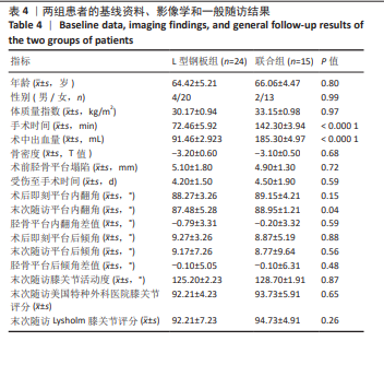
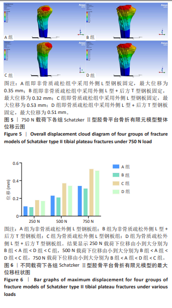
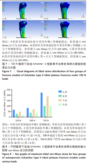
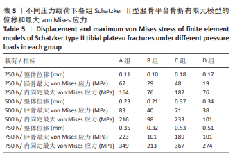
结果与结论:①临床试验结果:两组患者在年龄、性别、体质量指数、骨密度、术前胫骨平台塌陷情况以及受伤至手术时间方面无显著性差异(P > 0.05);L型钢板组手术时间及术中出血量均显著少于联合组(P < 0.000 1);术后即刻胫骨平台内翻角及后倾角组间比较无显著性差异(P > 0.05);末次随访时内翻角L型钢板组小于联合组(P=0.04),后倾角组间比较仍无显著性差异(P > 0.05);末次随访时膝关节活动度、美国特种外科医院评分、Lysholm评分组间均无显著性差异(P > 0.05)。②有限元结果:在相同载荷下,无论是非骨质疏松骨模型还是骨质疏松骨模型,采用T型钢板辅助固定(B/D组)比单纯L型钢板固定(A/C组)在整体位移和胫骨、内固定应力上具有更优异的性能。③提示对于骨质疏松性Schatzker Ⅱ型累及后外侧胫骨平台的骨折,单纯外侧L型钢板固定可获得满意的复位稳定性及功能恢复,其临床效果与双钢板技术相当;有限元分析表明双钢板固定力学性能更优,但考虑到单纯外侧L型钢板固定手术创伤显著更小且临床效果良好,无需常规加用后方T型钢板。


中国组织工程研究杂志出版内容重点:人工关节;骨植入物;脊柱;骨折;内固定;数字化骨科;组织工程

关键词: 骨质疏松, Schatzker Ⅱ型骨折, 胫骨平台骨折, 外侧L型钢板, 有限元分析

Abstract: BACKGROUND: Schatzke type II fractures often involve the posterolateral plateau, resulting in weakened bone strength, increased fixation challenges, and increased risk of postoperative collapse. Optimizing treatment strategies to balance stability and minimize trauma is urgently needed.
OBJECTIVE: To evaluate the clinical efficacy of simple lateral L-shaped plate fixation for osteoporotic Schatzker type II tibial plateau fractures involving the posterolateral aspect, and to compare its biomechanical properties with those of lateral L-shaped plate combined with posterior T-shaped plate fixation using finite element analysis.
METHODS: A retrospective analysis was conducted on 39 patients with osteoporotic Schatzker type II tibial plateau fractures involving the posterolateral aspect treated between January 2018 and December 2023. Patients were divided into L-shaped plate group (simple lateral L-shaped plate fixation, n=24) and combined group (lateral L-shaped plate combined with posterior T-shaped plate fixation, n=15). Key indicators were compared, including operative time, intraoperative blood loss, bone mineral density, preoperative tibial plateau collapse, time from injury to surgery, postoperative radiographic parameters (tibial plateau varus angle and posterior tilt angle), knee range of motion, and Hospital for Special Surgery and Lysholm scores. Simultaneously, finite element models of Schatzker type II tibial plateau fracture were established based on CT data from a healthy adult male. Four groups were constructed: non-osteoporotic bone (Group A: simple L-shaped plate; Group B: L-shaped + T-shaped plate) and osteoporotic bone (Group C: simple L-shaped plate; Group D: L-shaped + T-shaped plate). Under axial loads of 250, 500, and 750 N, the overall displacement, tibial stress, and internal fixation stress of the models were analyzed in each group.
RESULTS AND CONCLUSION: (1) Clinical trial results: The results showed no significant differences between the two groups in age, gender, body mass index, bone mineral density, preoperative tibial plateau collapse, or time from injury to surgery (P > 0.05). Operative time and intraoperative blood loss in L-shaped plate group were significantly lower than those in combined group (P < 0.000 1). Immediate postoperative tibial plateau varus angle and posterior tilt angle showed no significant differences between groups (P > 0.05). At the final follow-up, the varus angle in L-shaped plate group was smaller than in combined group (P=0.04), while the posterior tilt angle showed no significant difference (P > 0.05). At the final follow-up, no statistical differences were observed in knee range of motion, Hospital for Special Surgery scores, or Lysholm scores between groups (P > 0.05). (2) Finite element analysis results: Under the same loads, both non-osteoporotic and osteoporotic bone models with T-shaped plate-assisted fixation (Groups B/D) exhibited superior performance in terms of overall displacement and lower tibial stress compared with simple L-shaped plate fixation (Groups A/C). (3) These findings suggest that for osteoporotic Schatzker type II tibial plateau fractures involving the posterolateral aspect, simple lateral L-shaped plate fixation can achieve satisfactory reduction stability and functional recovery, with clinical outcomes comparable to dual-plate fixation. Finite element analysis indicates superior biomechanical performance with dual-plate fixation; however, considering the significantly reduced surgical trauma and favorable clinical outcomes with simple lateral L-shaped plate fixation, routine addition of a posterior T-shaped plate is not necessary.

Key words: osteoporosis, Schatzker type II fracture, tibial plateau fracture, lateral L-shaped plate, finite element analysis

中图分类号: