[1] STARK D, DENZINGER M, EBERT L, et al. Therapeutic approaches of diametaphyseal radius fractures in children. Arch Orthop Trauma Surg. 2024; 144(3):1179-1188.
[2] SNELLING PJ, JONES P, GILLESPIE A, et al. Point-of-Care Ultrasound Fracture-Physis Distance Association with Salter-Harris II Fractures of the Distal Radius in Children: The “POCUS 1-cm Rule”. Ultrasound Med Biol. 2023;49(2):520-526.
[3] WU X, MENG X, WANG S, et al. Ultrasound-guided closed reduction of distal radius Salter-Harris I and II fracture in children. Eur J Trauma Emerg Surg. 2025; 51(1):220.
[4] CEPELA DJ, TARTAGLIONE JP, DOOLEY TP, et al. Classifications In Brief: Salter-Harris Classification of Pediatric Physeal Fractures. Clin Orthop Relat Res. 2016; 474(11):2531-2537.
[5] LAURER H, SANDER A, WUTZLER S, et al. Therapy principles of distal fractures of the forearm in childhood. Chirurg. 2009;80(11):1042-1052.
[6] LIU DS, MURRAY MM, BAE DS, et al. Pediatric and Adolescent Distal Radius Fractures: Current Concepts and Treatment Recommendations. J Am Acad Orthop Surg. 2024;32(21):e1079-e1089.
[7] BERGKVIST A, LUNDQVIST E, PANTZAR-CASTILLA E. Distal radius fractures in children aged 5-12 years: a Swedish nationwide register-based study of 25 777 patients. BMC Musculoskelet Disord. 2023;24(1):560.
[8] GALÁN-OLLEROS M, MAYANS-SANESTEBAN J, MARTÍNEZ-ÁLVAREZ S, et al. Is reduction necessary in overriding metaphyseal distal radius fractures in children under 11 years: a systematic review and meta-analysis of comparative studies. Eur J Orthop Surg Traumatol. 2024;34(5):2225-2234.
[9] MESLIER QA, SHEFELBINE SJ. Using Finite Element Modeling in Bone Mechanoadaptation. Curr Osteoporos Rep. 2023;21(2):105-116.
[10] 祁艳梅,唐治,周晟.腕关节损伤的影像学检查研究进展[J].西北国防医学杂志,2020,41(9):589-594.
[11] 李永耀,程灏,赵勇,等.夹板固定治疗尺骨茎突骨折的三维有限元分析[J].中国组织工程研究,2018,22(11):1737-1742.
[12] 钦斌,黄永火,欧阳羽,等.轴向应力作用下的舟骨有限元分析[J].第三军医大学学报,2010,32(11):1213-1215.
[13] 李永耀,赵勇,程灏,等.夹板弹性固定桡骨远端FrykmanⅧ型骨折尺侧柱稳定性的有限元分析[J].中国组织工程研究,2020,24(30):4769-4774.
[14] LOTZ JC, CHEAL EJ, HAYES WC. Fracture prediction for the proximal femur using finite element models: Part II--Nonlinear analysis. J Biomech Eng. 1991; 113(4):361-365.
[15] LEWIS GS, MISCHLER D, WEE H, et al. Finite Element Analysis of Fracture Fixation. Curr Osteoporos Rep. 2021;19(4):403-416.
[16] DAILEY HL, KERSH ME, COLLINS CJ, et al. Mechanical Biomarkers in Bone Using Image-Based Finite Element Analysis. Curr Osteoporos Rep. 2023;21(3):266-277.
[17] CUI H, WEI W, SHAO Y, et al. Finite element analysis of fixation effect for femoral neck fracture under different fixation configurations. Comput Methods Biomech Biomed Engin. 2022;25(2):132-139.
[18] FERRE LS, DI NISIO FG, MENDONÇA CJA, et al. Comparative analysis of tibial plateau fracture osteosynthesis: A finite element study. J Mech Behav Biomed Mater. 2022;134:105392.
[19] CAO R, TAN J, ZHANG Y, et al. Finite element analysis of antegrade and retrograde internal intramedullary nailing for mid-shaft clavicle fracture. BMC Musculoskelet Disord. 2025;26(1):172.
[20] PRASIL L, ANDRAOS R, RISHMANY J, et al. Osteosynthesis of an extra-articular distal radius fracture using a palmar locking plate with 4 epiphyseal screws (Gold Standard) versus 2 epiphyseal screws: Finite element analysis. Injury. 2025;56(7):112360.
[21] MURAKAMI S, ZHAO Y, MIZUNO K, et al. Finite element analysis of hip fracture risk in elderly female: The effects of soft tissue shape, fall direction, and interventions. J Biomech. 2024;172:112199.
[22] GHEZELBASH F, SCHMIDT H, SHIRAZI-ADL A, et al. Internal load-sharing in the human passive lumbar spine: Review of in vitro and finite element model studies. J Biomech. 2020;102:109441.
[23] JOHNSON JE, ANDERSON DD. Influence of bone material behavior assumption on finite element-predicted tibia-implant micromotions in total ankle replacement. J Biomech. 2025;189:112832.
[24] HUANG F, TAN R, WANG MW, et al. Threedimensional finite element analysis: Anatomical splint fixation for Colles fractures. Exp Ther Med. 2024;27(3):98.
[25] XU C, WANG L, ZHANG M, et al. A clinical study on the application of three-dimensionally printed splints combined with finite element analysis in paediatric distal radius fractures. Front Pediatr. 2025;13:1559762.
[26] NGUYEN JC, CAINE D. The Immature Pediatric Appendicular Skeleton. Semin Musculoskelet Radiol. 2024;28(4):361-374.
[27] 马海龙,李阳,袁悦,等.超声联合关节造影辅助治疗儿童桡骨远端Salter-Harris Ⅲ型骨骺骨折的中短期疗效[J].中华小儿外科杂志,2024, 45(2):138-142.
[28] RAUER T, PAPE HC, GAMBLE JG, et al. Transitional fracture of the distal radius: a rare injury in adolescent athletes. Case series and literature review. Eur J Med Res. 2020;25(1):21.
[29] 赵纯,刘政,李静.郑氏手法整复结合夹板中立板外固定治疗儿童桡骨远端骨骺骨折[J].四川中医,2024,42(9):160-163.
[30] 刘琦,苏联彬,李楠竹.闭合复位经皮克氏针与石膏固定治疗儿童桡骨远端Salter-HarrisⅠ型/Ⅱ型骨骺骨折的临床效果[J].中国医学创新,2024, 21(14):1-5.
[31] 田野,侯婷婷.闭合复位交叉克氏针内固定治疗儿童桡骨远端Salter-Harris Ⅱ型骨折[J].临床骨科杂志,2024,27(5):694-697.
[32] 陈国富,姚灿,梁军波,等.闭合复位经皮克氏针及石膏固定治疗儿童桡骨远端骨折[J].临床骨科杂志,2023,26(1):83-86.
[33] STEVENS NM, PEAN C, NORRIS Z, et al. Clinical and Radiographic Comparison of Splinting Constructs for Distal Radius Fractures: An Effort to Free the Elbow. J Orthop Trauma. 2022;36(9):465-468.
[34] 朱晓飞,郑军,端磊.改良小夹板固定技术与传统石膏固定治疗桡骨远端骨折的疗效比较[J].外科研究与新技术,2022,11(3):156-160.
[35] PAKARINEN O, SAARINEN AJ, PONKILAINEN VT, et al. Soft bandage, splint or cast as the treatment of distal forearm torus fracture in children: a systematic review and meta-analysis. Sci Rep. 2024;14(1):21052.
[36] 谭景超,戴戈荣,王广伟,等.手法复位超腕关节夹板联合石膏托固定治疗Salter-HarrisⅡ型儿童桡骨远端骨骺骨折[J].中医正骨,2020,32(2):69-72.
[37] 陈元荣,江湧,朱秋贤,等.两种方法治疗桡骨远端骨骺骨折的临床疗效比较研究[J].中国中医骨伤科杂志,2020,28(9):34-39.
[38] 郝博川,谢克波,张君松,等.手法整复硬纸夹板外固定治疗儿童桡骨远端骨骺骨折26例[J].中国中医骨伤科杂志,2023,31(10):57-60.
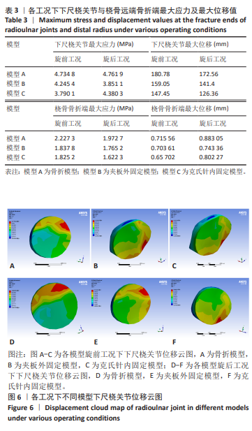
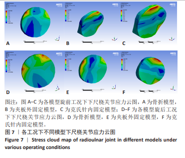
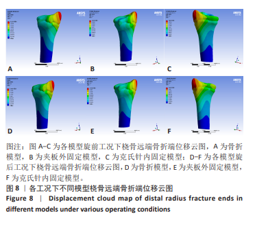
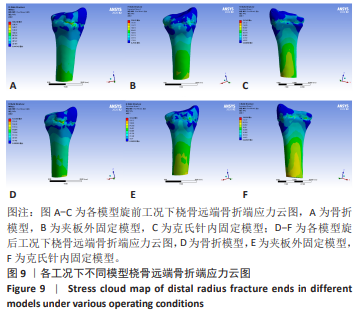
[39] 周云帆,孟祥奇,欧炳金,等.夹板治疗儿童桡骨远端骨骺骨折的有限元分析[J].中国当代医药,2023,30(17):82-86.
|