中国组织工程研究 ›› 2016, Vol. 20 ›› Issue (7): 940-946.doi: 10.3969/j.issn.2095-4344.2016.07.004

• 软骨组织构建 cartilage tissue construction • 上一篇    下一篇

MicroRNAs在缺氧诱导因子1α缺失椎间盘组织中的差异性表达

孟祥超,刘卓超,王 君,周 琦,齐 进,张兴凯   

  1. 上海交通大学医学院附属瑞金医院骨科,上海市伤骨科研究所,上海市 200025
  • 收稿日期:2015-08-07 出版日期:2016-02-12 发布日期:2016-02-12
  • 通讯作者: 张兴凯,博士,副主任医师,上海交通大学医学院附属瑞金医院骨科,上海市伤骨科研究所,上海市 200025
  • 作者简介:孟祥超,男,1990年生,山东省兰陵县人,汉族,上海交通大学医学院在读硕士,主要从事椎间盘退变机制的研究。
  • 基金资助:
    国家自然科学基金(81071502);上海科学技术委员会科研计划项目(15ZR1437600)

Differential expression of microRNAs in the intervertebral disc of hypoxia-inducible factor-1alpha deficient mice 

Meng Xiang-chao, Liu Zhuo-chao, Wang Jun, Zhou Qi, Qi Jin, Zhang Xing-kai   

  1. Department of Orthopedics, Ruijin Hospital, Shanghai Jiao Tong University School of Medicine, Shanghai Institute of Traumatology and Orthopaedics, Shanghai 200025, China
  • Received:2015-08-07 Online:2016-02-12 Published:2016-02-12
  • Contact: Zhang Xing-kai, M.D., Associate chief physician, Department of Orthopedics, Ruijin Hospital, Shanghai Jiao Tong University School of Medicine, Shanghai Institute of Traumatology and Orthopaedics, Shanghai 200025, China
  • About author:Meng Xiang-chao, Studying for master’s degree, Department of Orthopedics, Ruijin Hospital, Shanghai Jiao Tong University School of Medicine, Shanghai Institute of Traumatology and Orthopaedics, Shanghai 200025, China
  • Supported by:

    the National Natural Science Foundation of China, No. 81071502; the Scientific Research Project of Shanghai Science and Technology Commission, No. 15ZR1437600

摘要:

文章快速阅读:

文题释义:

缺氧诱导因子1α:是一种真核转录因子,由α和β两个亚基组成,其可通过诱导多种缺氧相关反应基因的转录,起到促进机体新血管生成、维持细胞能量代谢的重要作用。
microRNAs(miRNAs):是一种小的,类似于siRNA的分子,由高等真核生物基因组编码,miRNA通过和靶基因mRNA碱基配对引导沉默复合体(RISC)降解mRNA或阻碍其翻译。miRNAs在物种进化中相当保守,在植物、动物和真菌中发现的miRNAs只在特定的组织和发育阶段表达,miRNA组织特异性和时序性,决定组织和细胞的功能特异性,表明miRNA在细胞生长和发育过程的调节过程中起多种作用。


背景:MicroRNAs在椎间盘退变的疾病中起重要作用,缺氧诱导因子缺失可加速椎间盘的退变。
目的:检测缺氧诱导因子1α后小鼠椎间盘内microRNAs的变化情况,探究microRNAs与椎间盘退变的关系及缺氧诱导因子1α调控椎间盘退变机制和通路。
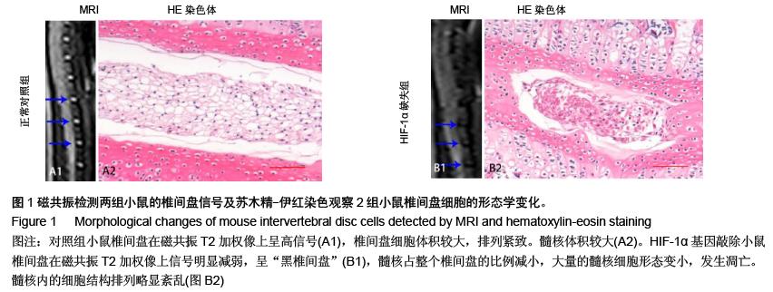
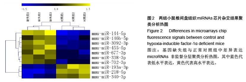
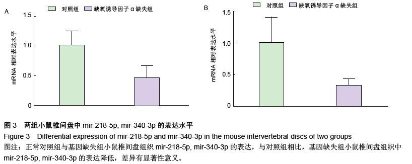
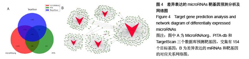
方法:通过前期构建的条件性敲除髓核细胞缺氧诱导因子1α基因的小鼠,分别取基因敲除组与正常对照组4周龄小鼠的椎间盘组织进行组织学染色观察;通过提取标本的总RNAs,从中分离microRNAs,荧光标记后与微阵列芯片杂交,扫描检测后数据经分析处理,筛选椎间盘组织中microRNA差异表达谱。采用实时荧光定量RT-PCR技术验证在椎间盘组织中均存在显著差异表达的microRNAs。分析差异表达microRNAs的靶基因及通路,预测其在椎间盘退变中的作用。
结果与结论:缺氧诱导因子1α基因缺失小鼠椎间盘髓核细胞数量减少,细胞形态变小,细胞质着色加深;两组小鼠椎间盘中的microRNAs的芯片筛选结果中,10个microRNAs发生了明显的差异表达,其中有7个microRNAs发生上调,3个microRNAs发生下调。结果提示缺氧诱导因子1α缺失后可能引起某些重要的microRNAs调节失衡,引起椎间盘髓核细胞大量的死亡,加速椎间盘的退变。 
ORCID: 0000-0002-0621-2499(张兴凯)

关键词: 组织构建, 组织工程, 缺氧诱导因子1α, 髓核细胞, 椎间盘退变, microRNAs, microRNA微阵列芯片, 基因敲除, 小鼠磁共振成像, 国家自然科学基金

Abstract:

BACKGROUND: It is confirmed that the absence of hypoxia-inducible factor-1α (HIF-1α) accelerates the degenerative process in the intervertebral discs, and microRNAs have an important role in degeneration of the intervertebral discs.
OBJECTIVE: To evaluate the changes of microRNAs in the intervertebral discs of HIF-1α-deficient (HIF-1α-/-) mice which may mediate the signaling pathway of HIF-1α in the intervertebral discs.
METHODS: As previously reported, HIF-1α-/- mice were established. HIF-1α-/- mice and HIF-1αflox/flox mice (control mice) aged 4 weeks were used. MRI and histological staining were used to evaluate the degeneration of the intervertebral discs. Total RNAs were extracted from the intervertebral discs tissues by Trizol, and the differential expression profile of microRNAs was harvested by significance analysis of microarrays and Cluster, based on microarray screening. Real-time quantitative reverse transcription-PCR was applied to verify the reliability of microRNA array results.
RESULTS AND CONCLUSION: The number of nucleus pulposus cells in the intervertebral discs of HIF-1α-/- mice was decreased, the cells presented with small size and the color deepened in the cytoplasm. Finally, differential expression profile of microRNAs (n=10) was obtained, seven of which were upregulated and three were downregulated. In conclusion, the loss of HIF-1α may cause the imbalance of some important miRNAs, which may result in a large amount of dead nuclear pulposus cells and mediate disc degeneration in HIF-1α-/- mice.