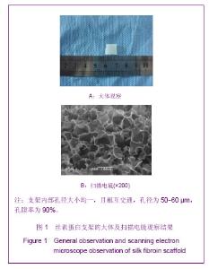
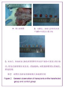
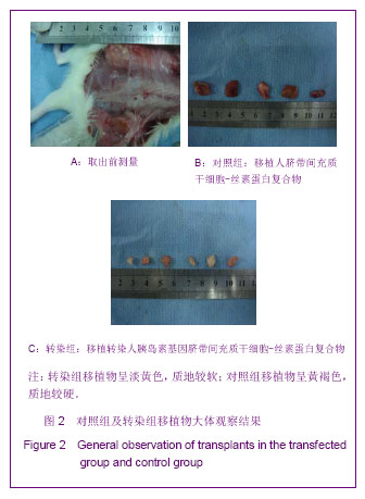
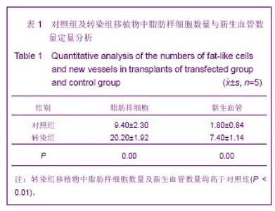
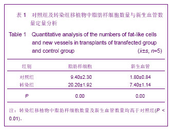
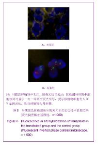
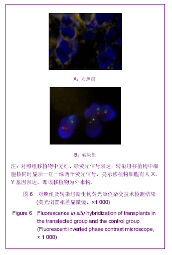
| [1] Lee KY, Halberstadt CR, Holder WD, et al. Breast reconstruction. In: Lanza RP, Langer R, Vacanti J, editors. Principles of tissue engineering. San Diego: Academic Press. 2000: 409-423.[2] Lidagoster MI, Cinelli PB, Levee EM, et al. Comparison of autologus fat transfer in fresh, refrigerated, and frozen specimens: an animal model. Ann Plast Surg. 2000; 44(5): 512-515. [3] Stashower M, Smith K, Williams J, et al. Stromalprogenitor cell present within liposuction and reduction abdominoplasty fat for auto logus transfer to aged skin. Dermatol Surg.1999; 25(12): 945-949.[4] Ricaurtej C, Murali R, Mandell W. Uncomplicated postoperative lipoid meningitis sencondary to auto-logus fat graft necrosis. Clin Infect Dis. 2000;30(3):613-615.[5] Liu Y. Beijing:Press of Military Medical Sciences. 2009: 227. 刘毅. 脂肪移植的基础与临床[M]. 北京:军事医学科学出版社, 2009:227.[6] Liu Y, Xue MS. Zhongguo Xiufu Chongjian Waike Zazhi. 2010; 24(7):822-827.刘毅,薛美思.携带人胰岛素基因慢病毒载体转染人脐带间充质干细胞的实验研究[J].中国修复重建外科杂志,2010,24(7): 822-827.[7] Liu Y, Xiao HT. Zhonghua Yixue Meixue Meirong Zazhi. 2010; 16(1):45-48.刘毅,肖宏涛.人脐带间充质干细胞与蚕丝素多孔支架的体外复合培养[J].中华医学美学美容杂志,2010,16(1):45-48.[8] Peng X, Zhang X, Zeng B. Locally administered lentivirus-mediated siRNA inhibits wear debris-induced inflammation in murine air pouch model. Biotechnol Lett. 2008;30:1923-1929. [9] Gordon D, Glover CP, Merrison AM, et al. Enhanced green fluorescent protein-expressing human mesenchymal stem cells retain neural marker expression. J Neuroimmunol. 2008;193(1-2): 59-67.[10] Ren G, Li T, Lan JQ, et al. Lentiviral RNAi-induced downregulation of adenosine kinase in human mesenchymal stem cell grafts: A novel perspective for seizure control. Exp Neurol. 2007;208(1):26-37. [11] Goomer RS. Repair of articular cartilage defects by ex-vivo gene therapy. Curr Opin Orthop. 2000 ;11(5):378-382.[12] Blanc MR, Anouassi A, Abed MA, et al. A new method to discriminate immunogen-specific heavy-chain homodimer from heterotetramer immunoglobulin G directly in immunized dromedary whole plasma proteins: Western ligand blotting. Vet Immunol Immunopathol. 2009;127(3-4):340-349. [13] Chassaigne H, Trégoat V, Nørgaard JV, et al. Resolution and identification of major peanut allergens using a combination of fluorescence two-dimensional differential gel electrophoresis, Western blotting and Q-TOF mass spectrometry. J Proteomics.2009;72(3): 511-526.[14] Tang J, Liu Y, Xu B. Zhonghua Yixue Meixue Meirong Zazhi. 2012;18(4):277-281.唐军,刘毅,徐斌.胰岛素基因转染的人脐带间充质干细胞与丝素蛋白支架构建组织工程脂肪的研究[J].中华医学美学美容杂志, 2012,18(4):277-281.[15] Wang HX, Li M. Zhongguo Xiufu Chongjian Waike Zazhi. 2008; 28(2):192-194.王宏昕,李敏.丝素蛋白作为组织工程生物材料的研究进展[J].中国修复重建外科杂志,2008,28(2):192-194.[16] Hofmann S, Hagenmuller H, Koch AM, et al. Control of in vitro tissue-engineered bone-like structures using human mesenchymal stem cells and porous silk scaffolds. Biomaterials. 2007;28(6):1152-1162.[17] Patrick CWJr, Chauvin PB, Hobl EYJ, et al. Integrin-mediated preadipocyte adhesion an migration on laminin-1. Ann Biomed Eng. 2003;31(5) :505-514.[18] Wang Y, Rudym DD, Walsh A, et al. In vivo degradation of three-dimensional silk fibroin scaffolds. Biomaterials. 2008; 29(24-25):3415-3428.[19] Liu Y,Xiao HT,Xue MS. Zhongguo Zuzhi Gongcheng Yanjiu yu Linchuang Kngfu. 2010;14(8):1361-1364.刘毅,肖宏涛,薛美思.适宜于构建组织工程化脂肪的蚕丝蛋白支架:最佳孔径筛选[J].中国组织工程研究与临床康复,2010,14(8): 1361-1364.[20] Tang J,Xu B,Liu Y. Zhongguo Meirong Yixue. 2011;20(8): 1238-1242.唐军,徐斌,刘毅.不同移植部位对组织工程化脂肪存活的影响[J].中国美容医学,2011,20(8):1238-1242.[21] Saito T, Kuang JQ, Bttira B, et al. Xenotansplant cardiac chimera:immune tolerance of adult stem cells. Ann Thorac Surg. 2002;74(l):19-24.[22] Unger EC. How can superparamagnetic iron oxides be used to monitor disease and treatment. Radiology. 2003;229(3): 615-616.[23] Li D, Cui L, Shu CF, et al. Zhonghua Chuangshang Guke Zazhi. 2004;6(3):311-314.李东,崔磊,舒朝锋,等.荧光活性染料DiI标记观察人骨髓基质干细胞与部分脱钙骨粘附的实验研究[J].中华创伤骨科杂志,2004, 6(3):311-314. |