Chinese Journal of Tissue Engineering Research ›› 2014, Vol. 18 ›› Issue (51): 8206-8211.doi: 10.3969/j.issn.2095-4344.2014.51.002
Previous Articles Next Articles
Primary culture and identification of kitten extraocular muscle myoblasts
Liu Tao1, Liu Gui-xiang1, Yin Ya-nan1, Zhou Juan2
- 1 Department of Ophthalmology, Affiliated Hospital of Qingdao University, Qingdao 266003, Shandong Province, China
2 Department of Endocrinology, Affiliated Hospital of Qinghai University, Xining 810001, Qinghai Province, China
-
Online:2014-12-10Published:2014-12-10 -
Contact:Liu Gui-xiang, M.D., Professor, Department of Ophthalmology, Affiliated Hospital of Qingdao University, Qingdao 266003, Shandong Province, China -
About author:Liu Tao, Studying for master’s degree, Department of Ophthalmology, Affiliated Hospital of Qingdao University, Qingdao 266003, Shandong Province, China -
Supported by:the Natural Science Foundation of Shandong Province, No. ZR2013HM014

CLC Number:
Cite this article
Liu Tao, Liu Gui-xiang, Yin Ya-nan, Zhou Juan. Primary culture and identification of kitten extraocular muscle myoblasts[J]. Chinese Journal of Tissue Engineering Research, 2014, 18(51): 8206-8211.
share this article
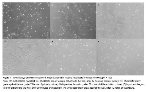
Separation and culture in vitro of myoblasts Myoblasts from the extraocular muscle were treated by two-step combined differential attachment (pancreatic enzyme and type II collagenase), and the cell survival rate was (91.0±2.1)% via trypan blue staining. After observation, cells cultured separately were greatly different from the myoblasts in the late phase, and would not merge into a myotube. The isolated original generation of myoblasts was in spherical shape under inverted microscope with strong refraction (Figure 1A). The cells began to be adhered to the wall after 12 hours of culture (Figure 1B), became fusiform or spindle shape gradually after 24 hours, and completely adhered after 72 hours (Figure 1C). The myoblasts were arranged parallel to the long axis. When myoblasts completely adhered and grew for a period of time, many myoblasts which were close with each other began to fuse. If cell fusion rate was more than 80% still without digestion and generation of the cells, myoblasts would appear to have widespread and irreversible fusion. On the 72nd hour of differentiation culture, a great mass of myotube cell formed (Figure 1D). After 30 minutes of subculture, the generated cells began to adhere to the wall and fully attached after 12 hours (Figures 1E, F)."

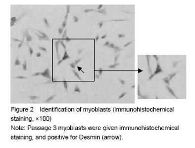
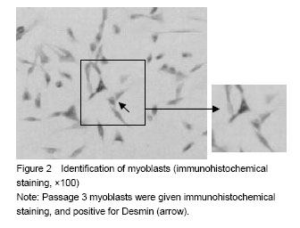
Identification and purity results of myoblasts Desmin filaments of myoblast antibody were stained. The cytoplasm was dyed brown or brownish red with one or two light-dyed cell nuclei. If more than 90% of the cells presented positive reaction, the cultured cells were extraocular muscle cells, as shown in Figure 2. After 8 days of primary culture, myoblasts were given immunochemical staining and then positive cells were counted. The cell purification rate was (95.0±3.8)%."
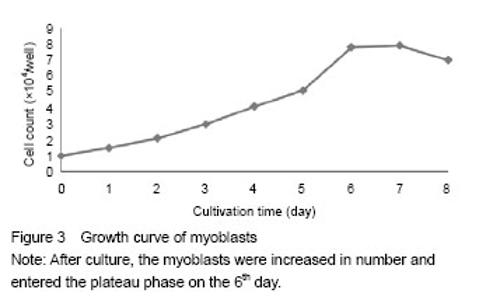
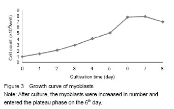
Proliferation ability of myoblasts Myoblasts seeded onto 24-well plates were adhered to the wall quickly, and completely adhered after 12 hours with a larger growth rate. The cells entered the logarithmic phase on the 2nd day, while the growth slowed significantly on the 6th day due to contact inhibition. The cell growth curve is shown in Figure 3."
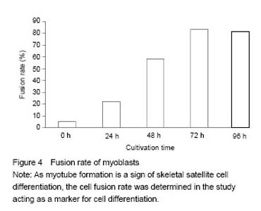
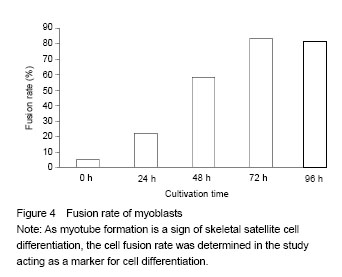
Differentiation capability of myoblasts The myoblasts were added with differentiation medium and fused in large amount. The fusion rate was increased significantly at 24 and 48 hours and reached the peak at 72 hours. After that, the variation tended to be stable, as shown in Figure 4. The myoblasts were arranged parallelly and mutually fused into myotubes. Multiple nuclei were arranged in the middle of the myotube (Figure 1D). Myotube formation is a sign of skeletal satellite cell differentiation[7]."
| [1]Chen BF. Pathology of Skeletal Muscle Disease Fuzhou: Fujian Science & Technology Publishing House. 1996:43. [2]Ekdawi NS, Nusz KJ, Diehl NN, et al. Postoperative outcomes in children with intermittent exotropia from a population-based cohort. J AAPOS. 2009;13(1):4-7. [3]Machida S, Spangenburg EE, Booth FW. Primary rat muscle progenitor cells have decreased proliferation and myotube formation during passages. Cell Prolif. 2004; 37(4):267-277. [4]Fujimaki S, Machida M, Hidaka R, et al. Intrinsic ability of adult stem cell in skeletal muscle: an effective and replenishable resource to the establishment of pluripotent stem cells. Stem Cells Int. 2013;2013:420164. [5]Yin H, Price F, Rudnicki MA. Satellite cells and the muscle stem cell niche. Physiol Rev. 2013;93(1):23-67. [6]The Ministry of Science and Technology of the People’s Republic of China. Guidance Suggestions for the Care and Use of Laboratory Animals. 2006-09-30. [7]Zhou F, Qin YW, Jing Q, et al. Primary culture of adult rat skeletal myoblasts. Zhonghua Laonian Xinnaoxueguan Bing Zazhi. 2001;3(1):43-46. [8]Wu H, Ren Y, Li S, et al. In vitro culture and induced differentiation of sheep skeletal muscle satellite cells. Cell Biol Int. 2012;36(6):579-587. [9]Freshney I. Culture of Animal Cells-A Manual of Basic Technique. 3rd ed. New York: Wiley-Liss, 1994:331. [10]Li FH, Hou LL, Ma YH, et al. Isolation, culture, identification and muscle differentiation of skeletal muscle satellite cells in Beijing fatty chicken. Zhongguo Nongye Kexue. 2010; 43(22):4725-4731. [11]Cao K, Cheng HB, Ying FW. Culture and identification of fibroblasts in human Tenon’s capsule. Zhongguo Yiyao Zhinan. 2013;11(9):87-88. [12]He YL, Wy YH, Quan FS, et al. Isolation and transfection of skeletal muscle satellite cells from Qinchuan fetal bovine. Xibei Nonglin Keji Daxue Xuebao: Ziran Kexue Ban. 2012; 40(6):1-6. [13]Wei Y, Li Y, Chen C, et al. Human skeletal muscle-derived stem cells retain stem cell properties after expansion in myosphere culture. Exp Cell Res. 2011;317(7):1016-1027. [14]Wang F, Zhao T, Zhang CP, et al. Hypoxia enhancing the proliferation of rat skeletal myoblasts in vitro and the effect of cobalt. Zhongguo Zuzhi Gongcheng Yanjiu yu Linchuang Kangfu. 2007;11(3):419-422. [15]Rada T, Gomes ME, Reis RL. A novel method for the isolation of subpopulations of rat adipose stem cells with different proliferation and osteogenic differentiation potentials. J Tissue Eng Regen Med. 2011;5(8):655-664. [16]Gu YL, Wang YB, Zhu WH, et al. Identification, purification and culture of myoblasts in vitro of adult beagle. Zhongguo Yundong Yixue Zazhi. 2014;33(3):224-228. [17]Gao P, Zhu XT, Yang YC, et al. The effects of glycyl-glutamine on the proliferation of in vitro cultivated porcine skeletal muscle satellite cells and medium IGF-1 level. Huanan Nongye Daxue Xuebao. 2005;26(2):100-102. [18]Rando TA, Blau HM. Primary mouse myoblast purification, characterization, and transplantation for cell-mediated gene therapy. J Cell Biol. 1994;125(6):1275-1287. [19]Xia JH, Xie AN, Xu L, et al. Culture of rat musclesatellite cell in vitro. Zhonghua Shiyan Waike Zazhi. 2005;22(2): 214-215. [20]Stratos I, Rotter R, Eipel C, et al. Granulocyte-colony stimulating factor enhances muscle proliferation and strength following skeletal muscle injury in rats. J Appl Physiol (1985). 2007;103(5):1857-1863. [21]Sanoudou D, Corbett MA, Han M, et al. Skeletal muscle repair in a mouse model of nemaline myopathy. Hum Mol Genet. 2006;15(17):2603-2612. [22]Menasché P. Skeletal muscle satellite cell transplantation. Cardiovasc Res. 2003;58(2):351-357. [23]Zhang LG, Wang CF, Huang XH. Proliferation and differentiation of extraocular muscle satellite cells in rats. Zhongguo Zuzhi Gongcheng Yanjiu yu Linchuang Kangfu. 2007;11(24):4702-4705. [24]Chen KQ, Dong SH, Luo LJ, et al. Effect of erythropoietin on the proliferation and differentiation of rat skeletal muscle satellite cells. Zhongguo Zuzhi Gongcheng Yanjiu yu Linchuang Kangfu. 2008;12(34):6687-6691. [25]Luo L, Liu ZQ, Yang L, et al. Culture and identification of primary atrial myocytes in neonatal rat. Guoji Xinxueguan Bing Zazhi. 2013;40(2):112-115. [26]Perruchot MH, Ecolan P, Sorensen IL, et al. In vitro characterization of proliferation and differentiation of pig satellite cells. Differentiation. 2012;84(4):322-329. [27]Bai C, Hou L, Li F, et al. Isolation and biological characteristics of Beijing Fatty chicken skeletal muscle satellite cells. Cell Commun Adhes. 2012;19(5-6):69-77. |
| [1] | Li Cai, Zhao Ting, Tan Ge, Zheng Yulin, Zhang Ruonan, Wu Yan, Tang Junming. Platelet-derived growth factor-BB promotes proliferation, differentiation and migration of skeletal muscle myoblast [J]. Chinese Journal of Tissue Engineering Research, 2021, 25(7): 1050-1055. |
| [2] | Gao Yang, Bai Shengchao, Chen Shengju, Wang Ruiyuan, Li Junping. Effect of exercise-induced muscle damage on desmin aggregates [J]. Chinese Journal of Tissue Engineering Research, 2020, 24(8): 1243-1248. |
| [3] | Li Miaomiao, Luo Jiong, Zhang Tingran, Ouyang Yiyi, Zhou Chenglin. Bone metabolism and exercise training: bone remodeling and osteoblast proliferation [J]. Chinese Journal of Tissue Engineering Research, 2019, 23(34): 5544-5549. |
| [4] | He Li, Zheng Xiaoli. Transplantation of allogeneic adipose-derived mesenchymal stem cells promotes the recovery of skeletal muscle function [J]. Chinese Journal of Tissue Engineering Research, 2019, 23(17): 2739-2745. |
| [5] | Gong Hewei, Liu Chengwei, Liang Bingsheng, Li Gang. Lentiviral vector-mediated overexpression of miRNA-1 in L6 myoblasts: cell proliferation, differentiation and histone deacetylase expression [J]. Chinese Journal of Tissue Engineering Research, 2019, 23(13): 2075-2080. |
| [6] | Chen You-bai, Zhang Wei, Li Li, Zhang Qi-xu, Han Yan. Adipose-derived stem cells: current progress and future perspectives regarding the myogenic differentiation and use in the repair of skeletal muscle defects [J]. Chinese Journal of Tissue Engineering Research, 2018, 22(1): 126-132. |
| [7] | He Yu-tong, Zhang Ma-hui, Song Chen, Ye Gen-lan, Yu Lei, Qiu Xiao-zhong, Wang Le-yu. MicroRNA expression profile in the process of cyclic mechanical stretch promoting C2C12 myogenesis [J]. Chinese Journal of Tissue Engineering Research, 2017, 21(28): 4505-4511. |
| [8] | Li Jing, Zhang Xiang, Wang Li-ping, Shi Lei, Mu Yu-zheng, Chang Qian-qian. Effects of cytokines on skeletal muscle cells and pancreatic beta cells [J]. Chinese Journal of Tissue Engineering Research, 2016, 20(37): 5594-5601. |
| [9] | Wei Yuan, Fang Chun-lu, Li Liang-ming, Xing Wen-hua, Yang Ze-yi. Establishing an animal model of delayed onset muscle soreness and its histomorphologic observation [J]. Chinese Journal of Tissue Engineering Research, 2015, 19(29): 4645-4651. |
| [10] | Meng Tian-tian, Li Shu-ren. C-kit+ cardiac stem cells for ischemic heart disease: research status, challenges and prospects [J]. Chinese Journal of Tissue Engineering Research, 2014, 18(50): 8196-8200. |
| [11] | Zhang Qiang, Wang Hong-ling, Ding Xian, Xia Chen-lei, Liu Li-juan, Wang Shuang-yu, Li Jian-ping, He Miao, Sun Wen-na, Yan Xiao, Liu Wen, Zhang Yue, Yao Ru-yong, Yuan Xiao. Effects of cyclic stretch on in vitro culture-tensile stimulation models of myoblasts [J]. Chinese Journal of Tissue Engineering Research, 2014, 18(5): 669-674. |
| [12] | Liu Qiang, Zhao Xiang-xuan, Pan Shi-nong, Guo Qi-yong. Damage to skeletal muscle cells leads to delayed muscle soreness: how to effectively improve muscle recovery both in recovery speed and quality [J]. Chinese Journal of Tissue Engineering Research, 2014, 18(38): 6189-6199. |
| [13] | Sun Hai-bo, Wei Jun-qiang, Chen Wei, Zhang Ying-ze, Zheng Zhan-le, Pan Jin-she. Prophylactic effects of edaravone on the extremity after limb replantation [J]. Chinese Journal of Tissue Engineering Research, 2014, 18(24): 3814-3820. |
| [14] | Gao Yan, Cheng Chen, Li Jing, Zhang Yue, Xiao Wei-fan, Pan Qiu-hui. Bone morphogenetic protein 2-induced C2C12 and MC3T3-E1 osteoblast differentiation and autophagy [J]. Chinese Journal of Tissue Engineering Research, 2014, 18(20): 3236-3241. |
| [15] | Wang Hong-ling, Liu Dong-xu, Yuan Xiao. Effect of cyclic stretch on the proliferation of C2C12 myoblasts [J]. Chinese Journal of Tissue Engineering Research, 2014, 18(20): 3242-3245. |
| Viewed | ||||||
|
Full text |
|
|||||
|
Abstract |
|
|||||