Chinese Journal of Tissue Engineering Research ›› 2014, Vol. 18 ›› Issue (30): 4797-4803.doi: 10.3969/j.issn.2095-4344.2014.30.007
Previous Articles Next Articles
Effect of Bio-Gide collagen membranes on the proliferation and osteogenetic differentiation of rabbit bone marrow mesenchymal stem cells
Xu Fan, Yang De-sheng
- Department of Stomatology, General Hospital of the Chinese People’s Armed Police Force, Beijing 100039, China
-
Revised:2014-05-28Online:2014-07-16Published:2014-08-08 -
Contact:Yang De-sheng, Master’s supervisor, Chief physician, Department of Stomatology, General Hospital of the Chinese People’s Armed Police Force, Beijing 100039, China -
About author:Xu Fan, Studying for master’s degree, Department of Stomatology, General Hospital of the Chinese People’s Armed Police Force, Beijing 100039, China -
Supported by:a grant from General Hospital of the Chinese People’s Armed Police Force, No. WZ2012008
CLC Number:
Cite this article
Xu Fan, Yang De-sheng. Effect of Bio-Gide collagen membranes on the proliferation and osteogenetic differentiation of rabbit bone marrow mesenchymal stem cells[J]. Chinese Journal of Tissue Engineering Research, 2014, 18(30): 4797-4803.
share this article
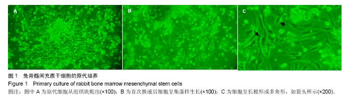
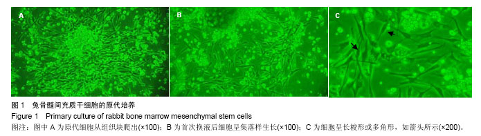
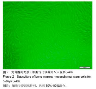
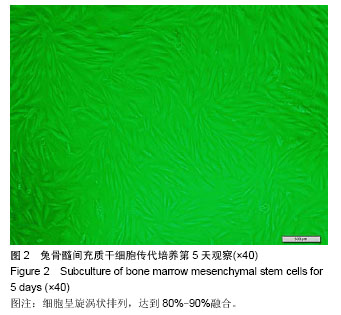
2.1 兔骨髓间充质干细胞的分离和培养 原代细胞在接种骨髓腔冲洗液的24 h后从组织块中爬出,倒置显微镜下可见在大量悬浮细胞下有少量呈梭形生长的贴壁细胞,在培养48 h后首次行全量换液去掉未贴壁的悬浮细胞,可见贴壁细胞数量增多且呈现集落样,经传代培养不断提纯贴壁细胞,细胞生长状态良好且分布均匀,倒置显微镜下观察细胞呈旋涡状排列,达到80%-90%融合,见图1,2。"
"

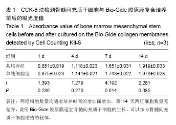
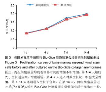
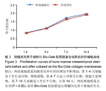
2.2 细胞增殖情况 CCK-8检测细胞吸光度值数据见表1,数据经两独立样本t 检验行统计学分析;以时间、A值为坐标,绘制细胞增殖曲线,见图3。图中可以看出,随着培养时间的增加,两组细胞数量均不断增加,第1-4天细胞处于生长适应期,第1,4天两组细胞数量比较无明显差异(P > 0.05);第4-7天细胞处于对数生长期,细胞大量增殖,在第7天两组细胞数量比较差异有显著性意义(P < 0.05),实验组细胞少于对照组;随着培养时间的增加,第7-14天细胞逐渐进入生长平台期,在第14天两组细胞数量比较差异无显著性意义(P > 0.05),说明Bio-Gide胶原膜适宜骨髓间充质干细胞的生长,可以作为骨髓间充质干细胞增殖的载体。"
"
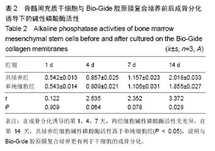
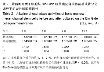
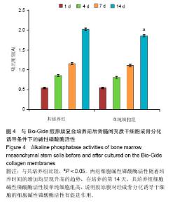
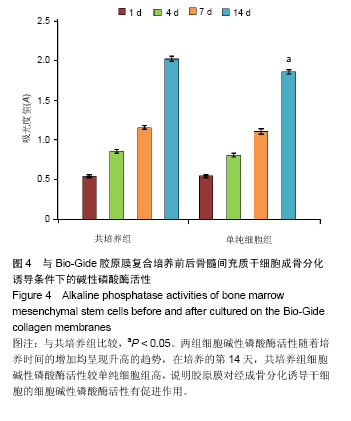
2.3 细胞碱性磷酸酶活性 碱性磷酸酶检测数据见表2,数据采用两独立样本t 检验行统计学分析;以时间、A值为坐标,绘制细胞碱性磷酸酶活性图,见图4。如图所示,两组细胞在成骨分化诱导液的诱导下,细胞向成骨方向分化,两组细胞碱性磷酸酶活性随着培养时间的增加不断升高,在成骨分化诱导的第1,4,7天,两组细胞碱性磷酸酶活性比差异无显著性意义(P > 0.05),但在第14天,实验组细胞碱性磷酸酶活性高于对照组(P < 0.05),说明在成骨分化诱导条件下,两组细胞均逐渐具有成骨分化能力,但实验组细胞成骨分化能力更强,说明与Bio-Gide胶原膜复合培养更有利于干细胞的成骨分化,随着培养时间的增加,Bio-Gide胶原膜对经成骨分化诱导干细胞的细胞碱性磷酸酶活性有促进作用。"
"
| [1] Nyman S,Lindhe J,Karring T,et al.New attachment following surgical treatment of human periodontal disease.J Clin Periodontol.1982;9(4):290-296. [2] Gottlow J,Nyman S,Karring T,et al.New attachment formation as the result of controlled tissue regeneration.J Clin Periodontol.1984;11(8):494-503. [3] 陈发明,金岩,吴织芬.生长因子复合生物膜引导牙周组织再生[J].牙体牙髓牙周病学杂志,2004,14(10):559-561. [4] Beresford JN.Osteogenic stem cells and the stromal system of bone and marrow. Clin Orthop Relat Res. 1989;(240): 270-280. [5] Takeuchi Y,Nakayama K,Matsunoto T.Differentiation and cell surface expression of transforming growth factor-beta receptors are regulated by interaction with matrix collagen in murine osteoblastic cells.J Boil Chem.1996;27(7): 3938-3944. [6] Behring J,Junker R,Frank X,et al.Toward guided tissue and bone regeneration: Morphology, attachment, proliferation, and migration of cells cultured on collagen barrier membranes: A systematic review.Odontology.2008;96(1):1-11. [7] Tonetti MS,Cortellini P,Lang NP,et al.Clinical outcomes following treatment of human intrabony defects with GTR/bone replacement material or access flap alone. A multicenter randomized controlled clinical trial.J Clin Periodontol. 2004;31(9):770-776. [8] Schwarz F,Bieling K,Latz T,et al.Healing of intrabony peri-implantitis defects following application of a nanocrystalline hydroxyapatite(Ostim) or a bovine-derived xenograft(Bio-Oss) in combination with a collagen membrane(Bio-Gide). A case Series.J Clin Periodontol. 2006; 33(7):491-499. [9] Sculean A,Schwarz F,Chiantella GC,et al.Five-year results of a prospective,randomized, controlled study evaluating treatment of intra-bony defects with a natural bone mineral and GTR. J Clin Periodontol. 2007;34(1):72-77. [10] Liu Q,Humpe A,Kletsas D,et al.Proliferation assessment of primary human mesenchymal stem cells on collagen membranes for guided bone regeneration.Int J Oral Maxillofac Implants. 2011;26(5):1004-1010. [11] 宋莉,曲丰江,汪泱,等.人骨髓间充质干细胞体外培养及与Bio-Gide胶原膜复合培养的实验研究[J].江西医学院学报, 2009, 49(5):15-18. [12] 马超,张丁,李平,等.成骨细胞在两种胶原支架材料上的生长特征[J].中国医学科学院学报,2011,33(5):538-542. [13] Nevins ML,Camelo M,Lynch SE,et al.Evaluation of periodontal regeneration following grafting intrabony defects with bio-oss collagen: a human histologic report.Int J Periodontics Restorative Dent.2003;23(1):9-17. [14] Rothamel D,Schwarz F,Fienitz T,et al.Biocompatibility and biodegradation of a nativeporcine pericardium membrane: results of in vitro and in vivo examinations.Int J Oral Maxillofac Implants.2012;27(1):146-154. [15] Caneva M,Botticelli D,Salata LA,et al.Collagen membranes at immediate implants: a histomorphometric study in dogs.Clin Oral Implants Res.2010;21(9):891-897. [16] 谢江涛,刘世清.骨髓基质细胞促进引导性骨再生的研究[J].中华试验外科杂志,2007,24(11):1417-1418. [17] Zubery Y,Goldlust A,Alves A,et al.Ossification of a novel cross-linked porcine collagen barrier in guided bone regeneration in dogs. J Periodontol.2007;78(1):112-121. [18] 王建,胡秀莲,林野.Bio-oss和Bio-oss骨胶原保持牙槽骨量的临床研究[J].现代口腔医学杂志,2009,23(1):4-6. [19] 柏树令,顾晓松,张传森.组织工程学教程[M].北京:人民军医出版社,2009:86-88. [20] Maeda S,Fujitomo T,Okabe T,et al.Shrinkage-free preparation of scaffold-free cartilage-like disk-shaped cell sheet using human bone marrow mesenchymal stem cells.J Biosci Bioeng. 2011;111(4):489-492. [21] Zhou W,Han C,Song Y,et al.The performance of bone marrow mesenchymal stem cell--implant complexes prepared by cell sheet engineering techniques. Biomaterials. 2010;31(12): 3212-3221. [22] 荆恒,陈欣,李宁毅.细胞片层技术的研究进展[J].国际口腔医学杂志,2010,37(1):71-73. [23] 王玲玲,李宁毅,樊功为,等.犬骨髓基质细胞片层在构建组织工程骨中的作用[J].中国口腔颌面外科杂志,2011,9(2):96-100. [24] Trabulsi M,Oh TJ,Eber R,et al.Effect of enamel matrix derivative on collagen guided tissue regeneration-based root coverage procedure.J Periodontol.2004;75(11):1446-1457. [25] Schwarz F,Rothamel D,Herten M,et al.Angiogenesis pattern of native and cross-linked collagen membranes: an immunohistochemical study in the rat. Clin Oral lmplants Res.2006;17(4):403-409. [26] Temenoff JS,Mikos AG.Review: tissue engineering for regeneration of articular cartilage. Biomaterials. 2000;21(5): 431-440. [27] 张铭杰,张庆文,何伟,等.成人骨髓间充质干细胞体外培养、鉴定与成骨诱导[J].中国组织工程研究,2013,17(45):7947-7953. [28] Nguyen MN,Lebarbe T,Zouani OF,et al.Impact of RGD nanopatterns grafted onto titanium on osteoblastic cell adhesion. Biomacromolecules.2012;13(3):896-904. [29] Takata T,Wang HL,Miyauchi M.Migration of osteoblastic cells on various guided bone regeneration membranes.Clin Oral Impants Res.2001;12(4):332-338. [30] Nur-E-Kamal A,Ahmed I,Kamal J,et al.Three-dimensional nanofibrillar surfaces promote self-renewal in mouse embryonic stem cells.Stem Cells.2006;24(2):426-433. [31] Chung CH,Golub EE,Forbe E,et al.Mechanism of action of beta-glycerophosphate on bone cell mineralization.Calaif Tissue Int.1992;51(4):305-311. [32] Bruder SP,Jaiswal N,Haynesworth SE.Growth kinetics, self-renewal, and the osteogenic potential of purified human mesenchymal stem cells during extensive subcultivation and following cryopreservation.J Cell Biochem. 1997;64(2): 278-294. [33] Tabata Y.Tissue regeneration based on tissue engineering technology. Congenit Anom(Kyoto).2004;44(3):111-124. [34] Herbertson A,Aubin JE.Cell sorting enriches osteogenic populations in rat bone marrow stromal cell culture. Bone.1997;21(6):491-500. [35] Li X,Jin L,Cui Q,et al.Steroid effects on osteogenesis through mesenchymal cell gene expression.Osteoporos Int.2005;16(1):101-108. [36] 谷子芽,翟强,吕秋峰,等.地塞米松诱导牙周膜干细胞的定向成骨分化及凋亡[J].中国组织工程研究,2013,17(40):7090-7095. [37] 邹慧儒,张兰成,秦宗长,等.地塞米松对人牙髓细胞增殖分化影响的研究[J].组织工程与重建外科杂志,2012,8(1):8-13. [38] Hidalgo-Bastida LA,Cartmell SH.Mesenchymal stem cells, osteoblasts and extracellular matrix proteins: enhancing cell adhesion and differentiation for bone tissue engineering. Tissue Eng Part B Rev.2010;16(4):405-412. [39] Wada K,Mizuno M,Komori T,et al.Extracellular inorganic phosphate regulates gibbon ape leukemia virus receptor-2/phosphate transporter mRNA expression in rat bone marrow stromal cells.J Cell Physiol.2004;198(1):40-47. [40] Linsley C,Wu B,Tawil B.The effect of fibrinogen, collagen type I, and fibronectin on mesenchymal stem cell growth and differentiation into osteoblasts. Tissue Eng Part A. 2013; 19(11-12):1416-1423. [41] Taguchi Y,Amizuka N,Nakadate M,et al. A histological evaluation for guided bone regeneration induced by a collagenous membrane.Biomaterials.2005;26(31): 6158-6166. [42] Mackie EJ.Osteoblasts:novel roles in orchestration of skeletal architecture. Int J Biochem Cell Biol.2003;35(9):1301-1305. [43] Anselme K.Osteoblast adhesion on biomaterials. Biomaterials. 2000;21(7):667-681. [44] Suzawa M,Takeuchi Y,Fukumoto S,et al.Extracellular Matrix-associated bone morphogenetic proteins are essential for differentiation of murine osteoblastic cells in vitro. Endocrinology. 1999;140(5):2125-2133. [45] Hsu FY,Chueh SC,Wang YJ.Microspheres of hydroxyapatite/ reconstituted collagen as supports for osteoblast cell growth. Biomaterials.1999;20(20):1931-1936. [46] Borkenhagen M,Clemence JF,Sigrist H,et al. Three-dimensional extracellular matrix engineering in the nervous system.J Biomed Mater Res.1998;40:392-400. |
| [1] | Dang Yi, Du Chengyan, Yao Honglin, Yuan Nenghua, Cao Jin, Xiong Shan, Zhang Dingmei, Wang Xin. Hormonal osteonecrosis and oxidative stress [J]. Chinese Journal of Tissue Engineering Research, 2023, 27(9): 1469-1476. |
| [2] | Xu Yan, Li Ping, Lai Chunhua, Zhu Peijun, Yang Shuo, Xu Shulan. Piezoelectric materials for vascularized bone regeneration [J]. Chinese Journal of Tissue Engineering Research, 2023, 27(7): 1126-1132. |
| [3] | Qiao Luhui, Ma Ziyu, Guo Haoyu, Hou Yudong. Comparison of puerarin and icariin on the biological properties of mouse preosteoblasts [J]. Chinese Journal of Tissue Engineering Research, 2023, 27(6): 872-877. |
| [4] | Li Xiaoyin, Yang Xiaoqing, Chen Shulian, Li Zhengchao, Wang Ziqi, Song Zhen, Zhu Daren, Chen Xuyi. Collagen/silk fibroin scaffold combined with neural stem cells in the treatment of traumatic spinal cord injury [J]. Chinese Journal of Tissue Engineering Research, 2023, 27(6): 890-896. |
| [5] | Zhang Min, Zhang Xiaoming, Liu Tongbin. Application potential of naringin in bone tissue regeneration [J]. Chinese Journal of Tissue Engineering Research, 2023, 27(5): 787-792. |
| [6] | Zhang Hui, Wang Jiayang, Wang Qian, Gan Hongquan, Wang Zhiqiang. Effects of hyaluronic acid combined with domestic porous tantalum on chondrocyte function under the dynamic environment [J]. Chinese Journal of Tissue Engineering Research, 2023, 27(3): 339-345. |
| [7] | Liu Huan, Li Han, Ma Yunhao, Zhong Weijian, Ma Guowu. Osteogenic capacity of partially demineralized dentin particles in the maxillary sinus lift [J]. Chinese Journal of Tissue Engineering Research, 2023, 27(3): 354-359. |
| [8] | Li Rui, Liu Zhen, Guo Zige, Lu Ruijie, Wang Chen. Aspirin-loaded chitosan nanoparticles and polydopamine modified titanium sheets improve osteogenic differentiation [J]. Chinese Journal of Tissue Engineering Research, 2023, 27(3): 374-379. |
| [9] | Zong Mingrui, Liu Haiyan, Li Bing, Wu Xiuping. Application of carboxymethyl chitosan in tissue engineering of stomatology [J]. Chinese Journal of Tissue Engineering Research, 2023, 27(3): 447-452. |
| [10] | Ma Rongxing, Li Ruifeng, Zhang Haoran, Xu Mingyou, Zhang Jingyu, Hu Yongcheng. Peracetic acid-ethanol treated tendons crosslinked with 1-ethyl-3-(3-dimethylaminopropyl)carbodiimide combined with N-hydroxysuccinimide: morphological features in vitro [J]. Chinese Journal of Tissue Engineering Research, 2023, 27(21): 3307-3313. |
| [11] | Cao Jin, Wang Ansu, Huang Nijiao, Wu Fujun, Chen Ping, Li Chengmei, Wang Xin. Role and mechanism of polyphosphate in bone tissue regeneration [J]. Chinese Journal of Tissue Engineering Research, 2023, 27(21): 3375-3381. |
| [12] | Bai Lei, Wang Yang, Tian Xiaojing, Wang Wenhang. Potential of collagen peptide to improve skin and the countermeasures to improve its bioavailability [J]. Chinese Journal of Tissue Engineering Research, 2023, 27(21): 3382-3390. |
| [13] | Niu Zihan, Yu Yang, Ai Jiang, Bu Panpan, Li Wenbo, Suriye·Reheman, Ma Shaolin. Total flavonoids of Hippophae rhamnoides L. interfere with the regression of hypertrophic scar tissue blocks in a rabbit ear model [J]. Chinese Journal of Tissue Engineering Research, 2023, 27(2): 258-263. |
| [14] | Zhang Jie, Tian Ai. Advances in the signaling pathway of M2 macrophages involved in bone regeneration [J]. Chinese Journal of Tissue Engineering Research, 2023, 27(2): 314-321. |
| [15] | Liang Weiye, Duan Qinghong. Correlation between femur bone morphogenetic protein 2 expression and bone fluoride content in fluorosis rabbits [J]. Chinese Journal of Tissue Engineering Research, 2023, 27(17): 2675-2680. |
| Viewed | ||||||
|
Full text |
|
|||||
|
Abstract |
|
|||||