Chinese Journal of Tissue Engineering Research ›› 2013, Vol. 17 ›› Issue (37): 6613-6619.doi: 10.3969/j.issn.2095-4344.2013.37.012
Previous Articles Next Articles
Zebrafish embryonic brain cell apoptosis and c-fos gene expression after hypoxia reperfusion
Chen Yan-chen, Zhao Dan, Qing Di, Cheng Dong-liang, Mao Jiao-yu, Wang Bin
- Center of Pediatrics, Zhujiang Hospital of Southern Medical University, Guangzhou 510282, Guangdong Province, China
-
Received:2013-02-28Revised:2013-04-25Online:2013-09-10Published:2013-09-10 -
Contact:Wang Bin, Associate professor, Chief physician, Doctoral supervisor, Center of Pediatrics, Zhujiang Hospital of Southern Medical University, Guangzhou 510282, Guangdong Province, China -
About author:Chen Yan-chen★, Master, Physician, Center of Pediatrics, Zhujiang Hospital of Southern Medical University, Guangzhou 510282, Guangdong Province, China hello-cyc@qq.com
CLC Number:
Cite this article
Chen Yan-chen, Zhao Dan, Qing Di, Cheng Dong-liang, Mao Jiao-yu, Wang Bin. Zebrafish embryonic brain cell apoptosis and c-fos gene expression after hypoxia reperfusion[J]. Chinese Journal of Tissue Engineering Research, 2013, 17(37): 6613-6619.
share this article
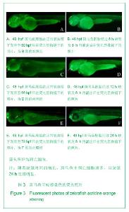
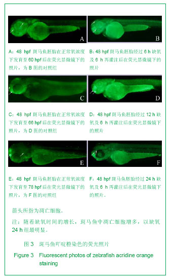
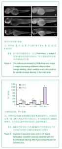
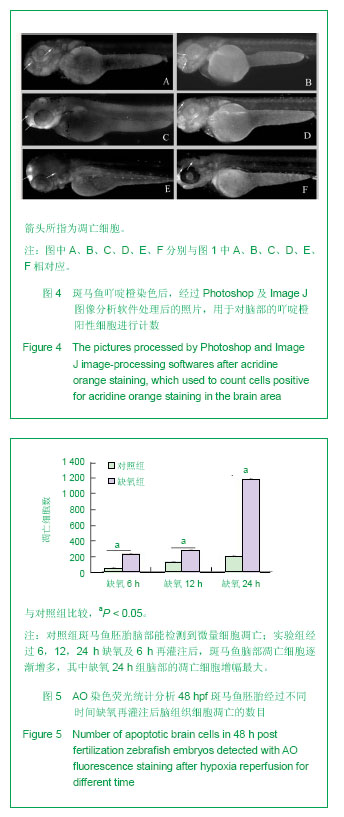
2.1 缺氧对斑马鱼胚胎脑部细胞凋亡的影响 在荧光显微镜下观察,凋亡细胞在吖啶橙染色后会显示绿色点状荧光。对照组斑马鱼胚胎脑部能检测到微量细胞凋亡;实验组经过6,12,24 h缺氧及6 h再灌注后,斑马鱼脑部凋亡细胞逐渐增多,其中缺氧24 h组脑部的凋亡细胞增幅最大(P < 0.05),见图3-5。"
"
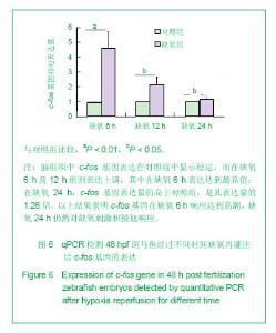
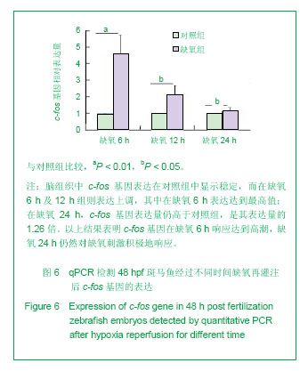
2.2 缺氧对斑马鱼脑组织c-fos基因表达的影响 脑组织中c-fos基因表达在对照组中显示稳定,而在缺氧6 h及12 h组则表达上调,其中在缺氧6 h表达达到最高值,其表达量是对照组表达量的4.48倍;在缺氧12 h,c-fos基因表达呈逐渐恢复的变化趋势,但其表达仍处于高于对照组的表达水平,是对照组的2.07倍;在缺氧24 h,c-fos基因表达量仍高于对照组,是其表达量的1.26倍。以上结果表明c-fos基因在缺氧6 h响应达到高潮,并在24 h仍然对缺氧刺激积极地响应,见图6。"
| [1] van Handel M, Swaab H, de Vries LS,et al. Long-term cognitive and behavioral consequences of neonatal encephalopathy following perinatal asphyxia: a review.Eur J Pediatr. 2007;166(7):645-654. [2] 黄晓红. 154例新生儿的窒息高危产科因素分析[J].中国医药指南, 2012, 10(29): 190-191. [3] 王彩华,姚丽萍,韩艳宾,等. 窒息新生儿缺氧缺血性脑病研究新进展[J].包头医学院学报, 2010,26(2): 112-113. [4] 刘国瑞,李仰康,郭岳霖,等.新生猪缺氧缺血脑损伤模型制备的研究[J]. 实用儿科临床杂志, 2005,20(3): 258-259, 291. [5] 周文浩,邵肖梅,李瑾,等. 新生猪缺氧缺血脑损伤模型制备的研究[J]. 中国当代儿科杂志, 2003, 5(2): 113-116. [6] 杨明峰,张颜波,管宏旭,等. 大鼠鼠胚海马神经元缺氧复氧致细胞凋亡模型的建立[J].泰山医学院学报,2008,29 (12):949-951. [7] 林霓阳,房晓祎,吴北燕,等. 新生儿缺氧缺血性脑病脑细胞凋亡研究[J]. 中国基层医药, 2004,11(3): 261-262. [8] Akins PT, Liu PK, Hsu CY. Immediate early gene expression in response to cerebral ischemia. Friend or foe?Stroke. 1996; 27(9):1682-1687. [9] Funahashi M, He YF, Sugimoto T,et al.Noxious tooth pulp stimulation suppresses c-fos expression in the rat hippocampal formation.Brain Res. 1999;827(1-2):215-220. [10] Giza CC, Prins ML, Hovda DA,et al.Genes preferentially induced by depolarization after concussive brain injury: effects of age and injury severity.J Neurotrauma. 2002; 19(4):387-402. [11] Gubits RM, Burke RE, Casey-McIntosh G,et al.Immediate early gene induction after neonatal hypoxia-ischemia.Brain Res Mol Brain Res. 1993;18(3):228-238. [12] Johansson IM, Wester P, Háková M,et al. Early and delayed induction of immediate early gene expression in a novel focal cerebral ischemia model in the rat.Eur J Neurosci. 2000; 12(10): 3615-3625. [13] Cui J, Liu PK. Neuronal NOS inhibitor that reduces oxidative DNA lesions and neuronal sensitivity increases the expression of intact c-fos transcripts after brain injury.J Biomed Sci. 2001;8(4):336-341. [14] Cullinan WE, Herman JP, Battaglia DF,et al.Pattern and time course of immediate early gene expression in rat brain following acute stress.Neuroscience. 1995;64(2):477-505. [15] Herrera DG, Robertson HA.Activation of c-fos in the brain. Prog Neurobiol. 1996;50(2-3):83-107. [16] Beattie DT, Smith JA.Serotonin pharmacology in the gastrointestinal tract: a review.Naunyn Schmiedebergs Arch Pharmacol. 2008;377(3):181-203. [17] 李红芳,卫杏利. c-fos基因与脑缺血损伤[J]. 长治医学院学报, 2009, 23(3): 233-235. [18] Westerfield M. The zebrafish book:A guide for the laboratory use of zebrafish (Danio rerio). 4th ed. Eugene:University of Oregon Press,2000. [19] Effects of hypoxia on development of the digestive system and metabolism in zebrafish (Danio rerio). Michelle Matozel. May, 2009. A thesis Presented to The Graduate Faculty of The University of Akron. [20] Tucker B, Lardelli M. A rapid apoptosis assay measuring relative acridine orange fluorescence in zebrafish embryos. Zebrafish. 2007;4(2):113-116. [21] Airhart MJ, Lee DH, Wilson TD,et al.Movement disorders and neurochemical changes in zebrafish larvae after bath exposure to fluoxetine (PROZAC).Neurotoxicol Teratol. 2007; 29(6):652-664. [22] Schmittgen TD, Livak KJ. Analyzing real-time PCR data by the comparative C(T) method.Nat Protoc. 2008;3(6): 1101-1108. [23] Giza CC, Prins ML, Hovda DA,et al.Genes preferentially induced by depolarization after concussive brain injury: effects of age and injury severity.J Neurotrauma. 2002; 19(4):387-402. [24] Morgan JI, Curran T.Stimulus-transcription coupling in the nervous system: involvement of the inducible proto-oncogenes fos and jun. Annu Rev Neurosci. 1991; 14:421-451. [25] 尚菁,史正刚.新生儿缺氧缺血性脑病与脑细胞凋亡及基因表达的关系[J].中国优生与遗传杂志, 2004,12(5):132-133. [26] Sheldon AL, Robinson MB.The role of glutamate transporters in neurodegenerative diseases and potential opportunities for intervention.Neurochem Int. 2007;51(6-7):333-355. [27] Shigeri Y, Seal RP, Shimamoto K. Molecular pharmacology of glutamate transporters, EAATs and VGLUTs. Brain Res Brain Res Rev. 2004;45(3):250-265. [28] Thöne-Reineke C, Neumann C, Namsolleck P,et al.The beta-lactam antibiotic, ceftriaxone, dramatically improves survival, increases glutamate uptake and induces neurotrophins in stroke.J Hypertens. 2008;26(12):2426-2435. [29] Takei N, Tanaka O, Endo Y,et al.BDNF and NT-3 but not CNTF counteract the Ca2+ ionophore-induced apoptosis of cultured cortical neurons: involvement of dual pathways. Neuropharmacology. 1999;38(2):283-288. [30] Malinski T, Bailey F, Zhang ZG,et al.Nitric oxide measured by a porphyrinic microsensor in rat brain after transient middle cerebral artery occlusion.J Cereb Blood Flow Metab. 1993; 13(3):355-358. [31] 杨卫忠,陈春美,王春华,等.一氧化氮和一氧化氮合酶在大鼠局灶性脑缺血中的表达特点[J].中国神经精神疾病杂志, 2007,33(6): 335-339. [32] Whitfield J, Neame SJ, Paquet L,et al.Dominant-negative c-Jun promotes neuronal survival by reducing BIM expression and inhibiting mitochondrial cytochrome c release.Neuron. 2001;29(3):629-643. [33] Northington FJ. Brief update on animal models of hypoxic-ischemic encephalopathy and neonatal stroke.ILAR J. 2006;47(1):32-38. [34] 殷梧, 邹苏琪,王光辉,等. 模式动物斑马鱼在神经系统疾病研究中的应用[J]. 生命科学, 2008,20(5): 773-778. [35] 全珊珊,吴新荣. 斑马鱼,人类疾病研究的理想模式动物[J]. 生命的化学, 2008,28(3): 260-263. [36] 王海涛. 科学奇葩——斑马鱼[J]. 生命世界, 2008,3: 22-25. [37] Dooley K, Zon LI. Zebrafish: a model system for the study of human disease.Curr Opin Genet Dev. 2000;10(3):252-256. [38] Paquet D, Bhat R, Sydow A,et al.A zebrafish model of tauopathy allows in vivo imaging of neuronal cell death and drug evaluation.J Clin Invest. 2009;119(5):1382-1395. [39] Lieschke GJ, Currie PD. Animal models of human disease: zebrafish swim into view.Nat Rev Genet. 2007;8(5):353-367. [40] Santoriello C, Zon LI.Hooked! Modeling human disease in zebrafish.J Clin Invest. 2012;122(7):2337-2343. [41] McGrath P, Li CQ. Zebrafish: a predictive model for assessing drug-induced toxicity. Drug Discov Today. 2008;13(9-10): 394-401. [42] Hill AJ, Teraoka H, Heideman W,et al.Zebrafish as a model vertebrate for investigating chemical toxicity.Toxicol Sci. 2005; 86(1):6-19. [43] Tucker NR, Middleton RC, Le QP,et al. HSF1 is essential for the resistance of zebrafish eye and brain tissues to hypoxia/reperfusion injury.PLoS One. 2011;6(7):e22268. [44] Stevenson TJ, Trinh T, Kogelschatz C,et al. Hypoxia disruption of vertebrate CNS pathfinding through ephrinB2 Is rescued by magnesium.PLoS Genet. 2012;8(4):e1002638. [45] Parente V, Balasso S, Pompilio G,et al. Hypoxia/reoxygenation cardiac injury and regeneration in zebrafish adult heart. PLoS One. 2013;8(1):e53748. [46] Santhakumar K, Judson EC, Elks PM,et al.A zebrafish model to study and therapeutically manipulate hypoxia signaling in tumorigenesis.Cancer Res. 2012;72(16):4017-4027. |
| [1] | Zeng Zhen, Hu Jingwei, Li Xuan, Tang Linmei, Huang Zhiqiang, Li Mingxing. Quantitative analysis of renal blood flow perfusion using contrast-enhanced ultrasound in rats with hemorrhagic shock during resuscitation [J]. Chinese Journal of Tissue Engineering Research, 2021, 25(8): 1201-1206. |
| [2] | Geng Qiudong, Ge Haiya, Wang Heming, Li Nan. Role and mechanism of Guilu Erxianjiao in treatment of osteoarthritis based on network pharmacology [J]. Chinese Journal of Tissue Engineering Research, 2021, 25(8): 1229-1236. |
| [3] | Pei Lili, Sun Guicai, Wang Di. Salvianolic acid B inhibits oxidative damage of bone marrow mesenchymal stem cells and promotes differentiation into cardiomyocytes [J]. Chinese Journal of Tissue Engineering Research, 2021, 25(7): 1032-1036. |
| [4] | Li Shibin, Lai Yu, Zhou Yi, Liao Jianzhao, Zhang Xiaoyun, Zhang Xuan. Pathogenesis of hormonal osteonecrosis of the femoral head and the target effect of related signaling pathways [J]. Chinese Journal of Tissue Engineering Research, 2021, 25(6): 935-941. |
| [5] | Xu Yinqin, Shi Hongmei, Wang Guangyi. Effects of Tongbi prescription hot compress combined with acupuncture on mRNA expressions of apoptosis-related genes,Caspase-3 and Bcl-2, in degenerative intervertebral discs [J]. Chinese Journal of Tissue Engineering Research, 2021, 25(5): 713-718. |
| [6] | Zhang Wenwen, Jin Songfeng, Zhao Guoliang, Gong Lihong. Mechanism by which Wenban Decoction reduces homocysteine-induced apoptosis of myocardial microvascular endothelial cells in rats [J]. Chinese Journal of Tissue Engineering Research, 2021, 25(5): 723-728. |
| [7] | Liu Qing, Wan Bijiang. Effect of acupotomy therapy on the expression of Bcl-2/Bax in synovial tissue of collagen-induced arthritis rats [J]. Chinese Journal of Tissue Engineering Research, 2021, 25(5): 729-734. |
| [8] | Xie Chongxin, Zhang Lei. Comparison of knee degeneration after anterior cruciate ligament reconstruction with or without remnant preservation [J]. Chinese Journal of Tissue Engineering Research, 2021, 25(5): 735-740. |
| [9] | Fan Junchao, Chen Yong, Song Junjie. Sevoflurance combined with xenon pretreatment protects against spinal cord ischemia-reperfusion injury in a rat model [J]. Chinese Journal of Tissue Engineering Research, 2021, 25(23): 3660-3665. |
| [10] | Zuo Zhenkui, Han Jiarui, Ji Shuling, He Lulu. Pretreatment with ginkgo biloba extract 50 alleviates radiation-induced acute intestinal injury in mice [J]. Chinese Journal of Tissue Engineering Research, 2021, 25(23): 3666-3671. |
| [11] | Zhang Liang, Ma Xiaoyan, Wang Jiahong. Regulatory mechanism of Shenshuai Yin on cell apoptosis in the kidney of chronic renal failure rats [J]. Chinese Journal of Tissue Engineering Research, 2021, 25(23): 3672-3677. |
| [12] | Xie Yang, Lü Zhiyu, Zhang Shujiang, Long Ting, Li Zuoxiao. Effects of recombinant adeno-associated virus mediated nerve growth factor gene transfection on oligodendrocyte apoptosis and myelination in experimental autoimmune encephalomyelitis mice [J]. Chinese Journal of Tissue Engineering Research, 2021, 25(23): 3678-3683. |
| [13] | Xu Bin, Yang Xiushu, Liu Xuan, Wang Zhenxing. Changes of intestinal epithelial cells and their apoptotic factors Caspase-3, Bax and Bcl-2 under urinary environment [J]. Chinese Journal of Tissue Engineering Research, 2021, 25(20): 3173-3177. |
| [14] | Song Shilei, Chen Yueping, Zhang Xiaoyun, Li Shibin, Lai Yu, Zhou Yi. Potential molecular mechanism of Wuling powder in treating osteoarthritis based on network pharmacology and molecular docking [J]. Chinese Journal of Tissue Engineering Research, 2021, 25(20): 3185-3193. |
| [15] | Liu Kun, Xie Lin, Cao Jun, Ding Ning, Xu Lingbo, Ma Shengchao, Li Guizhong , Jiang Yideng, Lu Guanjun. Increased FoxO1 DNA methylation level in homocysteine-induced podocyte apoptosis [J]. Chinese Journal of Tissue Engineering Research, 2021, 25(2): 269-273. |
| Viewed | ||||||
|
Full text |
|
|||||
|
Abstract |
|
|||||