Chinese Journal of Tissue Engineering Research ›› 2013, Vol. 17 ›› Issue (37): 6620-6627.doi: 10.3969/j.issn.2095-4344.2013.37.013
Previous Articles Next Articles
Constructing an artificial skin model of melanoma through seeding MV3 melanoma cells and HaCaT cells onto the de-epidermized dermis
Ye Teng, Lu Bin, Lu Hong-guang
- Department of Dermatology, Affiliated Hospital of Guiyang Medical College, Guiyang 550004, Guizhou Province, China
-
Received:2012-10-14Revised:2012-12-04Online:2013-09-10Published:2013-09-10 -
Contact:Lu Hong-guang, M.D., Professor, Department of Dermatology, Affiliated Hospital of Guiyang Medical College, Guiyang 550004, Guizhou Province, China hongguanglu@hotmail.com -
About author:Ye Teng★, Master, Physician, Department of Dermatology, Affiliated Hospital of Guiyang Medical College, Guiyang 550004, Guizhou Province, China hongguanglu@hotmail.com -
Supported by:Guizhou Provincial Scientific and Technological Projects, No. D2009-7*
CLC Number:
Cite this article
Ye Teng, Lu Bin, Lu Hong-guang. Constructing an artificial skin model of melanoma through seeding MV3 melanoma cells and HaCaT cells onto the de-epidermized dermis[J]. Chinese Journal of Tissue Engineering Research, 2013, 17(37): 6620-6627.
share this article
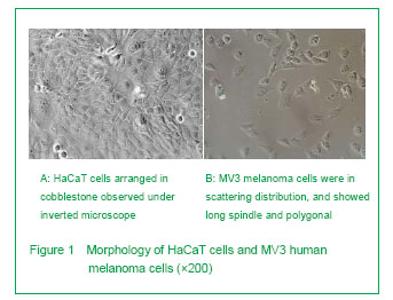
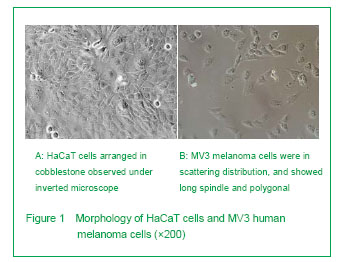
Observation of HaCaT cells and MV3 human melanoma cells under inverted microscope The HaCaT cells were in triangular or polygonal, and the cells were arranged tightly in cobblestone and connected into pieces with higher nucleus; the cytoplasm contained many particles, which similar with keratinocytes. There was a small amount of black dot cells with a small amount of low activity of the black dot cell, surrounded aperture shadow and surrounded with aperture shadow (Figure 1A). The MV3 human melanoma cells were in long spindle and polygonal and irregular in size and shape; the MVs human melanoma cells were large in cell body with abundant cytoplasm, prominent nucleoli and common karyokinesis (Figure 1B). "
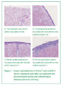
Histological observation with hematoxylin-eosin staining In the de-epidermized dermis without inoculation of HaCaT cells and MV3 human melanoma cells, there was no cell growth in the lumen (Figure 2A). Observation of hematoxylin-eosin stained sections with the ratio of MV3:HaCaT=1:10 showed the thin epidermis, the spinous layer cells were arranged like paving, the keratinocytes in some areas showed significant atypia, and showed pathological mitosis figures (Figure 2B). The observation of hematoxylin-eosin stained sections with the ratio of MV3:HaCaT=1:3 showed there was cell apoptosis and pathological mitosis figures in the superficial epidermis, the keratinocytes in some areas showed poloidal disorder with mild-moderate atypia, and the atypia was more significant in superficial epidermis (Figure 2C). Observation of hematoxylin-eosin stained sections with the ratio of MV3:HaCaT=1:1 showed there was no parakeratosis and hyperkeratosis in epidermis, cell apoptosis and pathological mitosis figures could be observed in the superficial epidermis, the keratinocytes in some areas showed poloidal disorder with mild-moderate atypia, and the atypia was more significant in superficial epidermis (Figure 2D)."

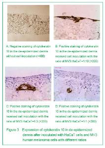
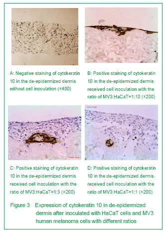
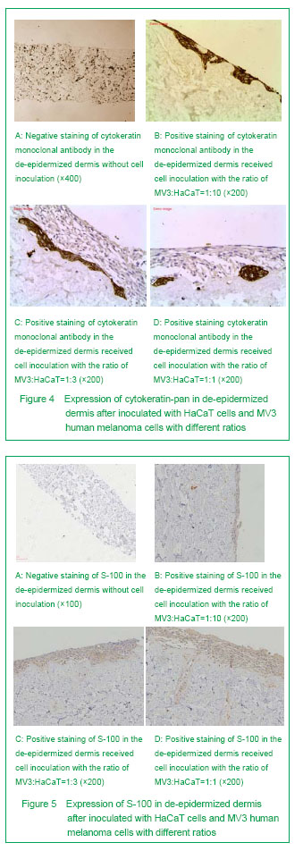
Immunohistochemical staining results Negative staining of cytokeratin 10 and cytokeratin-pan in the surface of de-epidermized dermis of the control group indicated that there were no expressions of cytokeratin 10 and cytokeratin-pan (Figures 3A and 4A). Immunohistological staining of cytokeratin 10 in the surface of de-epidermized dermis inoculated with two kinds of cells showed brownish yellow, which indicated that there was expression of cytokeratin 10, and suggested that HaCaT cells could differentiate into epidermal cells when growing on the surface of de-epidermized dermis (Figures 3B-3D). Immunohistological staining of cytokeratin monoclonal antibody showed the superficial cells were stained into brownish yellow completely, indicating there was expression of cytokeratin-pan (Figures 4B-4D). When the ratio of MV3:HaCaT=1:10, the positive cells of cytokeratin 10 and cytokeratin-pan were observed on the superficial layer, and distributed layer-by-layer. With the increasing of the ration of MV3:HaCaT, the positive cells of cytokeratin 10 and cytokeratin-pan were downward from the superficial layer to the middle and lower layer, and changed from layer-by-layer distribution to mass distribution. When the ratio of MV3:HaCaT=1:1, the positive cells of cytokeratin 10 and cytokeratin-pan were closely attached on the basement membrane of de-epidermized dermis. The staining of S-100 on the de-epidermized dermis of the control group was negative (Figure 5A). The protein staining of mixed inoculated S-100 specimen was positive (Figures 5B-5D). When the ratio of MV3:HaCaT=1:10, the number of MV3 melanoma cells was decreased and only formed 2-3 layer and all located on the surface of de-epidermized dermis; there was no significant positive stained cells in the de-epidermized dermis. With the increasing of the ratio of MV3:HaCaT, the number of MV3 melanoma cells was increased. When the ratio of MV3:HaCaT=1:1, the number of MV3 melanoma cells was increased, and the MV3 melanoma cells could be observed in the de-epidermized dermis."
"
| [1] | Chen Ziyang, Pu Rui, Deng Shuang, Yuan Lingyan. Regulatory effect of exosomes on exercise-mediated insulin resistance diseases [J]. Chinese Journal of Tissue Engineering Research, 2021, 25(25): 4089-4094. |
| [2] | Chen Yang, Huang Denggao, Gao Yuanhui, Wang Shunlan, Cao Hui, Zheng Linlin, He Haowei, Luo Siqin, Xiao Jingchuan, Zhang Yingai, Zhang Shufang. Low-intensity pulsed ultrasound promotes the proliferation and adhesion of human adipose-derived mesenchymal stem cells [J]. Chinese Journal of Tissue Engineering Research, 2021, 25(25): 3949-3955. |
| [3] | Yang Junhui, Luo Jinli, Yuan Xiaoping. Effects of human growth hormone on proliferation and osteogenic differentiation of human periodontal ligament stem cells [J]. Chinese Journal of Tissue Engineering Research, 2021, 25(25): 3956-3961. |
| [4] | Sun Jianwei, Yang Xinming, Zhang Ying. Effect of montelukast combined with bone marrow mesenchymal stem cell transplantation on spinal cord injury in rat models [J]. Chinese Journal of Tissue Engineering Research, 2021, 25(25): 3962-3969. |
| [5] | Gao Shan, Huang Dongjing, Hong Haiman, Jia Jingqiao, Meng Fei. Comparison on the curative effect of human placenta-derived mesenchymal stem cells and induced islet-like cells in gestational diabetes mellitus rats [J]. Chinese Journal of Tissue Engineering Research, 2021, 25(25): 3981-3987. |
| [6] | Hao Xiaona, Zhang Yingjie, Li Yuyun, Xu Tao. Bone marrow mesenchymal stem cells overexpressing prolyl oligopeptidase on the repair of liver fibrosis in rat models [J]. Chinese Journal of Tissue Engineering Research, 2021, 25(25): 3988-3993. |
| [7] | Liu Jianyou, Jia Zhongwei, Niu Jiawei, Cao Xinjie, Zhang Dong, Wei Jie. A new method for measuring the anteversion angle of the femoral neck by constructing the three-dimensional digital model of the femur [J]. Chinese Journal of Tissue Engineering Research, 2021, 25(24): 3779-3783. |
| [8] | Meng Lingjie, Qian Hui, Sheng Xiaolei, Lu Jianfeng, Huang Jianping, Qi Liangang, Liu Zongbao. Application of three-dimensional printing technology combined with bone cement in minimally invasive treatment of the collapsed Sanders III type of calcaneal fractures [J]. Chinese Journal of Tissue Engineering Research, 2021, 25(24): 3784-3789. |
| [9] | Qian Xuankun, Huang Hefei, Wu Chengcong, Liu Keting, Ou Hua, Zhang Jinpeng, Ren Jing, Wan Jianshan. Computer-assisted navigation combined with minimally invasive transforaminal lumbar interbody fusion for lumbar spondylolisthesis [J]. Chinese Journal of Tissue Engineering Research, 2021, 25(24): 3790-3795. |
| [10] | Hu Jing, Xiang Yang, Ye Chuan, Han Ziji. Three-dimensional printing assisted screw placement and freehand pedicle screw fixation in the treatment of thoracolumbar fractures: 1-year follow-up [J]. Chinese Journal of Tissue Engineering Research, 2021, 25(24): 3804-3809. |
| [11] | Shu Qihang, Liao Yijia, Xue Jingbo, Yan Yiguo, Wang Cheng. Three-dimensional finite element analysis of a new three-dimensional printed porous fusion cage for cervical vertebra [J]. Chinese Journal of Tissue Engineering Research, 2021, 25(24): 3810-3815. |
| [12] | Wang Yihan, Li Yang, Zhang Ling, Zhang Rui, Xu Ruida, Han Xiaofeng, Cheng Guangqi, Wang Weil. Application of three-dimensional visualization technology for digital orthopedics in the reduction and fixation of intertrochanteric fracture [J]. Chinese Journal of Tissue Engineering Research, 2021, 25(24): 3816-3820. |
| [13] | Sun Maji, Wang Qiuan, Zhang Xingchen, Guo Chong, Yuan Feng, Guo Kaijin. Development and biomechanical analysis of a new anterior cervical pedicle screw fixation system [J]. Chinese Journal of Tissue Engineering Research, 2021, 25(24): 3821-3825. |
| [14] | Lin Wang, Wang Yingying, Guo Weizhong, Yuan Cuihua, Xu Shenggui, Zhang Shenshen, Lin Chengshou. Adopting expanded lateral approach to enhance the mechanical stability and knee function for treating posterolateral column fracture of tibial plateau [J]. Chinese Journal of Tissue Engineering Research, 2021, 25(24): 3826-3827. |
| [15] | Zhu Yun, Chen Yu, Qiu Hao, Liu Dun, Jin Guorong, Chen Shimou, Weng Zheng. Finite element analysis for treatment of osteoporotic femoral fracture with far cortical locking screw [J]. Chinese Journal of Tissue Engineering Research, 2021, 25(24): 3832-3837. |
| Viewed | ||||||
|
Full text |
|
|||||
|
Abstract |
|
|||||