Chinese Journal of Tissue Engineering Research ›› 2023, Vol. 27 ›› Issue (6): 890-896.doi: 10.12307/2023.248
Previous Articles Next Articles
Collagen/silk fibroin scaffold combined with neural stem cells in the treatment of traumatic spinal cord injury
Li Xiaoyin1, Yang Xiaoqing2, Chen Shulian1, Li Zhengchao1, Wang Ziqi1, Song Zhen1, Zhu Daren1, Chen Xuyi1
- 1Chinese People’s Armed Police Force Special Medical Center, Tianjin Key Laboratory of Neurotrauma Repair, Tianjin 300162, China; 2Tianjin Anding Hospital, Tianjin 300222, China
-
Received:2022-01-18Accepted:2022-04-18Online:2023-02-28Published:2022-08-11 -
Contact:Chen Xuyi, MD, Associate chief physician, Doctoral supervisor, Chinese People’s Armed Police Force Special Medical Center, Tianjin Key Laboratory of Neurotrauma Repair, Tianjin 300162, China -
About author:Li Xiaoyin, Master, Research intern, Chinese People’s Armed Police Force Special Medical Center, Tianjin Key Laboratory of Neurotrauma Repair, Tianjin 300162, China Yang Xiaoqing, Master, Physician, Tianjin Anding Hospital, Tianjin 300222, China Li Xiaoyin and Yang Xiaoqing contributed equally to this article. -
Supported by:Key Project of National Natural Science Foundation of China, No. 11932013 (to CXY); National Science and Technology Key Research and Development Program, No. 2016YFC1101500 (to CXY)
CLC Number:
Cite this article
Li Xiaoyin, Yang Xiaoqing, Chen Shulian, Li Zhengchao, Wang Ziqi, Song Zhen, Zhu Daren, Chen Xuyi. Collagen/silk fibroin scaffold combined with neural stem cells in the treatment of traumatic spinal cord injury[J]. Chinese Journal of Tissue Engineering Research, 2023, 27(6): 890-896.
share this article
Add to citation manager EndNote|Reference Manager|ProCite|BibTeX|RefWorks
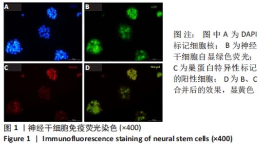
2.1 神经干细胞的免疫荧光染色 免疫荧光染色显示,新生绿色荧光蛋白小鼠海马区的神经细胞球内可见大部分细胞巢蛋白阳性,见图1,提示绝大多数细胞是神经干细胞。"

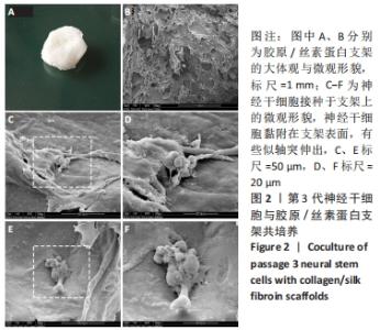
2.2 胶原/丝素蛋白支架上神经干细胞的生长情况 光镜下观察到,传到第3代神经干细胞接种于胶原/丝素蛋白支架后生长情况良好,细胞数量增加、体积增长正常,细胞形态良好。扫描电镜下可见,在胶原/丝素蛋白支架的三维孔洞和表面分布着不少类神经元样细胞,与支架的黏附效果良好,部分细胞有轴突伸出,见图2,提示胶原/丝素蛋白材质的支架给神经干细胞提供了一个良好的生长环境,利于其附着、伸展和分化,体现了该复合材料具有良好的生物相容性。"

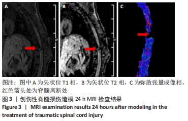
2.3 大鼠创伤性脊髓损伤造模后MRI和诱发电位检查结果 造模30只SD大鼠术后无死亡,全部参与了后续实验过程,进入结果分析。 2.3.1 MRI检查 造模后24 h行MRI检查,扫描矢状位T1、T2相及弥散张量成像后处理,T1和T2相中可见脊髓于T10段信号缺失;弥散张量成像通过检测组织中水分子向各方向的扩散来检测中枢神经系统的结构完整性,图像直观地显示出脊髓离断情况,见图3,影像学检查结果提示大鼠创伤性脊髓损伤模型制造成功,大鼠脊髓T10段完全离断。"
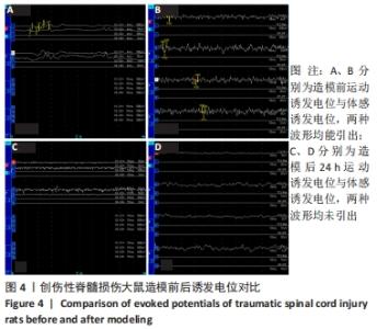
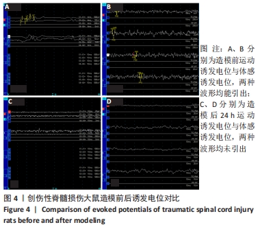
2.3.2 运动和体感诱发电位 运动和体感诱发电位波形曲线的宽度与峰值是反映神经传导速度和兴奋性轴突数量的重要参数,检测结果显示,造模前大鼠的运动和体感诱发电位波形均能引出,术后24 h两种波形均未引出,见图4,从神经电生理角度说明采用T10全脊髓离断术建立大鼠创伤性脊髓损伤模型成功。 "
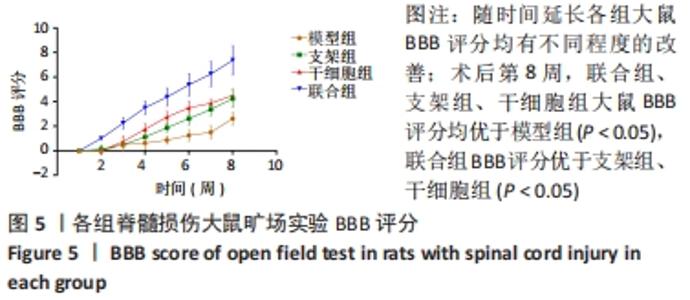
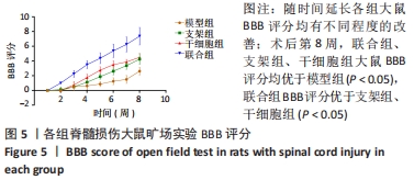
2.4 各组大鼠旷场实验BBB评分和斜坡实验 造模后,除正常组评分均为满分外,其余组损伤后的BBB评分为0分,随时间延长各组大鼠BBB评分均有不同程度的改善;术后第8周,联合组、支架组、干细胞组大鼠BBB评分均优于模型组(P < 0.05),联合组BBB评分优于支架组、干细胞组(P < 0.05),见图5。"
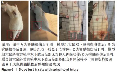
斜坡实验中,联合组大鼠双下肢及足部保持弯曲维持身体姿势和支撑身体质量,在斜坡实验中双下肢及足部能配合身体保持不下滑和姿势协调;模型组大鼠双下肢及足部则是软瘫的拖在身体后边,在同等坡度的斜坡上仅靠上肢艰难抓住铁丝网,双下肢和足部无任何支撑和抓握动作,见图6。上述结果反映出大鼠创伤性脊髓损伤造模后,干预组较单纯创伤性脊髓损伤的模型组运动功能恢复速度更快、恢复程度更大,尤以联合组最明显,说明该联合干预措施有助于大鼠创伤性脊髓损伤后运动功能的恢复。 "
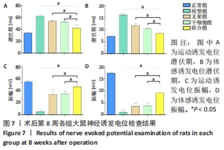
2.5 各组大鼠神经诱发电位检测结果 联合组、支架组、干细胞组大鼠运动诱发电位和体感诱发电位的潜伏期均低于模型组(P < 0.05),振幅高于模型组(P < 0.05);联合组大鼠运动诱发电位和体感诱发电位的潜伏期均低于支架组、干细胞组(P < 0.05),振幅高于支架组、干细胞组(P < 0.05),见图7。神经电生理检测结果提示,胶原/丝素蛋白支架和神经干细胞共同干预有助于创伤性脊髓损伤大鼠运动功能的恢复。"
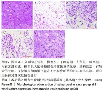
2.6 各组大鼠脊髓组织形态学观察 2.6.1 苏木精-伊红染色 与正常组相比,模型组大鼠脊髓损伤局部恢复情况差、结构混乱,有较大的空洞,支架组和细胞组也存在不同程度的结构破坏和小孔洞,联合组损伤局部恢复情况良好,见图8,提示创伤性脊髓损伤大鼠模型行胶原/丝素蛋白支架和神经干细胞共同移植干预有利于脊髓损伤局部组织恢复。"

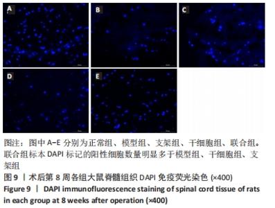
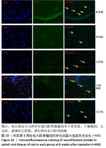
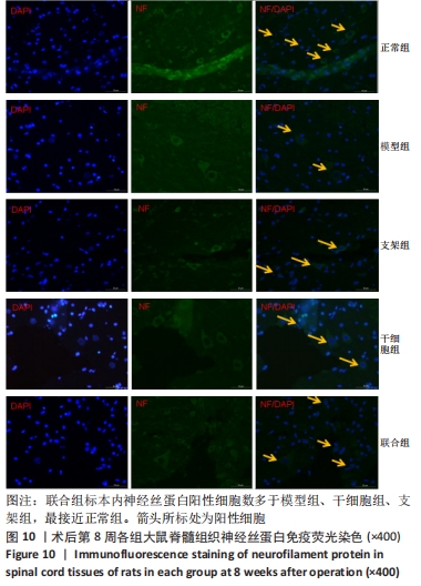
2.6.2 免疫荧光染色 联合组标本DAPI标记的阳性细胞数量明显多于模型组、干细胞组、支架组,提示在T10段移植的胶原/丝素蛋白支架利于神经干细胞存活,见图9。联合组标本内神经丝蛋白阳性细胞数多于模型组、干细胞组、支架组,最接近正常组,见图10,提示胶原/丝素蛋白支架+神经干细胞共同移植干预创伤性脊髓损伤大鼠模型不但提高了移植细胞的存活率,还有利于神经丝修复。"
"

2.7 胶原/丝素蛋白支架的生物相容性 由体外细胞接种实验与动物实验脊髓组织病理观察结果可知,胶原/丝素蛋白支架具有良好的生物相容性。"

| [1] ABU-BAKER NN, AL-ZYOUD NH, ALSHRAIFEEN A. Quality of life and self-care ability among individuals with spinal cord injury. Clin Nurs Res. 2021;30: 883-891. [2] DENISE GT, TRACEY W, GIULIA IL. Recommendations for evaluation of neurogenic bladder and bowel dysfunction after spinal cord injury and/or disease. J Spinal Cord Med. 2020;43(2):141-164. [3] JIANG L, SUN L, MENG QT. Identification and relationship of quality of life and self-care ability among Chinese patients with traumatic spinal cord injuries: a cross-sectional analysis. Braz J Med Biol Res. 2021;54(12):e11530. [4] Internationla Perspectives on Spinal Cord Injury. World Health Organization, 2013. [5] FISCHER I, DULIN JN, LANE MA. Transplanting neural progenitor cells to restore connectivity after spinal cord injury. Nat Rev Neurosci. 2020;21(7): 366-383. [6] QI MP, SI YC, QI JX, et al. Neuroinflammation and Scarring After Spinal Cord Injury: Therapeutic Roles of MSCs on Inflammation and Glial Scar. Front Immunol. 2021;12:751021. [7] LAI BQ, BAI YR, HAN WT, et al. Construction of a niche-specific spinal white matter-like tissue to promote directional axon regeneration and myelination for rat spinal cord injury repair. Bioact Mater. 2022;11:15-31. [8] ARSALAN A, SCOTT MD, SOHEILA KA. Traumatic Spinal Cord Injury: An Overview of Pathophysiology, Models and Acute Injury Mechanisms. Front Neurol. 2019;10:282. [9] WU HB, LI Y, WANG XF, et al. Long non-coding RNA TUG1 knockdown prevents neurons from death to alleviate acute spinal cord injury via the microRNA-338/BIK axis. Bioengineered. 2021;12(1):5566-5582. [10] ATOUSA ZM, MARYAM S, SEYED BJ, et al. Related Fluoxetine and Methylprednisolone Changes of TNF-α and IL-6 Expression in The Hypothyroidism Rat Model of Spinal Cord Injury. Cell J. 2021;23(7):763-771. [11] LIU S, XIE YY, WANG LD. A multi-channel collagen scaffold loaded with neural stem cells for the repair of spinal cord injury. Neural Regen Res. 2021;16(11): 2284-2292. [12] ATEFEH Z, SARA H, AYLIN G, et al. Spinal Cord Injury Management through the Combination of Stem Cells and Implantable 3D Bioprinted Platforms. Cells. 2021;10(11):3189. [13] POONGODI R, CHEN YL, YANG TH, et al. Bio-Scaffolds as Cell or Exosome Carriers for Nerve Injury Repair. Int J Mol Sci. 2021;22(24):13347. [14] YAMANE K, MAZAKI T, SHIOZAKI Y, et al. Collagen-Binding Hepatocyte Growth Factor(HGF)alone or with a Gelatin. furfurylamine Hydrogel Enhances Functional Recovery in Mice after Spinal Cord Injury. Sci Rep. 2018;8:917. [15] AIKEREMUJIANG M, LI S, JING L, et al. Sustained delivery of neurotrophic factors to treat spinal cord injury. Transl Neurosci. 2021;12(1):494-511. [16] MAŁGORZATA Z, ANNA K, KRZYSZTOF M, et al. Perspectives in the Cell-Based Therapies of Various Aspects of the Spinal Cord Injury-Associated Pathologies: Lessons from the Animal Models. Cells. 2021;10(11):2995. [17] ASSINCK P, DUNCAN GJ, HILTON BJ, et al. Cell transplantation therapy for spinal cord injury. Nat Neuro Sci. 2017;20:637-647. [18] HILTON BJ, MOULSON AJ, TETZLAFF W, et al. Neuroprotection and secondary damage following spinal cord injury: concepts and methods. Neuro Sci Lett. 2017;652:3-10. [19] MUNEHISA S, NARIHITO N, MASAYA N, et al. Mechanisms of Stem Cell Therapy in Spinal Cord Injuries. Cells. 2021;10(10):2676. [20] SUN W, GREGORY DA, TOMEH MA, et al. Silk Fibroin as a Functional Biomaterial for Tissue Engineering. Int J Mol Sci. 2021;22:1499. [21] GAO QQ, KIM BS, GAO G. Advanced Strategies for 3D Bioprinting of Tissue and Organ Analogs Using Alginate Hydrogel Bioinks. Mar Drugs. 2021;19(12):708. [22] 李晓寅,陈旭义,涂悦,等.精确显微技术条件下构建脊髓损伤模型的脊髓离断状态[J].中国组织工程研究,2014,18(27):4282-4286. [23] HUI QD, YI LP, CHEN XZ, et al. A novel, minimally invasive technique to establish the animal model of spinal cord injury. Ann Transl Med. 2021;9(10):881. [24] 曹宗锐,郑博,钟琳,等.胶原/硫酸肝素支架联合神经干细胞促进脊髓损伤后运动功能的恢复[J].中国组织工程研究,2019,23(34):5454-5461. [25] 郝定均,杨俊松,贺宝荣,等. “十三五”期间我国脊柱脊髓损伤临床诊疗研究亮点与进展[J].中华创伤杂志,2021,37(4):289-294. [26] HU XC, LU YB, YANG YN, et al. Progress in clinical trials of cell transplantation for the treatment of spinal cord injury: how many questions remain unanswered? Neural Regen Res. 2021;16(3):405-413. [27] HAN Q, SUN W, LIN H. Linear ordered collagen scaffolds loaded with collagen-binding brain-derived neurotrophic factor improve the recovery of spinal cord injury in rats. Tissue Eng Part A. 2009;15:2927-2935. [28] DENG WS, MA K, LIANG B, et al. Collagen scaffold combined with human umbilical cord-mesenchymal stem cells transplantation for acute complete spinal cord injury. Neural Regen Res. 2020;15(9):1686-1700. [29] 朱祥,陈旭义,李瑞欣,等.人工仿生脊髓导管的制备及性能分析[J].中国组织工程研究,2016,20(21):3045-3050. [30] CHEN MH, REN QX, YANG WF. Influences of HIF-lα on Bax/Bcl-2 and VEGF expressions in rats with spinal cord injury. Int J Clin Exp Pathol. 2013;6(11): 2312-2322. [31] YE X, CHEN YL , WANG JS, et al. Identification of Circular RNAs Related to Vascular Endothelial Proliferation, Migration, and Angiogenesis After Spinal Cord Injury Using Microarray Analysis in Female Mice. Front Neurol. 2021; 12:666750. |
| [1] | Zhong Yizheng, Huang Peizhen, Cai Qunbin, Zheng Liqin, He Xingpeng, Dong Hang. Microstructural indexes that determine the trabecular bone maximum stress of micro-finite element models [J]. Chinese Journal of Tissue Engineering Research, 2023, 27(9): 1313-1318. |
| [2] | Cao Sheng, Kong Lingwei, Xu Kun, Sun Zhijie. Correlation of cervical sagittal force line parameters with degenerative segment and Pfirrmann classification in patients with cervical intervertebral disc degeneration [J]. Chinese Journal of Tissue Engineering Research, 2023, 27(9): 1319-1324. |
| [3] | Ke Yuqi, Chen Changjian, Wu Hao, Zheng Lianjie. Comparison of 12-month follow-up results of primary total hip arthroplasty between modified direct anterior approach and direct anterior approach [J]. Chinese Journal of Tissue Engineering Research, 2023, 27(9): 1377-1382. |
| [4] | Zhang Lichuang, Gao Huali, Wang Jingchao, Lin Huijun, Wu Chonggui, Ma Yinghui, Huang Yunfei, Fang Xue, Zhai Weitao. Effect of tendon manipulation with equal emphasis on muscles and bones on accelerating the functional rehabilitation of quadriceps femoris after total knee arthroplasty [J]. Chinese Journal of Tissue Engineering Research, 2023, 27(9): 1383-1389. |
| [5] | Du Xueting, Zhang Xiaodong, Chen Yanjun, Wang Mei, Chen Wubiao, Huang Wenhua. Application of compressed sensing technology in two-dimensional magnetic resonance imaging of the ankle joint [J]. Chinese Journal of Tissue Engineering Research, 2023, 27(9): 1396-1402. |
| [6] | You Zhengqiu, Zhang Zhongzu, Wang Qunbo. Early symptomatic intervertebral disc pseudocysts after discectomy detected on MRI [J]. Chinese Journal of Tissue Engineering Research, 2023, 27(9): 1403-1409. |
| [7] | Li Chao, Zhang Peipei, Xu Mengting, Li Linlin, Ding Jiangtao, Liu Xihua, Bi Hongyan. Respiratory training improves morphological changes of the multifidus muscle in patients with chronic nonspecific lower back pain assessed by musculoskeletal ultrasound [J]. Chinese Journal of Tissue Engineering Research, 2023, 27(9): 1417-1421. |
| [8] | He Yinhao, Li Xiaosheng, Chen Hongwen, Chen Tiezhu. 3D printed porous tantalum metal in the treatment of developmental dysplasia of the hip: current status and application prospect [J]. Chinese Journal of Tissue Engineering Research, 2023, 27(9): 1455-1461. |
| [9] | Jiang Xiaocheng, Shi Lu, Wang Yinbin, Li Qiujiang, Xi Chuangzhen, Ma Zefeng, Cai Lijun. Systematical evaluation of bone fusion rate after interbody fusion in patients with osteoporosis and lumbar degenerative disease treated with teriparatide [J]. Chinese Journal of Tissue Engineering Research, 2023, 27(9): 1427-1433. |
| [10] | Li Mengfei, Zhang Hong, Zhao Shaojian, Yin Guanghao, Wang Qibao. Expression of forkhead box protein 3 in refractory periapical periodontitis in rats with Enterococcus faecalis infection [J]. Chinese Journal of Tissue Engineering Research, 2023, 27(8): 1187-1192. |
| [11] | Ruan Ling, Wang Guanghua, Wu Rongping, Jin Zhan, Lyu Zhenqing, Zhang Nan, Li Shoubang. Correlation between exercise intensity and lipid metabolism disorder and oxidative stress in a high-diet rat model [J]. Chinese Journal of Tissue Engineering Research, 2023, 27(8): 1149-1155. |
| [12] | Lian Shilin, Zhang Yan, Jiang Qiang, Zhang Hanshuo, Li Tusheng, Ding Yu. Interventional effects of whole blood and platelet-rich plasma with different preparation methods on nucleus pulposus cells [J]. Chinese Journal of Tissue Engineering Research, 2023, 27(8): 1199-1204. |
| [13] | Wu Dongzhe, Gao Xiaolin, Li Chuangtao, Wang Hao. Constructing the prediction model of maximal oxygen uptake by back-propagation neural network based on the cardiorespiratory optimal point [J]. Chinese Journal of Tissue Engineering Research, 2023, 27(8): 1224-1231. |
| [14] | Yang Jiujie, Li Zhi, Wang Shujie, Tian Ye, Zhao Wei. Intraoperative neurophysiological monitoring of functional changes following durotomy with decompression for acute spinal cord injury [J]. Chinese Journal of Tissue Engineering Research, 2023, 27(8): 1232-1236. |
| [15] | Liu Xiaolin, Mu Xinyue, Ma Ziyu, Liu Shutai, Wang Wenlong, Han Xiaoqian, Dong Zhiheng. Effect of hydrogel-loaded simvastatin microspheres on osteoblast proliferation and differentiation [J]. Chinese Journal of Tissue Engineering Research, 2023, 27(7): 998-1003. |
| Viewed | ||||||
|
Full text |
|
|||||
|
Abstract |
|
|||||