[1] 凌均棨,韦曦,刘红艳. 难治性根尖周炎的病因及防治策略[J]. 中华口腔医学杂志,2010,45(1):52-57.
[2] ZHANG C, DU J, PENG Z. Correlation between Enterococcus faecalis and Persistent Intraradicular Infection Compared with Primary Intraradicular Infection: A Systematic Review. J Endod. 2015;41(8):1207-1213.
[3] PERSOON IF, ÖZOK AR. Definitions and Epidemiology of Endodontic Infections. Curr Oral Health Rep. 2017;4(4):278-285.
[4] 张昱佳,钱友坤,朱一成,等.FOXP3~+调节性T细胞与非感染性炎症疾病[J].生命科学,2020,32(9):879-889.
[5] 严妍,那迪娜·帕尔哈提,车千纪,等.FOXP3~+调节性T细胞与人体传染性疾病[J].生命科学,2020,32(11):1159-1175.
[6] SAKAGUCHI S, MIKAMI N, WING JB, et al. Regulatory T Cells and Human Disease. Annu Rev Immunol. 2020;38:541-566.
[7] 伊刚,赵毅,解丰,等.单细胞RNA测序揭露人FOXP3+调节性T细胞稳定性的关键调控分子(英文)[J]. Science Bulletin. 2020,65(13): 1114-1124.
[8] YOSHIDA H, JONTELL M, SUNDQVIST G,et al. Effect of sonicated material from Fusobacterium nucleatum on the functional capacity of accessory cells derived from dental pulp. Oral Microbiol Immunol. 1995;10(4):208-212.
[9] 张文娟,谭昭,蔡如佳,等.调节性T细胞在慢性牙周炎小鼠模型中的作用[J].中国医科大学学报,2021,50(2):135-140.
[10] 李宝坤,陈晓涛,杨柯,等.慢性牙周炎患者外周血CD4~+CD25~+调节性T细胞表达[J].中华实用诊断与治疗杂志,2019,33(5):487-490.
[11] 吕广应,刘乙臻,张秀梅,等.叉头样转录因子3和白细胞介素10在慢性根尖周炎组织中的表达研究[J].实用口腔医学杂志,2019, 35(5):653-657.
[12] YANG S, ZHU L, XIAO L, et al. Imbalance of interleukin-17+ T-cell and Foxp3+ regulatory T-cell dynamics in rat periapical lesions. J Endod. 2014;40(1):56-62.
[13] WANG L, JIN H, AO X, et al. JAK2-STAT3 signaling pathway is involved in rat periapical lesions induced by Enterococcus faecalis. Oral Dis. 2019;25(7):1769-1779.
[14] STUART CH, SCHWARTZ SA, BEESON TJ, et al. Enterococcus faecalis: its role in root canal treatment failure and current concepts in retreatment. J Endod. 2006;32(2):93-98.
[15] ZHANG C, DU J, PENG Z. Correlation between Enterococcus faecalis and Persistent Intraradicular Infection Compared with Primary Intraradicular Infection: A Systematic Review. J Endod. 2015;41(8):1207-1213.
[16] 李莹雪,王雨霏,张凌琳.粪肠球菌在牙髓根尖周病中致病机制及抗菌治疗的研究进展[J].口腔疾病防治,2019,27(8):535-540.
[17] GOMES BPFA, HERRERA DR. Etiologic role of root canal infection in apical periodontitis and its relationship with clinical symptomatology. Braz Oral Res. 2018;32(suppl 1):e69.
[18] KINANE DF, LAPPIN DF, KOULOURI O, et al. Humoral immune responses in periodontal disease may have mucosal and systemic immune features. Clin Exp Immunol. 1999;115(3):534-541.
[19] GRAVES DT, OATES T, GARLET GP. Review of osteoimmunology and the host response in endodontic and periodontal lesions. J Oral Microbiol. 2011;3:10.3402/jom.v3i0.5304.
[20] COLIĆ M, GAZIVODA D, VUCEVIĆ D,et al. Proinflammatory and immunoregulatory mechanisms in periapical lesions. Mol Immunol. 2009;47(1):101-113.
[21] DE CARVALHO FRAGA CA, ALVES LR, DE SOUSA AA, et al. Th1 and Th2-like protein balance in human inflammatory radicular cysts and periapical granulomas. J Endod. 2013;39(4):453-455.
[22] NAUFEL AO, AGUIAR MCF, MADEIRA FM, et al. Treg and Th17 cells in inflammatory periapical disease: a systematic review. Braz Oral Res. 2017;31:e103.
[23] HORI S, NOMURA T, SAKAGUCHI S. Pillars Article: Control of Regulatory T Cell Development by the Transcription Factor Foxp3. Science. 2003; 299:1057-1061.
[24] VAN DER VEEKEN J, GLASNER A, ZHONG Y, et al. The Transcription Factor Foxp3 Shapes Regulatory T Cell Identity by Tuning the Activity of trans-Acting Intermediaries. Immunity. 2020;53(5):971-984.e5.
[25] 周欣. Th17细胞和Treg细胞在大鼠实验性牙周炎牙周组织中的分布和意义[D].石家庄:河北医科大学,2018.
[26] STADHOUDERS R, LUBBERTS E, HENDRIKS RW. A cellular and molecular view of T helper 17 cell plasticity in autoimmunity. J Autoimmun. 2018; 87:1-15.
[27] LI Y, WEI C, XU H, et al. The Immunoregulation of Th17 in Host against Intracellular Bacterial Infection. Mediators Inflamm. 2018;2018: 6587296.
[28] ROWE JH, ERTELT JM, WAY SS. Foxp3(+) regulatory T cells, immune stimulation and host defence against infection. Immunology. 2012; 136(1):1-10.
[29] PENG G, GUO Z, KINIWA Y, et al. Toll-like receptor 8-mediated reversal of CD4+ regulatory T cell function. Science. 2005;309(5739):1380-1384.
[30] FORWARD NA, FURLONG SJ, YANG Y, et al. Signaling through TLR7 enhances the immunosuppressive activity of murine CD4+CD25+ T regulatory cells. J Leukoc Biol. 2010;87(1):117-125.
[31] 纪周新,贺德.粪肠球菌脂磷壁酸激活Toll样受体2抑制胰腺导管癌BxPC-3细胞的增殖、侵袭和迁移[J].中国肿瘤生物治疗杂志, 2021,28(5):477-481.
[32] JOHANNS TM, ERTELT JM, ROWE JH, et al. Regulatory T cell suppressive potency dictates the balance between bacterial proliferation and clearance during persistent Salmonella infection. PLoS Pathog. 2010; 6(8):e1001043.
[33] 税钰森,吕潇颖,李静雅,等.粪肠球菌在口腔及全身系统性疾病中的致病相关因素及其机制的研究进展[J].国际口腔医学杂志, 2020,47(2):225-234.
[34] 孟磊,刘雪,张蕾,等.压力状态下粪肠球菌培养上清液诱导人牙周膜细胞炎症反应的研究[J].中华口腔医学杂志,2021,56(4):335-341.
[35] DONG M, JIN H, ZUO M, et al. The potential effect of Bruton’s tyrosine kinase in refractory periapical periodontitis. Biomed Pharmacother. 2019;112:108710.
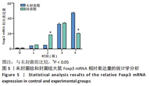
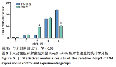
|