Chinese Journal of Tissue Engineering Research ›› 2023, Vol. 27 ›› Issue (12): 1870-1876.doi: 10.12307/2023.017
Previous Articles Next Articles
Effects of quercetin sustained release system on osteogenic properties of MC3T3-E1 cells
Ma Ziyu1, Zhang Bin2, Zhang Yuntao2, Liu Xiaolin1, Bian Zhihong1, Qiao Luhui3, Hou Yudong4
- 1Binzhou Medical College, Binzhou 256600, Shandong Province, China; 2Affiliated Hospital of Binzhou Medical College, Binzhou 256600, Shandong Province, China; 3Yantai Stomatological Hospital, Yantai 264010, Shandong Province, China; 4School of Stomatology, Binzhou Medical College, Yantai 264010, Shandong Province, China
-
Received:2021-11-25Accepted:2022-01-05Online:2023-04-28Published:2022-07-30 -
Contact:Hou Yudong, Master, Professor, School of Stomatology, Binzhou Medical College, Yantai 264010, Shandong Province, China Zhang Bin, Master, Affiliated Hospital of Binzhou Medical College, Binzhou 256600, Shandong Province, China -
About author:Ma Ziyu, Master candidate, Binzhou Medical College, Binzhou 256600, Shandong Province, China
CLC Number:
Cite this article
Ma Ziyu, Zhang Bin, Zhang Yuntao, Liu Xiaolin, Bian Zhihong, Qiao Luhui, Hou Yudong. Effects of quercetin sustained release system on osteogenic properties of MC3T3-E1 cells[J]. Chinese Journal of Tissue Engineering Research, 2023, 27(12): 1870-1876.
share this article
Add to citation manager EndNote|Reference Manager|ProCite|BibTeX|RefWorks
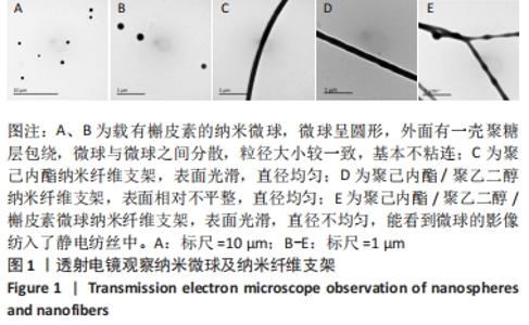
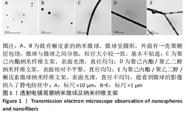
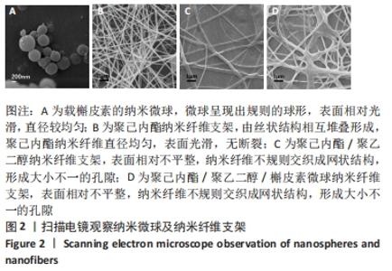
2.1 微球与纳米纤维支架的微观形貌 见图1,2。"
"
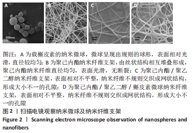
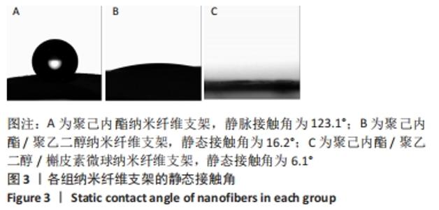
透射电镜观察结果:载有槲皮素的纳米微球呈圆形,外面有一壳聚糖层包绕,微球与微球之间分散,粒径大小较一致,基本不粘连。聚己内酯纳米纤维表面光滑,直径均匀。聚己内酯/聚乙二醇纳米纤维由于聚乙二醇的加入表面相对不平整,直径均匀。聚己内酯/聚乙二醇/槲皮素微球纳米纤维表面光滑,直径不均匀,能看到微球的影像纺入了静电纺丝中。 扫描电镜观察结果:载药纳米微球呈现出规则的球形,表面相对光滑,直径较均匀。聚己内酯纳米纤维支架由丝状结构相互堆叠形成,聚己内酯纳米纤维直径均匀,表面光滑,无断裂。聚己内酯/聚乙二醇和聚己内酯/聚乙二醇/槲皮素微球纳米纤维表面相对不平整,可能是聚乙二醇与微球的加入所导致,纳米纤维不规则交织成网状结构,形成大小不一的孔隙,增加了支架材料的比表面积,且能便于营养物质与代谢产物的进出,为细胞的附着与生存提供了足够的空间和良好的环境。 2.2 纳米纤维支架的静态接触角 见图3。 "
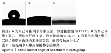
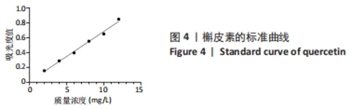
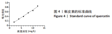
图3为去离子水滴在纳米纤维支架薄膜上2 s后所拍摄的图片,聚己内酯组2 s后的静态接触角为123.1°,聚己内酯/聚乙二醇组2 s后的静态接触角为16.2°,聚己内酯/聚乙二醇/槲皮素微球组2 s后的静态接触角为6.1°。可以看出,聚己内酯/聚乙二醇组的静态接触角相对于聚己内酯组显著减小,说明液滴大部分渗透入纤维材料中,证明聚乙二醇发挥了它的亲水性的优点,显著增加了材料的亲水性能;聚己内酯/聚乙二醇/槲皮素微球组的静态接触角小于聚己内酯/聚乙二醇组,可能微球的加入增大了纤维的粗糙程度或孔隙率,从而进一步改善了材料的亲水性。 2.3 槲皮素标准曲线及回归方程 以无水乙醇溶液作为空白对照组,归零后,测定每种槲皮素乙醇溶液在375 nm处的吸光度值,以Y轴作为吸光度值,X轴作为浓度,绘制标准曲线图,如图4所示,并得出回归方程为:Y=0.067 33×X+0.011 53,R2=0.991 9,表明药物质量浓度在2-12 mg/L之内,呈现良好的线性关系。 "
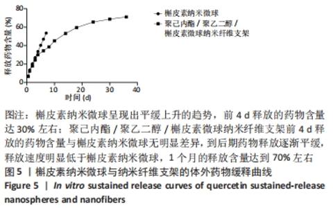
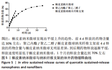
2.4 微球包封率及材料体外释药曲线 经计算得出,槲皮素纳米微球的包封率约为75.08%,槲皮素可以有效包裹于微球内。 通过体外释药曲线可见,槲皮素纳米微球呈现出平缓上升的趋势,前4 d释放的药物含量达到30%左右;聚己内酯/ 聚乙二醇/槲皮素微球纳米纤维支架前4 d释放的药物含量与槲皮素纳米微球无明显差异,到后期药物释放逐渐平缓,释放速度明显低于槲皮素纳米微球,1个月的释放含量达到70%左右,见图5。 "
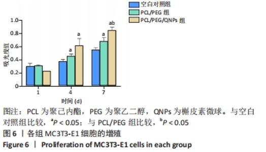
2.5 纳米纤维支架的细胞相容性 2.5.1 细胞增殖实验结果 见图6。"


培养第1天,3组之间细胞增殖比较差异无显著性意义(P > 0.05);培养第4,7天,聚己内酯/聚乙二醇组、聚己内酯/聚乙二醇/槲皮素微球组细胞增殖吸光度值高于空白对照组(P < 0.05),表明聚己内酯/聚乙二醇、聚己内酯/聚乙二醇/槲皮素微球纳米纤维支架都能够保持MC3T3-E1细胞的活性并促使其增殖;聚己内酯/聚乙二醇/槲皮素微球培养第7天的细胞增殖吸光度值高于聚己内酯/聚乙二醇组(P < 0.05),说明槲皮素的加入进一步促进了细胞的增殖。 2.5.2 碱性磷酸酶活性检测结果 见图7。 "
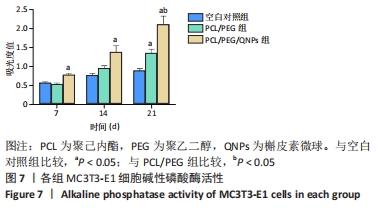
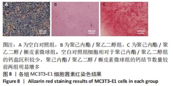
诱导培养第7,14天时,聚己内酯/聚乙二醇组与空白对照组细胞碱性磷酸酶活性比较差异无显著性意义(P > 0.05);诱导培养第7,14,21天时,聚己内酯/聚乙二醇/槲皮素微球组细胞碱性磷酸酶活性高于空白对照组(P < 0.05);诱导培养第21天时,聚己内酯/聚乙二醇/槲皮素微球组细胞碱性磷酸酶活性高于聚己内酯/聚乙二醇组(P < 0.05),说明聚己内酯/聚乙二醇、聚己内酯/聚乙二醇/槲皮素微球纤维支架能够促进细胞碱性磷酸酶的表达。 2.5.3 茜素红染色结果 见图8。 "

可以看出,空白对照组细胞相对于聚己内酯/聚乙二醇组的钙盐沉积较少,聚己内酯/聚乙二醇/槲皮素微球组的钙结节数量较前两组明显增多,说明聚己内酯/聚乙二醇纳米纤维支架材料与槲皮素微球均能够促进细胞的矿化。 由细胞增殖实验与成骨性能实验结果可知,聚己内酯/聚乙二醇/槲皮素微球纳米纤维支架具有良好的细胞相容性。 "

| [1] 刘相杰,宋科官.生物支架材料及间充质干细胞在骨组织工程中的研究与应用[J].中国组织工程研究,2018,22(10):1618-1624. [2] KONG D, SHI Y, GAO Y, et al. Preparation of BMP-2 loaded MPEG-PCL microspheres and evaluation of their bone repair properties. Biomed Pharmacother. 2020;130:110516. [3] INJAMURI S, RAHAMAN MN, SHEN Y, et al. Relaxin enhances bone regeneration with BMP-2-loaded hydroxyapatite microspheres. J Biomed Mater Res A. 2020;108(5):1231-1242. [4] SONG H, ZHANG Y, ZHANG Z, et al. Hydroxyapatite/NELL-1 Nanoparticles Electrospun Fibers for Osteoinduction in Bone Tissue Engineering Application. Int J Nanomedicine. 2021;16:4321-4332. [5] LAI K, XI Y, DU X, et al. Activation of Nell-1 in BMSC Sheet Promotes Implant Osseointegration Through Regulating Runx2/Osterix Axis. Front Cell Dev Biol. 2020;8:868. [6] HUANG K, CHEN C, CHANG C, et al. The synergistic effects of quercetin- containing 3D-printed mesoporous calcium silicate/calcium sulfate/poly-ε-caprolactone scaffolds for the promotion of osteogenesis in mesenchymal stem cells. J Formos Med Assoc. 2021;120(8):1627-1634. [7] HUANG Y, WANG Z, DENG L, et al. Oral Administration of Quercetin or Its Derivatives Inhibit Bone Loss in Animal Model of Osteoporosis. Oxid Med Cell Longev. 2020;2020:1-21. [8] WONG SK, CHIN K, IMA-NIRWANA S. Quercetin as an Agent for Protecting the Bone: A Review of the Current Evidence. Int J Mol Sci. 2020;21(17): 6448. [9] WANG N, WANG L, YANG J, et al. Quercetin promotes osteogenic differentiation and antioxidant responses of mouse bone mesenchymal stem cells through activation of the AMPK/SIRT1 signaling pathway. Phytother Res. 2021;35(5):2639-2650. [10] ZHANG Q, CHANG B, ZHENG G, et al. Quercetin stimulates osteogenic differentiation of bone marrow stromal cells through miRNA-206/connexin 43 pathway. Am J Transl Res. 2020;12(5):2062-2070. [11] ZHANG W, JIA L, ZHAO B, et al. Quercetin reverses TNFα induced osteogenic damage to human periodontal ligament stem cells by suppressing the NFκB/NLRP3 inflammasome pathway. Int J Mol Med. 2021;47(4):1-11. [12] ANGELLOTTI G, MURGIA D, CAMPISI G, et al. Quercetin-Based Nanocomposites as a Tool to Improve Dental Disease Management. Biomedicines. 2020;8(11):504. [13] ZHOU Y, WU Y, MA W, et al. The effect of quercetin delivery system on osteogenesis and angiogenesis under osteoporotic conditions. Journal of materials chemistry. J Mater Chem B. 2017;5(3):612-625. [14] JÓHANNESSON G, STEFÁNSSON E, LOFTSSON T. Microspheres and nanotechnology for drug delivery. Dev Ophthalmol. 2016;55(3):93-103. [15] DEFRATES K, MARKIEWICZ T, GALLO P, et al. Protein polymer- based nano-particles: Fabrication and medical Applications. Int J Mol Sci. 2018;19(6):1717. [16] CHA C, JEONG JH, KONG H. Poly(ethylene glycol)-poly(lactic- co-glycolic acid) core–shell microspheres with enhanced controllability of drug encapsulation and release rate. J Biomater Sci Polym Ed. 2015; 26(13):828-840. [17] DELDAR Y, ZARGHAMI F, PILEHVAR-SOLTANAHMADI Y, et al. Antioxidant effects of chrysin-loaded electrospun nanofibrous mats on proliferation and stemness preservation of human adipose-derived stem cells. Cell Tissue Bank. 2017;18(4):475-487. [18] SIDDIQUI N, ASAWA S, BIRRU B, et al. PCL-Based Composite Scaffold Matrices for Tissue Engineering Applications. Mol Biotechnol. 2018; 60(7):506-532. [19] ENGEBRETSON B, SIKAVITSAS VI. Long-term in vivo effect of PEG bone tissue engineering scaffolds. J Long Term Eff Med Implants. 2012; 22(3):211. [20] 傅娜,罗晓丁,焦铁军,等.骨组织工程中应用的聚己内酯-聚乙二醇-聚己内酯静电纺丝支架[J].中国组织工程研究,2019,23(22): 3445-3450. [21] 潘璐,程亭亭,徐岚.聚己内酯/聚乙二醇大孔径纳米纤维膜的制备及其在组织工程支架中的应用[J].纺织学报,2020,41(9):167-173. [22] 彭兰兰.静电纺丝法制备PCL/PEG组织工程支架的研究[D].上海:东华大学,2009. [23] LOBO AO, AFEWERKI S, DE PAULA MMM, et al. Electrospun nanofiber blend with improved mechanical and biological performance. Int J Nanomedicine. 2018;13:7891-7903. [24] 王乐,惠敏,董西玲,等.缓释阿托伐他汀钙纳米纤维支架对细胞黏附增殖的影响[J].中国组织工程研究,2020,24(28):4492-4497. [25] BHARATHALA S, KOTARKONDA LK, SINGH VP, et al. In silico and experimental studies of bovine serum albumin-encapsulated carbenoxolone nanoparticles with reduced cytotoxicity. Colloids Surf B Biointerfaces. 2021;202:111670. [26] ARRIAGADA F, GÜNTHER G, ZABALA I, et al. Development andcharacterization offlorfenicol-loaded BSAnanoparticles ascontrolledreleasecarrier. AAPS PharmSciTech. 2019;20(5):202. [27] ZHANG Y, DONG R, PARK Y, et al. Controlled release of NELL-1 protein from chitosan/hydroxyapatite- modified TCP particles. Int J Pharm. 2016;511(1):79-89. [28] GIZAW M, THOMPSON J, FAGLIE A, et al. Electrospun Fibers as a Dressing Material for Drug and Biological Agent Delivery in Wound Healing Applications. Bioengineering. 2018;5(1):9. [29] LEONÉS A, MUJICA-GARCIA A, ARRIETA MP, et al. Organic and Inorganic PCL-Based Electrospun Fibers. Polymers. 2020;12(6):1325. [30] PAGANGA G, RICE-EVANS CA. The identification of flavonoids as glycosides in human plasma. FEBS Lett. 1997;401(1):78-82. [31] 周宇宁.中药槲皮素应用于骨缺损修复的初步研究[D].上海:上海交通大学,2016:75. [32] WONG RWK, RABIE ABM. Effect of quercetin on preosteoblasts and bone defects. Open Orthop J. 2008;2(1):27-32. [33] LI B, YOSHII T, HAFEMAN AE, et al. The effects of rhBMP-2 released from biodegradable polyurethane/microsphere composite scaffolds on new bone formation in rat femora. Biomaterials. 2009;30(35):6768-6779. [34] 关德强,于波,李欣,等.中药促进骨再生的研究进展[J].时珍国医国药,2018,29(4):956-958. [35] 陈述祥,康乐.中药促成骨细胞增殖和分化的机制与作用[J].中国组织工程研究,2012,16(7):1299-1302. [36] 潘慧欣,崔元璐.中药诱导骨髓间充质干细胞成软骨及成骨分化的研究进展[J].天津中医药,2018,35(10):794-797. [37] WANG XC, ZHAO NJ, GUO C, et al. Quercetin reversed lipopolysaccharide-induced inhibition of osteoblast differentiation through the mitogenactivated protein kinase pathway in MC3T3-E1 cells. Mol Med Rep. 2014;10(6):3320-3326. [38] PANG X, CONG Y, BAO N, et al. Quercetin Stimulates Bone Marrow Mesenchymal Stem Cell Differentiation through an Estrogen Receptor-Mediated Pathway. Biomed Res Int. 2018;2018:1-11. [39] WEI J, ZHANG X, LI Y, et al. Novel application of bergapten and quercetin with anti-bacterial, osteogenesis-potentiating, and anti-inflammation tri-effects. Acta Biochim Biophys Sin. 2021;53(6):683-696. [40] ZENG Y, NIKITKOVA A, ABDELSALAM H, et al. Activity of quercetin and kaemferol against Streptococcus mutans biofilm. Arch Oral Biol. 2019; 98:9-16. |
| [1] | Liu Xiaolin, Mu Xinyue, Ma Ziyu, Liu Shutai, Wang Wenlong, Han Xiaoqian, Dong Zhiheng. Effect of hydrogel-loaded simvastatin microspheres on osteoblast proliferation and differentiation [J]. Chinese Journal of Tissue Engineering Research, 2023, 27(7): 998-1003. |
| [2] | Gao Ting, Ma Xiaohong, Li Xiaorong. Extraction and identification of exosomes from three different sources of ovarian granulosa cells [J]. Chinese Journal of Tissue Engineering Research, 2023, 27(6): 860-865. |
| [3] | Tian Qinyu, Tian Xinggui, Tian Zhuang, Sui Xiang, Liu Shuyun, Lu Xiaobo, Guo Quanyi. Protection of manganese oxide nanoparticles for bone marrow mesenchymal stem cell spreading against oxidative stress [J]. Chinese Journal of Tissue Engineering Research, 2023, 27(6): 821-826. |
| [4] | Li Rui, Liu Zhen, Guo Zige, Lu Ruijie, Wang Chen. Aspirin-loaded chitosan nanoparticles and polydopamine modified titanium sheets improve osteogenic differentiation [J]. Chinese Journal of Tissue Engineering Research, 2023, 27(3): 374-379. |
| [5] | Li Yue, Lyu Yan, Feng Wanying, Song Yang, Yan Yu, Guan Yongge. Preparation of hyperoside nanoparticles to repair endometrial injury [J]. Chinese Journal of Tissue Engineering Research, 2023, 27(3): 360-366. |
| [6] | Li Zhen, Liu Hongbao. Influencing factors and mechanism of nanoparticle renal targeting [J]. Chinese Journal of Tissue Engineering Research, 2023, 27(3): 453-460. |
| [7] | Peng Kun. Properties of force growth factor E peptide bionic bone matrix with polyethylene glycol derivative hydrogel as a carrier [J]. Chinese Journal of Tissue Engineering Research, 2023, 27(12): 1811-1816. |
| [8] | Ye Xuwen, Gu Yong, Chen Liang. Curcumin loaded injectable microspheres retard progression of intervertebral disc degeneration [J]. Chinese Journal of Tissue Engineering Research, 2023, 27(12): 1884-1891. |
| [9] | Hu Jinlong, Quan Huahong, Wang Jingcheng, Zhang Pei, Zhang Jiale, Chen Pengtao, Liang Yuan. Effect of copper sulfide nanoparticles loaded thermosensitive hydrogel Pluronic F127 on infected wound healing in rats [J]. Chinese Journal of Tissue Engineering Research, 2023, 27(12): 1927-1931. |
| [10] | Gan Tian, Wang Wenyuan, Yan Shujin, Hao Lan, Ran Haitao, Wang Zhigang, Xia Jizhu. Near infrared photoresponsive nanoparticles loaded with LXR agonists for photothermal immunotherapy [J]. Chinese Journal of Tissue Engineering Research, 2023, 27(12): 1863-1869. |
| [11] | Li Zhiyi, He Pengcheng, Bian Tianyue, Xiao Yuxia, Gao Lu, Liu Huasheng. Bibliometric and visualized analysis of ferroptosis mechanism research [J]. Chinese Journal of Tissue Engineering Research, 2022, 26(8): 1202-1209. |
| [12] | Gao Cangjian, Yang Zhen, Liu Shuyun, Li Hao, Fu Liwei, Zhao Tianyuan, Chen Wei, Liao Zhiyao, Li Pinxue, Sui Xiang, Guo Quanyi. Electrospinning for rotator cuff repair [J]. Chinese Journal of Tissue Engineering Research, 2022, 26(4): 637-642. |
| [13] | Cao Fei, Hui Min, Dong Xiling, Wang Le, Wang Zuxu, Zhang Min, Zhang Xiaoming, Liu Tongbin. Preparation of silver-loaded nanohydroxyapatite/polycaprolactone composite nanofiber scaffold and its osteogenic and antibacterial properties [J]. Chinese Journal of Tissue Engineering Research, 2022, 26(34): 5461-5467. |
| [14] | Lin Lingqi, Chen Jin, Qian Kun, Zhao Liang, Shi Yijie. Preparation and in vitro release of manganese-based metal-organic framework materials loaded with baicalin [J]. Chinese Journal of Tissue Engineering Research, 2022, 26(34): 5475-5481. |
| [15] | Wang Xiao, Liu Qing, Hu Yaorui, Gu Chengxu, Guo Qixuan, Zhu Yonglin, Zhang Luping. Preparation of wogonoside polycaprolactone-polyethylene glycol micelles delivered by adipose stem cells [J]. Chinese Journal of Tissue Engineering Research, 2022, 26(31): 4996-5001. |
| Viewed | ||||||
|
Full text |
|
|||||
|
Abstract |
|
|||||