Chinese Journal of Tissue Engineering Research ›› 2022, Vol. 26 ›› Issue (34): 5461-5467.doi: 10.12307/2022.456
Previous Articles Next Articles
Preparation of silver-loaded nanohydroxyapatite/polycaprolactone composite nanofiber scaffold and its osteogenic and antibacterial properties
Cao Fei, Hui Min, Dong Xiling, Wang Le, Wang Zuxu, Zhang Min, Zhang Xiaoming, Liu Tongbin
- Department of Prosthodontics, Affiliated Hospital of Binzhou Medical University, Binzhou 256600, Shandong Province, China
-
Received:2021-05-22Accepted:2021-07-03Online:2022-12-08Published:2022-04-15 -
Contact:Zhang Xiaoming, Master, Chief physician, Master’s supervisor, Department of Prosthodontics, Affiliated Hospital of Binzhou Medical University, Binzhou 256600, Shandong Province, China Liu Tongbin, Master, Department of Prosthodontics, Affiliated Hospital of Binzhou Medical University, Binzhou 256600, Shandong Province, China -
About author:Cao Fei, Master candidate, Department of Prosthodontics, Affiliated Hospital of Binzhou Medical University, Binzhou 256600, Shandong Province, China -
Supported by:the Medical and Health Science and Technology Development Plan Project of Shandong Province, No. 2016WS0121 (to LTB); the Science and Technology Program of Binzhou Medical University, No. BY2017KJ05 (to LTB)
CLC Number:
Cite this article
Cao Fei, Hui Min, Dong Xiling, Wang Le, Wang Zuxu, Zhang Min, Zhang Xiaoming, Liu Tongbin. Preparation of silver-loaded nanohydroxyapatite/polycaprolactone composite nanofiber scaffold and its osteogenic and antibacterial properties[J]. Chinese Journal of Tissue Engineering Research, 2022, 26(34): 5461-5467.
share this article
Add to citation manager EndNote|Reference Manager|ProCite|BibTeX|RefWorks
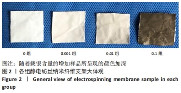
2.1 各组纳米纤维支架的微观形貌与元素组成 图2为4组样品的大体观,随着载银含量的增加样品所呈现的颜色加深。"
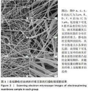
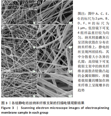
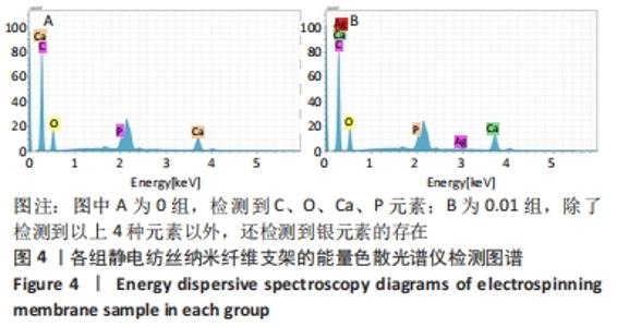
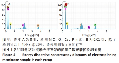
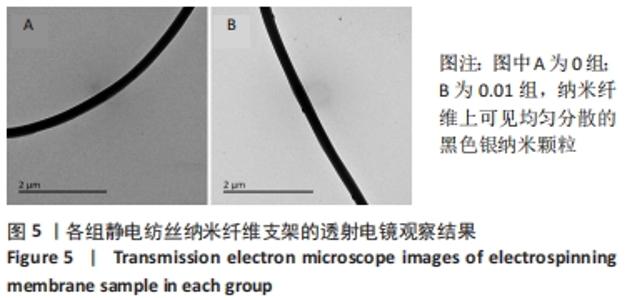
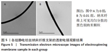
4组样品的扫描电镜观察结果,见图3,低倍镜下可见4组样品较为连续,直径较为均匀,纳米羟基磷灰石呈团块状散在分布在纳米纤维上,未见明显的大液滴及杂质的混入,静电纺丝随机乱序交错排列,呈现网状结构,其中分散着大小各异的孔隙;高倍镜下可见载银支架中的纳米纤维表面散在轻微凸起的金属银颗粒,并随着载银量的增加在纳米纤维上呈现增多的趋势。0组、0.001组、0.01组和0.1组电纺丝纤维的平均直径分别为(538±99),(542±96),(693±139),(775±180) nm。4组样品的元素组成,见图4,未载银组(0组)检测到C、O、Ca、P元素,说明聚己内酯支架中成功混入纳米羟基磷灰石;载银组(以0.01组为例)除了检测到以上4种元素以外,还检测到银元素的存在,说明金属银成功载入了静电纺丝样品中。各组样品的透射电镜观察结果,见图5,载银组(以0.01组为例)纳米纤维上可见均匀分散的黑色颗粒(即银纳米颗粒),颗粒直径为(46±24) nm,说明负载到纳米纤维支架上的银离子成功转化为银纳米颗粒。"
"
"
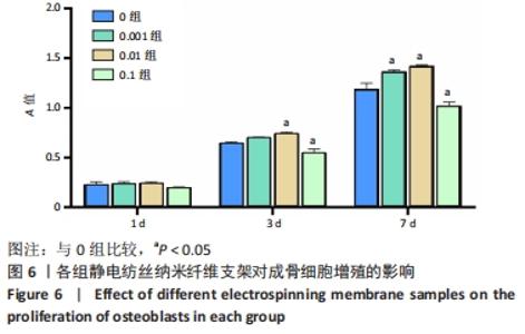
2.2 各组纳米纤维支架的的细胞相容性 各组样品对成骨细胞增殖的影响,见图6。细胞培养的第1天,各组间细胞A值比较差异无显著性意义(P > 0.05);培养第3天,0.01组细胞A值高于0组(P < 0.05),0.1组细胞A值低于0组(P < 0.05),0.001组细胞A值与0组比较差异无显著性意义(P > 0.05);培养第7天,0.001组、0.01组细胞A值高于0组(P < 0.05),0.1组细胞A值低于0组(P < 0.05),说明0.1组表现为较强的细胞毒性,进而抑制了细胞增殖。通过图6还可以发现,随着培养天数逐渐增加,0.1组细胞A值增高,但细胞活性明显低于0组,提示纳米羟基磷灰石所发挥的积极效果明显小于过量银所产生的细胞毒性。"

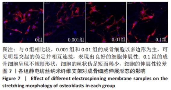
各组样品对成骨细胞伸展形态的影响,见图7。0组、0.001组和0.01组的成骨细胞呈多边形,与0组相比较,0.001组和0.01组的成骨细胞以多边形为主,可见明显突起的伪足并相互连接,表现出良好的细胞伸展性,具有典型的成骨细胞的生物学形态特征;0.1组的成骨细胞呈现不规则形状,细胞的丝状伪足短而稀少,甚至没有,细胞的伸展性较差。"

2.3 各组纳米纤维支架的成骨性能 各组样品对成骨细胞成骨能力的影响,见图8。"
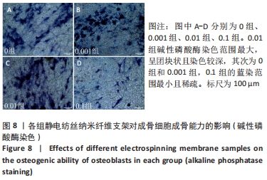
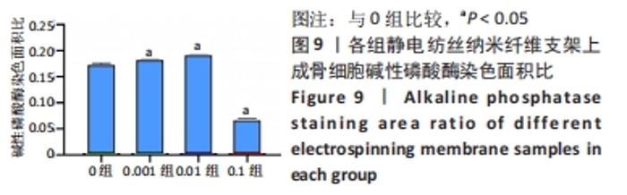
图8可见蓝染的范围代表成骨细胞的相关活力。0.01组碱性磷酸酶染色的范围最大,呈团块状且染色较深,其次为0组和0.001组,也可见染色区域,0.1组的蓝染范围最小且稀疏。通过对染色面积的比值计算分析发现,0.01组的染色面积比最高,0.1组的染色面积比最低,0.001组、0.01组、0.1组与0组比较差异均有显著性意义(P < 0.05),见图9。"

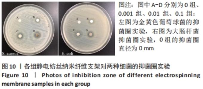
2.4 各组纳米纤维支架的抗菌性能 各组样品的抑菌圈实验见图10所示,只有0组的抑菌圈直径为0 mm,提示纳米羟基磷灰石/聚己内酯支架对金黄色葡萄球菌与大肠杆菌没有抗菌活性。"
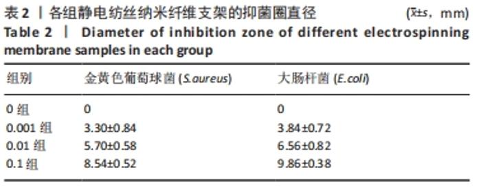
各组样品的抑菌圈直径,见表2,随着载银浓度的增加,样品的抑菌圈直径逐渐增大,表明复合支架的抗菌性能增强。"
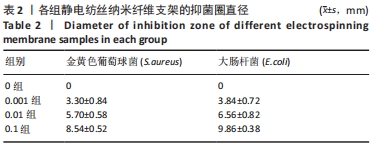
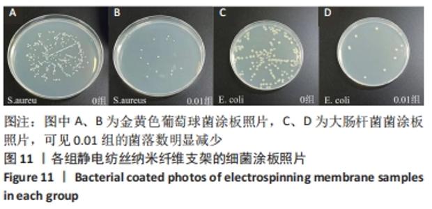
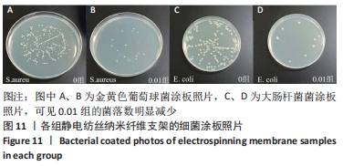
图11所示为未载银组(0组)和载银组(以0.01组为例)与两种细菌的涂板照片,可见0.01组的菌落数明显减少,通过抑菌率公式计算得出,0.01组对金黄色葡萄球菌和大肠杆菌的抑菌率分别为94.91%和94.79%。"

2.5 纳米纤维支架的细胞相容性 由CCK8实验与细胞核及细胞骨架染色结果可知,载银(0,0.001,0.01 mol/L)的纳米羟基磷灰石/聚己内酯复合纳米纤维支架具有良好的细胞相容性。"

| [1] YANG JW, HIROKI U, MISHINA Y. Energy metabolism: A newly emerging target of BMP signaling in bone homeostasis. Bone. 2020;138:115467. [2] MASTERS EA, TROMBETTA RP, DE MESY BENTLEY KL, et al. Evolving concepts in bone infection: redefining “biofilm”, “acute vs. chronic osteomyelitis”, “the immune proteome” and “local antibiotic therapy”. Bone Res. 2019;7:20. [3] KARYGIANNI L, REN Z, KOO H, et al. Biofilm Matrixome: Extracellular Components in Structured Microbial Communities. Trends Microbiol. 2020;28(8):668-681. [4] MALIKMAMMADOV E, TANIR TE, KIZILTAY A, et al. PCL and PCL-based materials in biomedical applications. J Biomater Sci Polym Ed. 2018;29: 863-893. [5] CHOU PY, CHOU YC, LAI YH, et al. Fabrication of Drug-Eluting Nano-Hydroxylapatite Filled Polycaprolactone Nanocomposites Using Solution-Extrusion 3D Printing Technique. Polymers (Basel). 2021;13(3):318. [6] PARK S, KIM JE, HAN J, et al. 3D-Printed Poly(ε-Caprolactone)/Hydroxyapatite Scaffolds Modified with Alkaline Hydrolysis Enhance Osteogenesis In Vitro. Polymers (Basel). 2021;13(2):257. [7] BARNES CP, SELL SA, BOLAND ED, et al. Nanofiber technology: Designing the next generation of tissue engineering scaffolds. Adv Drug Deliv Rev. 2007;59(14):1413-1433. [8] MIRICĂ IC, FURTOS G, LUCACIU O, et al. Electrospun Membranes Based on Polycaprolactone, Nano-Hydroxyapatite and Metronidazole. Materials (Basel). 2021;14(4):931. [9] EL-HABASHY S, ELTAHER H, GABALLAH A, et al. Biomaterial-Based Nanocomposite for Osteogenic Repurposing of Doxycycline. Int J Nanomedicine. 2021;16:1103-1126. [10] LIU HM, DU YY, YANG GJ, et al. Delivering Proangiogenic Factors from 3D-Printed Polycaprolactone Scaffolds for Vascularized Bone Regeneration. Adv Healthc Mater. 2020;9(23):2000727. [11] HE YZ, JIN YH, WANG XM, et al. An Antimicrobial Peptide-Loaded Gelatin/Chitosan Nanofibrous Membrane Fabricated by Sequential Layer-by-Layer Electrospinning and Electrospraying Techniques. Nanomaterials (Basel). 2018;8(5):327. [12] JUNG WK, KOO HC, KIM KW, et al. Antibacterial Activity and Mechanism of Action of the Silver Ion in Staphylococcus aureus and Escherichia coli. Appl Environ Microbiol. 2008;74(7):2171-2178. [13] SILVER S. Bacterial silver resistance: molecular biology and uses and misuses of silver compounds. FEMS Microbiol Rev. 2003;27(2-3): 341-353. [14] WYPIJ M, JĘDRZEJEWSKI T, OSTROWSKI M, et al. Biogenic Silver Nanoparticles: Assessment of Their Cytotoxicity, Genotoxicity and Study of Capping Proteins. Molecules. 2020;25(13):3022. [15] Vik H, Andersen KJ, Julshamn K, et al. Neuropathy caused by silver absorption from arthroplasty cement. Lancet. 1985;1:872. [16] QIAN YZ, ZHOU XF, ZHANG FM, et al. Triple PLGA/PCL Scaffold Modification Including Silver Impregnation, Collagen Coating, and Electrospinning Significantly Improve Biocompatibility, Antimicrobial, and Osteogenic Properties for Orofacial Tissue Regeneration. ACS Appl Mater Interfaces. 2019;11(41):37381-37396. [17] PATERSON TE, SHI R, TIAN JJ, et al. Electrospun Scaffolds Containing Silver-Doped Hydroxyapatite with Antimicrobial Properties for Applications in Orthopedic and Dental Bone Surgery. J Funct Biomater. 2020;11(3):58. [18] 王乐,惠敏,董西玲,等.缓释阿托伐他汀钙纳米纤维支架对细胞黏附增殖的影响[J].中国组织工程研究,2020,24(28):4492-4497. [19] BALLESTEROS CAS, CORREA DS, ZUCOLOTTO V. Polycaprolactone nanofiber mats decorated with photoresponsive nanogels and silver nanoparticles: Slow release for antibacterial control. Mater Sci Eng C Mater Biol Appl. 2020;107:110334. [20] HASSAN AA, RADWAN HA, ABDELAAL SA, et al. Polycaprolactone based electrospun matrices loaded with Ag/hydroxyapatite as wound dressings: Morphology, cell adhesion and antibacterial activity. Int J Pharm. 2021;593:120143. [21] 刘丹丹.静电纺丝法制备纳米级银颗粒EVOH导向纤维及其性能研究[D].西安:西安科技大学,2019. [22] BHULLAR SK, RUZGAR DG, FORTUNATO G, et al. A Facile Method for Controlled Fabrication of Hybrid Silver Nanoparticle-Poly(ε-Caprolactone) Fibrous Constructs with Antimicrobial Properties. J Nanosci Nanotechnol. 2019;19(11):6949-6955. [23] ALI W, VALBONE S, MATTHIAS L, et al. Electrical conductivity of silver nanoparticle doped carbon nanofibres measured by CS-AFM. RSC Adv. 2019;9(8):4553-4562. [24] QUIRÓS J, BORGES JP, BOLTES K, et al. Antimicrobial electrospun silver-, copper- and zinc-doped polyvinylpyrrolidone nanofibers. J Hazard Mater. 2015;299:298-305. [25] PASTORIZA-SANTOS I, SERRA-RODRÍGUEZ C, LIZ-MARZÁN LM. Self-Assembly of Silver Particle Monolayers on Glass from Ag(+) Solutions in DMF. J Colloid Interface Sci. 2000;221(2):236-241. [26] CAO HL, ZHANG WJ, MENG FH, et al. Osteogenesis Catalyzed by Titanium-Supported Silver Nanoparticles. ACS Appl Mater Interfaces. 2017;9(6):5149-5157. [27] NING CY, WANG XL, LI LH, et al. Concentration Ranges of Antibacterial Cations for Showing the Highest Antibacterial Efficacy but the Least Cytotoxicity against Mammalian Cells: Implications for a New Antibacterial Mechanism. Chem Res Toxicol. 2015;28(9):1815-1822. [28] SHIMABUKURO M. Antibacterial Property and Biocompatibility of Silver, Copper, and Zinc in Titanium Dioxide Layers Incorporated by One-Step Micro-Arc Oxidation: A Review. Antibiotics (Basel). 2020:9(10):716. [29] LÜTHJE FL, JENSEN LK, JENSEN HE, et al. The inflammatory response to bone infection - a review based on animal models and human patients. APMIS. 2020;128:275-286. [30] GUNDTOFT PH, PEDERSEN AB, SCHØNHEYDER HC, et al. One-year incidence of prosthetic joint infection in total hip arthroplasty: a cohort study with linkage of the Danish Hip Arthroplasty Register and Danish Microbiology Databases. Osteoarthritis Cartilage. 2017;25(5):685-693. [31] SCHIERHOLZ JM, BEUTH J. Implant infections: a haven for opportunistic bacteria. J Hosp Infect. 2001;49(2):87-93. [32] DURÁN N, DURÁN M, DE JESUS MB, et al. Silver Nanoparticles: A New View on Mechanistic Aspects on Antimicrobial Activity. Nanomedicine. 2016;12:789-799. [33] YAN L, XIANG Y, YU J, et al. Fabrication of Antibacterial and Antiwear Hydroxyapatite Coatings via In Situ Chitosan-Mediated Pulse Electrochemical Deposition. ACS Appl Mater Interfaces. 2017;9(5):5023-5030. [34] RIEGER KA, CHO HJ, YEUNG HF, et al. Antimicrobial Activity of Silver Ions Released from Zeolites Immobilized on Cellulose Nanofiber Mats. ACS Appl Mater Interfaces. 2016;8(5):3032-3040. [35] FENG QL, WU J, CHEN GQ, et al. A mechanistic study of the antibacterial effect of silver ions on Escherichia coli and Staphylococcus aureus. J Biomed Mater Res. 2000;52(4):662-668. [36] DAI GY, TANG SC, WANG TL. A novel approach to prepare high antibacterial polylactic acid surface encapsulated nano-silver through stereocomplexation. Mater Res Express. 2019;6(5):55310. [37] SHEIKH FA, BARAKAT NA, KANJWAL MA, et al. Electrospun titanium dioxide nanofibers containing hydroxyapatite and silver nanoparticles as future implant materials. J Mater Sci Mater Med. 2010;21:2551-2559. |
| [1] | Wang Jing, Xiong Shan, Cao Jin, Feng Linwei, Wang Xin. Role and mechanism of interleukin-3 in bone metabolism [J]. Chinese Journal of Tissue Engineering Research, 2022, 26(8): 1260-1265. |
| [2] | Gao Cangjian, Yang Zhen, Liu Shuyun, Li Hao, Fu Liwei, Zhao Tianyuan, Chen Wei, Liao Zhiyao, Li Pinxue, Sui Xiang, Guo Quanyi. Electrospinning for rotator cuff repair [J]. Chinese Journal of Tissue Engineering Research, 2022, 26(4): 637-642. |
| [3] | Zhang Shengmin, Cao Changhong, Wang Ningning, Wang Jing, Li Zhangyi. Desferrioxamine-loaded polylactic-co-glycolic acid/hydroxyapatite composite scaffold: vascularization and osteogenesis [J]. Chinese Journal of Tissue Engineering Research, 2022, 26(34): 5413-5418. |
| [4] | Long Zhisheng, Xiong Long, Gong Feipeng, Li Jingtang, Zeng Jianhua, Deng Ying, Lan Min, Kong Weihao, Chen Gang. Effect of artificial bone with multi-scale hydroxyapatite/chitosan microtubule structure on rabbit bone defect repair and angiogenesis [J]. Chinese Journal of Tissue Engineering Research, 2022, 26(34): 5436-5441. |
| [5] | Wu Chengcong, Wang Fang, Wan Jianshan, Wu Zheng, Sun Rong, Huang Hefei, Qian Xuankun, Ou Hua, Ren Jing. Adenovirus-mediated bone morphogenetic protein 2 induces osteogenic differentiation of rabbit bone marrow mesenchymal stem cells [J]. Chinese Journal of Tissue Engineering Research, 2022, 26(30): 4757-4761. |
| [6] | Shen Enpu, Huang Ba, Liu Danping, Qi Hui, Wu Zhiwen, Li Beibei. Exosomes derived from melatonin-modified bone marrow mesenchymal stem cells promote osteogenesis of bone marrow mesenchymal stem cells [J]. Chinese Journal of Tissue Engineering Research, 2022, 26(30): 4800-4805. |
| [7] | Jia Qiyu, Huang Xiaoxia, Guo Jian, Huang Jinyong, Guo Xiaobin, Abdussalam·Alimujiang, Wu Tong, Ma Chuang. Integrin-targeted peptide promotes proliferation of bone marrow mesenchymal stem cells in SD rats [J]. Chinese Journal of Tissue Engineering Research, 2022, 26(30): 4780-4786. |
| [8] | Lu Yunan, Zhang Xinzhao, Lin Binbin, Xu Gan, Chen Jingdi, Chen Shunyou. Naringin-chitosan/hydroxyapatite composite scaffold in repair of rat skull defect [J]. Chinese Journal of Tissue Engineering Research, 2022, 26(28): 4441-4445. |
| [9] | Wu Yuchong, Peng Xu, Yu Xixun. Osteogenesis, angiogenesis and anti-aseptic loosening of europium-doped calcium polyphosphate bone tissue engineering scaffold [J]. Chinese Journal of Tissue Engineering Research, 2022, 26(28): 4458-4465. |
| [10] | Xue Xuexin, Liu Zhepeng. Electrospun fiber-based nerve tissue engineering scaffold: material, function and structure design strategy [J]. Chinese Journal of Tissue Engineering Research, 2022, 26(28): 4575-4580. |
| [11] | Hu Qiuyu, Yang Long, Yang Yong, Song Shenchao. Aligned poly(butylene adipate-co-terephthalate)/type I collagen fibers promote tendon-bone healing after anterior cruciate ligament rupture [J]. Chinese Journal of Tissue Engineering Research, 2022, 26(27): 4314-4319. |
| [12] | Dong Xiling, Hui Min, Cao Fei, Lin Peng, Zhou Han, Wang Le, Zhang Xiaoming, Liu Tongbin. Preparation of copper loaded coating on polydopamine-modified polycaprolactone electrospun membrane and antibacterial and cellular properties evaluation [J]. Chinese Journal of Tissue Engineering Research, 2022, 26(27): 4272-4278. |
| [13] | Zhao Doudou, Lin Kaili. Application of multicellular construction of vascularized tissue engineered bone in bone repair [J]. Chinese Journal of Tissue Engineering Research, 2022, 26(27): 4386-4392. |
| [14] | Zhang Zihan, Wang Wenli, Li Jinnuo, Li Yourui. Carvacrol: antibacterial activity, bone repair, and prevention and treatment in oral diseases [J]. Chinese Journal of Tissue Engineering Research, 2022, 26(26): 4252-4257. |
| [15] | Chen Chichi, Zhang Yu, He Jiachen, Shi Qin. Osteogenic differentiation of bone marrow mesenchymal stem cells in obese mice [J]. Chinese Journal of Tissue Engineering Research, 2022, 26(24): 3846-3851. |
| Viewed | ||||||
|
Full text |
|
|||||
|
Abstract |
|
|||||