Chinese Journal of Tissue Engineering Research ›› 2022, Vol. 26 ›› Issue (30): 4826-4833.doi: 10.12307/2022.762
Previous Articles Next Articles
Human umbilical cord mesenchymal stem cell transplantation for myocardial hypertrophy in mice
Xie Liying1, Liu Siqi1, Wu Mingrui1, Gao Erhe2, Han Zhibo3, Zuo Lin1
- 1Key Laboratory of Cellular Physiology, Ministry of Education, and the Department of Physiology, Shanxi Medical University, Taiyuan 030001, Shanxi Province, China; 2Center for Translational Medicine, Temple University School of Medicine, Philadelphia, PA, USA; 3Hematology Hospital/Institute of Hematology, Chinese Academy of Medical Sciences; Tianjin Oncay Cell Genetic Engineering Co., Ltd./National Engineering Research Center for Cell Products, Tianjin 300041, China
-
Received:2021-01-29Accepted:2021-03-18Online:2022-10-28Published:2022-03-29 -
Contact:Zuo Lin, MD, Associate professor, Key Laboratory of Cellular Physiology, Ministry of Education, and the Department of Physiology, Shanxi Medical University, Taiyuan 030001, Shanxi Province, China -
About author:Xie Liying, Master candidate, Key Laboratory of Cellular Physiology, Ministry of Education, and the Department of Physiology, Shanxi Medical University, Taiyuan 030001, Shanxi Province, China -
Supported by:the Shanxi Youth Fund, No. 201601D202106 (to ZL); the Scientific Research Fund for Returned Overseas Students in Shanxi Province, No. 2014-036 (to ZL); the Shanxi “1331 Project” Key Subjects Construction, Nos. 1331KSC, XK201708 (to ZL)
CLC Number:
Cite this article
Xie Liying, Liu Siqi, Wu Mingrui, Gao Erhe, Han Zhibo, Zuo Lin. Human umbilical cord mesenchymal stem cell transplantation for myocardial hypertrophy in mice[J]. Chinese Journal of Tissue Engineering Research, 2022, 26(30): 4826-4833.
share this article
Add to citation manager EndNote|Reference Manager|ProCite|BibTeX|RefWorks

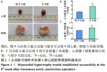
2.1 实验动物数量分析 参加实验80只C57BL/6J小鼠,有2只假手术组小鼠由于操作不当术中出血后死亡,给予重新补充,最后8只小鼠进入造模评估结果分析,假手术组和手术组各4只,剩余72只小鼠进入后续实验,假手术组、PBS组、hUC-MSCs组各24只。 2.2 小鼠心肌肥厚模型建立成功 为评判小鼠在主动脉弓缩窄术后心肌肥厚模型是否建立成功,对假手术组(4只)和手术组(4只)小鼠的左室质量/体质量比以及心质量/肺质量比进行了测量。实验结果显示:术后第4周,手术组小鼠心脏、肺脏质量明显增加,见图1A;与假手术组(3.06±0.21)相比,手术组左室质量/体质量比(5.58±0.47)明显增加(P < 0.001),见图1B;手术组小鼠的心质量/肺质量比(1.00±0.04)也明显高于假手术组(0.78±0.01,P < 0.01),见图1C。以上结果提示,手术组在术后第4周时已出现明显左心室肥厚,提示压力负荷型心肌肥厚模型造模成功。"
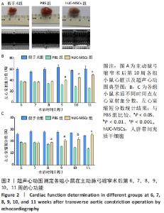
2.3 hUC-MSCs尾静脉注射治疗可显著改善心肌肥厚小鼠的心功能 hUC-MSCs注射治疗后,每周对小鼠心功能进行动态监测。超声心动图结果证实:经hUC-MSCs治疗后,小鼠心功能明显改善,见图2A;术后第10周,与同期PBS组(28.92±0.64)%相比,hUC-MSCs组小鼠左心室射血分数(45.23±1.18)%显著升高(P < 0.001),见图2B;与同期PBS组(14.85±0.22)%相比,hUC-MSCs组小鼠在第10周左心室缩短分数(21.91±1.47)%明显升高(P < 0.01),见图2C;以上结果提示,hUC-MSCs尾静脉注射治疗可以明显改善主动脉弓缩窄术后小鼠心功能,有效延缓心肌肥厚向心衰恶化的进程。"
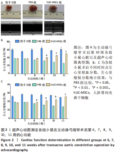
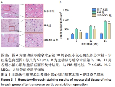
2.4 hUC-MSCs尾静脉注射治疗可明显减轻肥厚型心肌病小鼠心肌细胞的肥大水平 术后第9,10,11周收取各组小鼠心脏组织标本,通过苏木精-伊红染色来分析心肌组织结构以及心肌细胞肥大情况,研究结果发现:相较于PBS组,hUC-MSCs组小鼠心肌纤维排列整齐,心肌细胞大小、形态接近正常,见图3A;术后第10周,hUC-MSCs组小鼠心肌细胞面积(519.8±41.71) μm2明显低于同期PBS组(667.4±34.64) μm2,差异有显著性意义(P < 0.05),见图3B。以上结果提示,hUC-MSCs尾静脉注射治疗后,小鼠心肌细胞肥大、间质增加及肌纤维断裂等情况均明显减轻。"
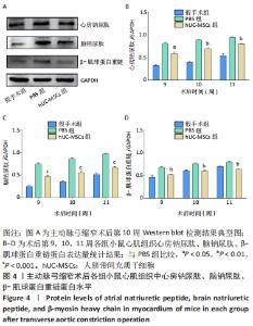
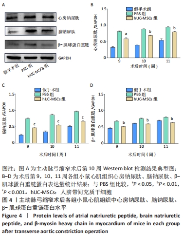
2.5 hUC-MSCs尾静脉注射治疗可明显减少肥厚型心肌病小鼠心肌组织中相关肥大蛋白水平 采用Western blot检测术后第9,10,11周各组小鼠心肌组织相关肥大蛋白水平,见图4A。从第9周开始,与同期PBS组比较(心房钠尿肽:0.811±0.02;脑钠尿肽:0.75±0.02;β-肌球蛋白重链:0.68±0.02),hUC-MSCs组小鼠心肌组织中心房钠尿肽蛋白(0.58±0.03)、脑钠尿肽蛋白(0.47±0.02)以及β-肌球蛋白重链蛋白水平(0.52±0.01)均明显下降(P < 0.01),见图4B-D。从结果可以看出,hUC-MSCs经由尾静脉注射到小鼠体内,可以明显降低主动脉弓缩窄术后小鼠心肌组织中肥大相关蛋白心房钠尿肽、脑钠尿肽、β-肌球蛋白重链的表达。"

2.6 hUC-MSCs可明显缓解异丙肾上腺素诱导的H9c2细胞肥大 培养48 h后,通过F-actin染色测量H9c2细胞面积大小,见图5A。与异丙肾上腺素组比较(7 151.10±33.53) μm2,hUC-MSCs组细胞面积(4 915.93±75.18) μm2明显减小(P < 0.01),见图5B。通过qRT-PCR检测各组H9c2细胞中相关肥大基因mRNA表达量,与异丙肾上腺素组比较(心房钠尿肽:1.26±0.05;脑钠尿肽:1.66±0.04;β-肌球蛋白重链:1.43±0.03),与hUC-MSCs共培养后,H9c2细胞中心房钠尿肽(1.07±0.02)、脑钠尿肽(1.52±0.13)以及β-肌球蛋白重链(1.31±0.06)的mRNA水平均明显降低(P < 0.05),见图5C。"
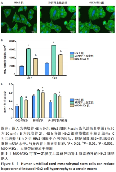

以上实验结果提示,hUC-MSCs可以通过降低H9c2细胞中肥大相关基因的转录,从而减轻异丙肾上腺素诱导的细胞肥大。 2.7 小鼠注射hUc-MSCs的生物相容性检测结果 经由尾静脉注射hUc-MSCs治疗后,小鼠存活率为100%,未出现由于注射细胞而引发的死亡情况,直至收取标本时未发现明显靶器官损害症状,并且小鼠体温均在37 ℃左右,未出现发热等免疫反应现象。以上结果说明尾静脉注射hUc-MSCs不会导致小鼠出现机体免疫过敏反应。"

| [1] 陈伟伟,高润霖,刘力生,等.《中国心血管病报告2017》概要[J].中国循环杂志,2018,33(1):1-8. [2] YILDIZ M, OKTAY AA, STEWART MH, et al. Left ventricular hypertrophy and hypertension. Prog Cardiovasc Dis. 2020;63(1):10-21. [3] LIANG Y, IDREES E, SZOJKA ARA, et al. Chondrogenic differentiation of synovial fluid mesenchymal stem cells on human meniscus-derived decellularized matrix requires exogenous growth factors. Acta Biomater. 2018;80:131-143. [4] MIYAGAWA I, NAKAYAMADA S, KONDO M, et al. Regulatory Mechanism of The Induction of Regulatory T Cells through Growth Factors Released by Human Mesenchymal Stem Cells. Crit Rev Immunol. 2018; 38(6):471-478. [5] WANG F, TANG H, ZHU J, et al. Transplanting Mesenchymal Stem Cells for Treatment of Ischemic Stroke. Cell Transplant. 2018;27(12):1825-1834. [6] LI T, XIA M, GAO Y, et al. Human umbilical cord mesenchymal stem cells: an overview of their potential in cell-based therapy. Expert Opin Biol Ther. 2015;15(9):1293-1306. [7] LIN Z, HE H, WANG M, et al. MicroRNA-130a controls bone marrow mesenchymal stem cell differentiation towards the osteoblastic and adipogenic fate. Cell Prolif. 2019;52(6):e12688. [8] CHO PS, MESSINA DJ, HIRSH EL, et al. Immunogenicity of umbilical cord tissue derived cells. Blood. 2008;111(1):430-438. [9] BATSALI AK, PONTIKOGLOU C, KOUTROULAKIS D, et al. Differential expression of cell cycle and WNT pathway-related genes accounts for differences in the growth and differentiation potential of Wharton’s jelly and bone marrow-derived mesenchymal stem cells. Stem Cell Res Ther. 2017;8(1):102. [10] RIORDAN NH, MORALES I, FERNÁNDEZ G, et al. Clinical feasibility of umbilical cord tissue-derived mesenchymal stem cells in the treatment of multiple sclerosis. J Transl Med. 2018; 16(1):57. [11] MATAS J, ORREGO M, AMENABAR D, et al. Umbilical Cord-Derived Mesenchymal Stromal Cells (MSCs) for Knee Osteoarthritis: Repeated MSC Dosing Is Superior to a Single MSC Dose and to Hyaluronic Acid in a Controlled Randomized Phase I/II Trial. Stem Cells Transl Med. 2019;8(3):215-224. [12] NI J, LIU X, YIN Y, et al. Exosomes Derived from TIMP2-Modified Human Umbilical Cord Mesenchymal Stem Cells Enhance the Repair Effect in Rat Model with Myocardial Infarction Possibly by the Akt/Sfrp2 Pathway. Oxid Med Cell Longev. 2019;2019:1958941. [13] SUN Y, LIU J, XU Z, et al. Matrix stiffness regulates myocardial differentiation of human umbilical cord mesenchymal stem cells. Aging (Albany NY). 2020;13(2):2231-2250. [14] MUSHAHARY D, SPITTLER A, KASPER C, et al. Isolation, cultivation, and characterization of human mesenchymal stem cells. Cytometry A. 2018;93(1):19-31. [15] KHAN I, ALI A, AKHTER MA, et al. Preconditioning of mesenchymal stem cells with 2,4-dinitrophenol improves cardiac function in infarcted rats. Life Sci. 2016;162:60-69. [16] SHEU JJ, LEE FY, YUEN CM, et al. Combined therapy with shock wave and autologous bone marrow-derived mesenchymal stem cells alleviates left ventricular dysfunction and remodeling through inhibiting inflammatory stimuli, oxidative stress & enhancing angiogenesis in a swine myocardial infarction model. Int J Cardiol. 2015;193:69-83. [17] MAUMUS M, PERS YM, RUIZ M, et al. Mesenchymal stem cells and regenerative medicine: future perspectives in osteoarthritis. Med Sci (Paris). 2018;34(12):1092-1099. [18] Berry JM, Naseem RH, Rothermel BA, et al. Models of cardiac hypertrophy and transition to heart failure. Drug Discovery Today: Disease Models. 2008;4(4):197-206. [19] YU Y, HU Z, LI B, et al. Ivabradine improved left ventricular function and pressure overload-induced cardiomyocyte apoptosis in a transverse aortic constriction mouse model. Mol Cell Biochem. 2019;450(1-2): 25-34. [20] CAI B, TAN X, ZHANG Y, et al. Mesenchymal Stem Cells and Cardiomyocytes Interplay to Prevent Myocardial Hypertrophy. Stem Cells Transl Med. 2015;4(12):1425-1435. [21] DAS S, GE P, OZTUG DURER ZA, et al. D-loop Dynamics and Near-Atomic-Resolution Cryo-EM Structure of Phalloidin-Bound F-Actin. Structure. 2020 ;28(5):586-593.e3. [22] MILLS KT, STEFANESCU A, HE J. The global epidemiology of hypertension. Nat Rev Nephrol. 2020;16(4):223-237. [23] NAKAMURA M, SADOSHIMA J. Mechanisms of physiological and pathological cardiac hypertrophy. Nat Rev Cardiol. 2018;15(7):387-407. [24] HUANG S, ZOU X, ZHU JN, et al. Attenuation of microRNA-16 derepresses the cyclins D1, D2 and E1 to provoke cardiomyocyte hypertrophy. J Cell Mol Med. 2015;19(3):608-619. [25] SHAO JS, ALY ZA, LAI CF, et al. Vascular Bmp Msx2 Wnt signaling and oxidative stress in arterial calcification. Ann N Y Acad Sci. 2007; 1117:40-50. [26] LOU G, CHEN Z, ZHENG M, et al. Mesenchymal stem cell-derived exosomes as a new therapeutic strategy for liver diseases. Exp Mol Med. 2017;49(6):e346. [27] LI Z, GUO J, CHANG Q, et al. Paracrine role for mesenchymal stem cells in acute myocardial infarction. Biol Pharm Bull. 2009;32(8):1343-1346. [28] MIYAHARA Y, NAGAYA N, KATAOKA M, et al. Monolayered mesenchymal stem cells repair scarred myocardium after myocardial infarction. Nat Med. 2006;12(4):459-465. [29] UCCELLI A, MORETTA L, PISTOIA V. Mesenchymal stem cells in health and disease. Nat Rev Immunol. 2008;8(9):726-736. [30] LEE S, KIM S, AHN J, et al. Membrane-bottomed microwell array added to Transwell insert to facilitate non-contact co-culture of spermatogonial stem cell and STO feeder cell. Biofabrication. 2020; 12(4):045031. [31] WANG QQ, JING XM, BI YZ, et al. Human Umbilical Cord Wharton’s Jelly Derived Mesenchymal Stromal Cells May Attenuate Sarcopenia in Aged Mice Induced by Hindlimb Suspension. Med Sci Monit. 2018;24: 9272-9281. [32] TAVAKOLI R, NEMSKA S, JAMSHIDI P, et al. Technique of Minimally Invasive Transverse Aortic Constriction in Mice for Induction of Left Ventricular Hypertrophy. J Vis Exp. 2017;(127):56231. [33] LI CX, SONG J, LI X, et al. Circular RNA 0001273 in exosomes derived from human umbilical cord mesenchymal stem cells (UMSCs) in myocardial infarction. Eur Rev Med Pharmacol Sci. 2020;24(19): 10086-10095. [34] SUN J, SHEN H, SHAO L, et al. HIF-1α overexpression in mesenchymal stem cell-derived exosomes mediates cardioprotection in myocardial infarction by enhanced angiogenesis. Stem Cell Res Ther. 2020;11(1): 373. [35] FORTE M, MADONNA M, SCHIAVON S, et al. Cardiovascular Pleiotropic Effects of Natriuretic Peptides. Int J Mol Sci. 2019;20(16):3874. [36] ALANAZI WA, AL-HARBI NO, IMAM F, et al. Role of carnitine in regulation of blood pressure (MAP/SBP) and gene expression of cardiac hypertrophy markers (α/β-MHC) during insulin-induced hypoglycaemia: Role of oxidative stress. Clin Exp Pharmacol Physiol. 2021;48(4):478-489. [37] DAI H, JIA G, LIU X, et al. Astragalus polysaccharide inhibits isoprenaline-induced cardiac hypertrophy via suppressing Ca²⁺-mediated calcineurin/NFATc3 and CaMKII signaling cascades. Environ Toxicol Pharmacol. 2014;38(1):263-271. [38] PASCHOS NK, BROWN WE, ESWARAMOORTHY R, et al. Advances in tissue engineering through stem cell-based co-culture. J Tissue Eng Regen Med. 2015;9(5):488-503. [39] LOSS H, ASCHENBACH JR, EBNER F, et al. Inflammatory Responses of Porcine MoDC and Intestinal Epithelial Cells in a Direct-Contact Co-culture System Following a Bacterial Challenge. Inflammation. 2020;43(2):552-567. [40] LIUFU R, SHI G, HE X, et al. The therapeutic impact of human neonatal BMSC in a right ventricular pressure overload model in mice. Stem Cell Res Ther. 2020;11(1):96. [41] QIU J, XIAO H, ZHOU S, et al. Bone marrow mesenchymal stem cells inhibit cardiac hypertrophy by enhancing FoxO1 transcription. Cell Biol Int. 2021;45(1):188-197. |
| [1] | Zhu Bingbing, Deng Jianghua, Chen Jingjing, Mu Xiaoling. Interleukin-8 receptor enhances the migration and adhesion of umbilical cord mesenchymal stem cells to injured endothelium [J]. Chinese Journal of Tissue Engineering Research, 2022, 26(7): 1045-1050. |
| [2] | Lin Xuchen, Zhu Hainian, Wang Zengshun, Qi Tengmin, Liu Limin, Suonan Angxiu. Effect of xanthohumol on inflammatory factors and articular cartilage in a mouse mode of osteoarthritis [J]. Chinese Journal of Tissue Engineering Research, 2022, 26(5): 676-681. |
| [3] | Zhao Xinxin, Yang Xiaoqing, Zhang Yuquan. Umbilical cord mesenchymal stem cells improve pregnancy outcomes of spontaneous abortion mouse models by promoting vascular endothelial growth factor expression in decidua [J]. Chinese Journal of Tissue Engineering Research, 2022, 26(30): 4793-4799. |
| [4] | Zeng Chunrong, Liu Menglan, Xie Yang, Li Zuoxiao. Preventive and therapeutic effects of naringin on experimental autoimmune encephalomyelitis via regulating microglial polarization [J]. Chinese Journal of Tissue Engineering Research, 2022, 26(26): 4127-4135. |
| [5] | Shi Yao, Han Shufeng, Yuan Yitong, Du Ruochen, Jing Zhijie, Zhao Bichun, Zhang Ruxin, Zhang Yujuan, Wang Chunfang. Efficacy and safety of human umbilical cord mesenchymal stem cells in the treatment of spinal cord injury: a meta-analysis [J]. Chinese Journal of Tissue Engineering Research, 2022, 26(25): 4093-4100. |
| [6] | Liu Xingyu, Hu Xiaofang, Xu Guangli, Wang Rui, Li Peiyao, Wang Mengyuan, Peng Li, Zhu Xiangying. Human umbilical cord mesenchymal stem cells in endometrial injury repair [J]. Chinese Journal of Tissue Engineering Research, 2022, 26(24): 3921-3927. |
| [7] | Li Jinglu, Wang Sainan, Wang Yangyang, Fu Guiqiang, Wang Ying, Hu Jianguo, Tang Jie, Lyu Hezuo. CRID3, a blocker of apoptosis-associated speck-like protein containing a card, influences local gene transcription in mice with acute spinal cord injury: a transcriptomic analysis [J]. Chinese Journal of Tissue Engineering Research, 2022, 26(23): 3620-3632. |
| [8] | Liu Yue, Jiang Ziyi, Li Jingjing, Meng Kai, Zhao Huijing. Cell co-culture and in vivo biocompatibility of poly(L-lactic caprolactone)/silk fibroin small-diameter artificial blood vessels [J]. Chinese Journal of Tissue Engineering Research, 2022, 26(22): 3505-3513. |
| [9] | Zhang Haiyang, Bi Shengli, Feng Jingru, Li Fan, Wang Jing, Zhao Na, Li Xinjun. Effects of silent information regulator 1 on chronic heart failure in a postmenopausal mouse model and its mechanism [J]. Chinese Journal of Tissue Engineering Research, 2022, 26(20): 3184-3189. |
| [10] | Liu Yulu, Jia Weiwei, Dai Yaling, Xu Wenshan, Ding Yanyi, Liang Shengxiang, Liu Weilin, Chen Lidian. Effects of time-specific AMP-activated protein kinase alpha1/2 gene knockout on hippocampal energy metabolism and synaptic plasticity in mice [J]. Chinese Journal of Tissue Engineering Research, 2022, 26(20): 3230-3235. |
| [11] | Wang Jiajia, Liu Jie, Wang Min. Establishing a murine model of experimental apical periodontitis induced by Fusobacterium nucleatum [J]. Chinese Journal of Tissue Engineering Research, 2022, 26(2): 176-181. |
| [12] | Shan Zhengming, Tao Shuchun, Hu Chunmei, Zhang Zhiyuan, Ding Yinan, He Mengcheng, Tang Qiusha. Extraction, identification and proteomic analysis of exosomes derived from human umbilical cord mesenchymal stem cells [J]. Chinese Journal of Tissue Engineering Research, 2022, 26(19): 3036-3042. |
| [13] | Yang Qin, Wang Min. Constructing a mouse model of periodontitis with occlusal trauma [J]. Chinese Journal of Tissue Engineering Research, 2022, 26(17): 2667-2672. |
| [14] | Zhu Yaping, Li Xiang, Chen Zhiwen, Liu Ying, Zhao Wenzhuo, Ma Ketao, Gu Qiang. Calcium-sensitive receptors influence pulmonary vascular remodeling in neonatal mice with persistent pulmonary hypertension [J]. Chinese Journal of Tissue Engineering Research, 2022, 26(17): 2708-2712. |
| [15] | Yin Liang, Zhang Mingxue, Li Jianan, Jiang Feng. Inducing macrophage polarization to M2 anti-inflammatory type reduces oxidative damage in diabetic retinopathy mice [J]. Chinese Journal of Tissue Engineering Research, 2022, 26(17): 2685-2689. |
| Viewed | ||||||
|
Full text |
|
|||||
|
Abstract |
|
|||||