Chinese Journal of Tissue Engineering Research ›› 2022, Vol. 26 ›› Issue (17): 2685-2689.doi: 10.12307/2022.535

Previous Articles     Next Articles

Inducing macrophage polarization to M2 anti-inflammatory type reduces oxidative damage in diabetic retinopathy mice

Yin Liang, Zhang Mingxue, Li Jianan, Jiang Feng   

  1. General Hospital of Tianjin Medical University, Tianjin 300014, China
  • Received:2021-06-15 Revised:2021-06-16 Accepted:2021-07-24 Online:2022-06-18 Published:2021-12-24
  • Contact: Yin Liang, General Hospital of Tianjin Medical University, Tianjin 300014, China
  • About author:Yin Liang, Master, Attending physician, General Hospital of Tianjin Medical University, Tianjin 300014, China
  • Supported by:
    the National Natural Science Foundation of China, No. 81800853 (to JF)

Abstract: BACKGROUND: One of the main signs in diabetic retinopathy is the continuous formation of new blood vessels, and tumor necrosis factor ligand superfamily member 15 (TNFSF15), as an angiogenesis inhibitor, can significantly inhibit angiogenesis.
OBJECTIVE: To investigate the mechanism by which TNFSF15 reduces oxidative damage in diabetic retinopathy mice by inducing macrophage polarization to M2 anti-inflammatory type. 
METHODS: Forty male CD-1 mice were randomly divided into four groups (n=10 per group): a normal group, a model group, a TNFSF15 group, and a vascular endothelial growth factor group. Except for the normal group, a diabetic retinopathy model was established in all the groups by using streptozotocin and fundus angiography respectively. After the success of the modeling, mice in the TNFSF15 group were intraperitoneally injected with 250 mg/L TNFSF15, and mice in the vascular endothelial growth factor group were intraperitoneally injected with 250 mg/L vascular endothelial growth factor. Mice in the normal group and the model group were given the same volume of normal saline simultaneously. After the experiment was completed, the mouse retina was taken, and macrophages were extracted for in vitro culture. The cells were cultured with TNFSF15 in observation group or with phosphate buffered saline in control group. Hematoxylin-eosin staining was used to detect the pathological morphology of mouse omentum. Enzyme linked immunosorbent assay was used to detect serum levels of TNFSF15, vascular endothelial growth factor, malondialdehyde, and superoxide dismutase. Western blot was used to detect the protein expression of inducible nitric oxide synthase, Ym1, CD206, and CD163 in mouse retinal tissue. In vitro macrophage polarization to M2 anti-inflammatory type was also observed. This study protocol was approved by the Experimental Animal Ethics Committee of Tianjin Medical University (approval No. TJYKDX2021025).
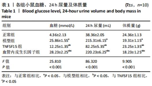
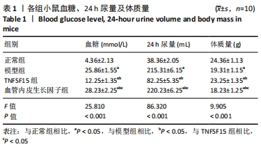
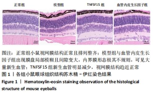
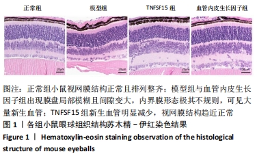
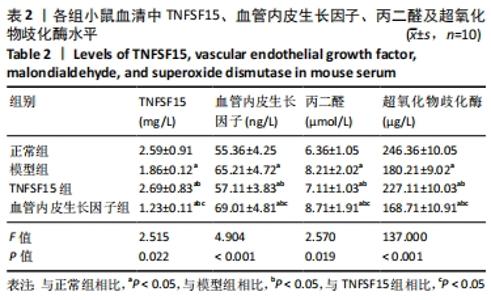
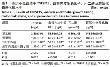
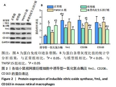
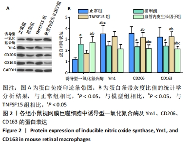
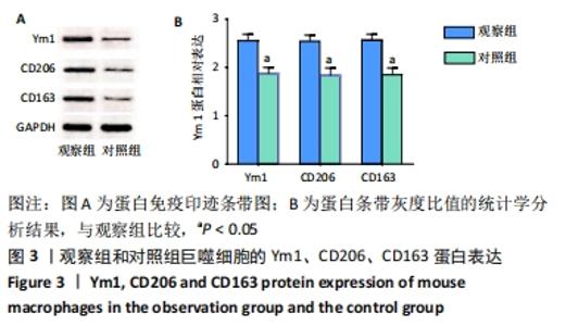
RESULTS AND CONCLUSION: Compared with the normal group, the blood glucose level and 24-hour urine volume, the serum level of vascular endothelial growth factor and malondialdehyde, and the protein expression of inducible nitric oxide synthase in retinal macrophages were significantly increased in model group (P < 0.05). While the above indicators in the TNFSF15 group were significantly lower than those in the model group (P < 0.05), and those in the vascular endothelial growth factor group were significantly higher than those in the TNFSF15 group (P < 0.05). Compared with the normal group, the body mass, serum levels of TNFSF15 and superoxide dismutase, and protein expression of YM1, CD206 and CD163 were significantly decreased in the model group (P < 0.05), while the above indicators in the TNFSG15 group were significantly higher than those in the model group (P < 0.05), and these indicators in the vascular endothelial growth factor group were significantly lower than those in the TNFSF15 group (P < 0.05). The mouse retina in the normal group had normal structure that was in alignment, and obvious retinal lesions were observed in the model group and vascular endothelial growth factor group. Compared with the model group and vascular endothelial growth factor group, there were fewer new blood vessels in the TNFSR15 group and its retinal structure intended to be normal. In the cell experiment, the protein expression of Ym1, CD206, and CD163 was increased significantly in the observation group compared with the control group (P < 0.05). Therefore, TNFSF15 may reduce oxidative damage in diabetic retinopathy mice and improve retinopathy by inducing macrophage polarization to M2 anti-inflammatory type.

Key words: tumor necrosis factor ligand superfamily member 15, macrophage, M2 polarization, diabetic retinopathy, oxidative damage, mouse

CLC Number: