Chinese Journal of Tissue Engineering Research ›› 2022, Vol. 26 ›› Issue (26): 4173-4179.doi: 10.12307/2022.820
Previous Articles Next Articles
Exercise effects on myocardial type I, III collagen and angiotensin II/transforming growth factor beta1/Smad2 pathway in diabetic myocardial fibrosis rats
Liu Ya1, 2, Liu Xia1, Deng Penghui1, Ji Wei1, Li Jianping1
- 1Hunan Provincial Key Laboratory of Physical Fitness and Sports Rehabilitation, School of Physical Education of Hunan Normal University, Changsha 410012, Hunan Province, China; 2Hunan International Economics University, Changsha 410205, Hunan Province, China
-
Received:2021-06-29Accepted:2021-09-18Online:2022-09-18Published:2022-03-08 -
Contact:Liu Xia, Professor, Master’s supervisor, Hunan Provincial Key Laboratory of Physical Fitness and Sports Rehabilitation, School of Physical Education of Hunan Normal University, Changsha 410012, Hunan Province, China -
About author:Liu Ya, Master, Hunan Provincial Key Laboratory of Physical Fitness and Sports Rehabilitation, School of Physical Education of Hunan Normal University, Changsha 410012, Hunan Province, China; Hunan International Economics University, Changsha 410205, Hunan Province, China -
Supported by:Hunan Provincial Social Science Foundation, No. 19YBA238 (to LX); Hunan Provincial Department of Education Key Project, No. 19A303 (LX)
CLC Number:
Cite this article
Liu Ya, Liu Xia, Deng Penghui, Ji Wei, Li Jianping. Exercise effects on myocardial type I, III collagen and angiotensin II/transforming growth factor beta1/Smad2 pathway in diabetic myocardial fibrosis rats[J]. Chinese Journal of Tissue Engineering Research, 2022, 26(26): 4173-4179.
share this article
Add to citation manager EndNote|Reference Manager|ProCite|BibTeX|RefWorks
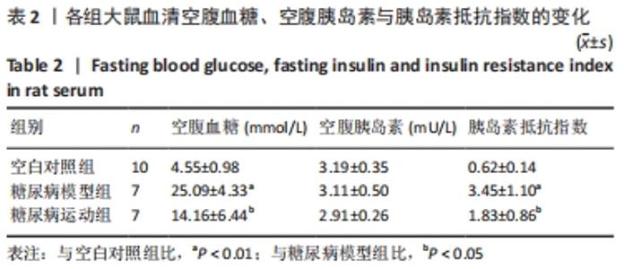
2.1 实验动物数量分析 建模和游泳过程中死亡大鼠16只,尸体已按要求处理,最后进入结果分析24只,其中空白对照组10只、糖尿病模型组7只、糖尿病运动组7只。 2.2 各组大鼠血清空腹血糖与空腹胰岛素检测结果 与空白对照组相比,糖尿病模型组大鼠血清空腹血糖、胰岛素抵抗指数增加(P < 0.01);与糖尿病模型组相比,糖尿病运动组大鼠血清空腹血糖、胰岛素抵抗指数减少(P < 0.05),见表2。"
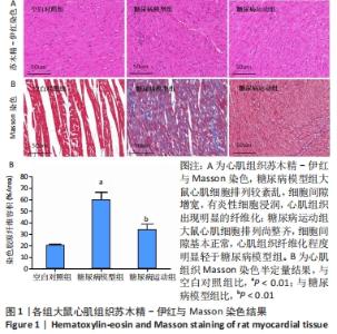
2.3 各组大鼠心肌组织病理观察结果 见图1。"
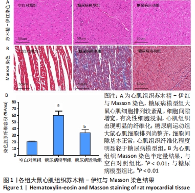
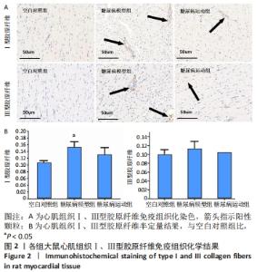
苏木精-伊红染色:空白对照组大鼠心肌细胞排列整齐,细胞形态、大小、间隙正常,细胞核均匀分布;糖尿病模型组大鼠心肌细胞排列较紊乱,细胞形态、大小异常,细胞间隙增宽,有炎性细胞浸润;糖尿病运动组大鼠心肌细胞排列尚整齐,细胞形态、大小异常,细胞间隙基本正常。 Masson染色:空白对照组间质蓝染面积小,纤维化不明显;糖尿病模型组间质蓝染面积明显增加,有明显的纤维化;糖尿病运动组间质蓝染面积减少,有少许纤维化改变。Masson染色半定量结果显示,与空白对照组相比,糖尿病模型组大鼠心肌纤维胶原容积分数增加(P < 0.01);与糖尿病模型组相比,糖尿病运动组大鼠心肌纤维胶原容积分数减少(P < 0.01)。 2.4 各组大鼠心肌组织Ⅰ、Ⅲ型胶原纤维免疫组织化染色结果 见图2。"


大鼠心肌组织Ⅰ、Ⅲ型胶原纤维阳性染色区域主要位于细胞间质,与空白对照组相比,糖尿病模型组大鼠心肌组织Ⅰ型胶原纤维增加(P < 0.05),Ⅲ型胶原纤维无明显变化(P > 0.05);与糖尿病模型组相比,糖尿病运动组大鼠心肌组织的Ⅰ、Ⅲ型胶原纤维无明显变化(P > 0.05)。 2.5 各组大鼠心肌组织相关基因表达量检测结果 见图3。"
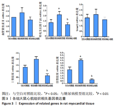

与空白对照组相比,糖尿病模型组大鼠心肌组织Ⅲ型胶原纤维和转化生长因子β1 mRNA表达量增加,(P < 0.05),心肌组织Ⅰ型胶原纤维、血管紧张素Ⅱ、Smad2 mRNA表达量无明显变化(P > 0.05);与糖尿病模型组相比,糖尿病运动组大鼠心肌组织Ⅰ、Ⅲ型胶原纤维和转化生长因子β1 mRNA表达量均减少(P < 0.01),血管紧张素Ⅱ、Smad2 mRNA表达量无明显变化(P > 0.05)。"

| [1] CHO NH, SHAW JE, KARURANGA S, et al. IDF diabetes at las:global estimates of diabetes prevalenc e for 2017 and projections for 2045. Diabetes Res Clin Pract. 2018;138(4):271. [2] XU Y, WANG L, HE J, et al. Prevalence and control of diabetes in Chinese adults. JAM A. 2013;310(9):948-959. [3] SAMUEL CS, HEWITSON TD, ZHANG Y, et al.Relaxin ameliorates fibrosis in experimental diabetic cardiomyopathy. Endocrinology. 2008;149(7): 3286-3293. [4] RUSSO I, FRANGOGIANNIS NG. Diabetes-associated cardiac fibrosis: Cellular effectors,molecular mechanisms and therapeutic opportunities. J Mol Cell Cardiol. 2016;90:84-93. [5] 郑海清,应苗法,顾胜龙,等.心肌纤维化的信号转导机制及新型抑制剂的研究进展[J].中国细胞生物学学报,2019,41(2):268-274. [6] 王禹川.不同因子致心肌纤维化分子学机制[J].医学综述,2012, 18(17):2736-2740. [7] LEASK A. Potential therapeutic targets for cardiac fibrosis:TGF-β, angiotensin, endothelin, CCN2, and PDGF, partners in fibroblast activation. Circ Res. 2010;106(11):1675-1680. [8] 魏峰涛,方宁远,杨巍维,等.体外模拟局部肾素血管紧张素系统对心肌纤维化的影[J].上海交通大学学报(医学版),2007,27(9): 1115-1118. [9] COPAJA SOTO M, VALENZUELA R, SALDANA A, et al. Early expression of monocyte chemoattractant Protein-1 correlates with the onset of isoproterenol-induced cardiac fibrosis in rats with distinct Angiotensin-converting enzyme polymorphism. J Renin Angiotensin Aldosterone Sys. 2008;9(3):154-162. [10] MENG XM, TANG PM, LI J, et al. TGF-β/Smad signaling in re-nal fibrosis. Front Physiol. 2015;6:82. [11] MEJIAS-PENA Y, ESTEBANEZ B, RODRIGUEZ-MIGUELEZ P, et al. Impact of resistance training on the autophagy inflammation apoptosis crosstalk in elderly subjects. Aging (Albany NY). 2017;9(2):408-418. [12] CHEN TI, TU WC. Exercise attenuates intermittent hypoxia-induced cardiac fibrosis associated with sodium-hydrogen exchanger-1 in rats. Front Physiol. 2016;7:462. [13] 侯改霞,肖国强.运动训练对糖尿病大鼠心肌纤维化的改善作用及其可能机制[J].体育学刊,2013(2):130-134. [14] 黄昶签,肖谦,赵柯湘,等.2型糖尿病心肌病变模型建立方法的实验研究[J].重庆医科大学学报,2007,32(1):15-17. [15] 孙昌亮.TGF-β1/Smad及其相关miRNA在运动干预糖尿病心肌纤维化形成中的调节作用[D].扬州:扬州大学,2018. [16] 杨四宝,高永建.转化生长因子β与microRNAs的串话效应在心肌纤维化中的研究进展[J].中国循环杂志,2013,28(7):552-554. [17] 孙帅锋,刘巍.心肌纤维化病理机制及诊疗策略进展[J].国际心血管病杂志,2020,47(5):264-267. [18] LIU JC, ZHOU L, WANG F, et al. Osthole decreases collagenⅠ/III contents and their ratio in TGF-β1-overexpressed mouse cardiac fibroblasts through regulating the TGF-β/Smad signaling pathway. Chin J Nat Med. 2018;16:321-329. [19] DA SILVA E, NATALI A, DA SILVA MF, et al. Swimming training attenuates the morphological reorganization of the myocardium and local inflammation in the left ventricle of growing rats with untreated experimental diabetes. Pathol Res Pract. 2016;212(4):325-334. [20] NOVOA U, ARAUNA D, MORAN M, et al. High-intensity exercise reduces cardiac fibrosis and hypertrophy but does not restore the nitroso-redox imbalance in diabetic cardiomyopathy. Oxid Med Cell Longev. 2017;2017:7921363. [21] SACRE JW, JELLIS CL, JENKINS C, et al. A six-month exercise intervention in subclinical diabetic heart disease:effects on exercise capacity, autonomic and myocardial function. Metabolism. 2014;63(9):1104-1114. [22] SILVA FS, BORTOLIN RH, ARAUJO DN, et al. Exercise training ameliorates matrix metalloproteinases 2 and 9messenger RNA expression and mitigates adverse left ventricular remodeling in streptozotocin-induced diabetic rats. Cardiovasc Pathol. 2017;29:37-44. [23] VEERANKI S, GIVVIMANI S, KUNDU S, et al. Moderate intensity exercise prevents diabetic cardiomyopathy associated contractile dysfunction through restoration of mitochondrial function and connexin 43 levels in db/db mice. J Mol Cell Cardiol. 2016;92:163-173. [24] SILVA E, NATALI AJ, SILVA MF, et al. Ventricular remodeling in growing rats with experimental diabetes: The impact of swimming training. Pathol Res Pract. 2013;209(10):618. [25] LI S, LIU J, TAN J, et al. Inhibition of raf1 ameliorates bleomycin induced pulmonary fibrosis through attenuation of TGFβ1 signaling. Am J Physiol Lung Cell Mol Physiol. 2018;315(2):241-247. [26] MA ZG, YUAN YP, WU HM, et al. Cardiac fibrosis: new insights into the pathogenesis. Int J Biol Sci. 2018;14(12):1645-1657. [27] WANG SQ, LI D, YUAN Y. Long-term moderate intensity exercise alleviates myocardial fibrosis in type 2 diabetic rats via inhibitions of oxidative stress and TGF-β1/Smad pathway. J Physiol Sci. 2019;69(6): 861-873. [28] 沈豪,郭霜,刘秀芬,等.TGF-β1/Smad信号通路对糖尿病大鼠心肌纤维化中的作用[J].中国药理学通报,2018,34(4):522-527. [29] YUE Y, MENG K, PU Y, et al.Transforming growth factor beta (TGF-beta) mediates cardiac fibrosis and induces diabetic cardiomyopathy. Diabetes Res Clin Pract. 2017;133:124-130. [30] GUAN J, LIU WQ, XING MQ, et al. Elevated expression of periostin in diabetic cardiomyopathy and the effect of valsartan. BMC Cardiovasc Disord. 2015;15:90. [31] 王秋岩,田鹤,穆长征.TGF-β1在猪糖尿病心肌组织的表达及通心络的干预作用[J].中国体视学与图像分析,2014,19(2):204-209. [32] EL-RAHMAN SA, FAYED HM. Targeting AngII/AT1R signaling pathway by Perindopril inhibits ongoing liver fibrosis in rat. J Tissue Eng Regen Med. 2019;13(12):2131-2141. [33] PAN XC, LIU Y, CEN YY, et al. Dual Role of Triptolide in Interrupting the NLRP3 Inflammasome Pathway to Attenuate Cardiac Fibrosis. Int J Mol Sci. 2019;20(2):360. [34] DING J, TANG Q, LUO B, et al. Klotho inhibits angiotensin II-induced cardiac hypertrophy, fibrosis, and dysfunction in mice through suppression of transforming growth factor-β1 signaling pathway. Eur J Pharmacol. 2019;859:172549. [35] SILVA RB, MESQUITA FF, ANDREO M, et al.Effect of gestational protein restriction on left ventricle hypertrophy and heart angiotensin II signaling pathway in adult offspring rats. Health. 2013;5:78-84. [36] TSUTSUI H, MATSUSHIMA S, KINUGAWA S, et al. Angiotensin II type 1 receptor blocker attenuates myocardial remodeling and preserves diastolic function in diabetic heart. Hypertens Res. 2007;30(5):439-449. [37] LIU X, XU Q, WANG X, et al. Irbesartan ameliorates diabetic cardiomyopathy by regulating protein kinase D and ER stress activation in a type 2 diabetes rat model. Pharmacol Res. 2015;93:43-51. [38] ROSIN NL, FALKENHAM A, SOPEL MJ, et al. Regulation and role of connective tissue growth factor in AngII-induced myocardial fibrosis. Am J Pathol. 2013;182(3):714-726. [39] HE JG, CHEN SL, HUANG YY, et al. The nonpeptide AVE0991 attenuates myocardial hypertrophy as induced by angiotensin II through downregulation of transforming growth factor-beta1/Smad2 expression. Heart Vessels. 2010;25(5):438-443. [40] LIU Y, LV H, TAN R, et al. Platelets Promote Ang II (Angiotensin II)-Induced Atrial Fibrillation by Releasing TGF-β1 (Transforming Growth Factor-β1) and Interacting With Fibroblasts. Hypertension. 2020;76(6): 1856-1867. [41] LI J, WANG S, ZHANG YL, et al. Immunoproteasome Subunit β5i Promotes Ang II (Angiotensin II)-Induced Atrial Fibrillation by Targeting ATRAP (Ang II Type I Receptor-Associated Protein) Degradation in Mice. Hypertension. 2019;73(1):92-101. [42] ZHANG Y, SHAO L, MA A, et al. Telmisartan delays myocardial fibrosis in rats with hypertensive left ventricular hypertrophy by TGF-β1/Smad signal pathwa. Hypertens Res. 2013;37:43-49. [43] GONZÁLEZ-RAMOS M, CALLEROS L, LÓPEZ-ONGIL S, et al. HSP70 increases extracellular matrix production by human vascular smooth muscle through TGF-β1 up-regulation.Int J Biochem Cell Biol. 2013; 45(2):232-242. [44] 叶春芳,李树民,王晓丽,等.糖尿病肾病合并高血压患者血清转化生长因子及相关指标的变化[J].江苏医药,2010,36(9):1035-1037. [45] GLENN DJ, CARDEMA MC, NI W, et al. Cardiac steatosis potentiates angiotensin Ⅱ effects in the heart. Am J Physiol Heart Circ Physiol. 2015;30(4):H339-350. [46] CHEN RR, FAN XH, CHEN G, et al. Irisin attenuates angiotensin II-induced cardiac fibrosis via Nrf2 mediated inhibition of ROS/ TGFβ1/Smad2/3 signaling axis. Chem Biol Interact. 2019;302:11-21. [47] MA N, LIU HM, XIA T, et al. Chronic aerobic exercise training alleviates myocardial fibrosis in aged rats through restoring bioavailability of hydrogen sulfide. Can J Physiol Pharmacol. 2018;96(9):902-908. [48] HAFSTAD AD, BOARDMAN NT, AASUM E. How Exercise May Amend Metabolic Disturbances in Diabetic Cardiomyopathy. Antioxid Redox Signal. 2015;22:1587-1605. [49] ZHANG Y, ZHANG Y. Toll-like receptor-6 (TLR6) deficient mice are protected from myocardial fibrosis induced by high fructose feeding through anti-oxidant and inflammatory signaling pathway. Biochem Biophys Res Commun. 2016;473(2):388-395. [50] MOUSA TM, LIU D, CORNISH KG, et al. Exercise training enhances baroreflex sensitivity by an angiotensin II-dependent mechanism in chronic heart failure. J Appl Physiol. 2008;104(3):616-624. [51] GU Q, WANG B, ZHANG XF, et al. Contribution of renin-angiotensin system to exercise-induced attenuation of aortic remodeling and improvement of endothelial function in spontaneously hypertensive rats. Cardiovasc Pathol. 2014;23(5):298-305. [52] 贺慧杰.糖尿病心肌病大鼠NO、AngⅡ和TGF-β1的表达水平及心肌细胞凋亡的实验研究[D].天津:天津医科大学,2005. |
| [1] | Gao Lei, Qin Xinyuan, Nie Xin, Wang Lei, Wang Jiangning. Extracorporeal circulation compression perfusion in the reconstruction of limb microcirculation from the mechanism of mechanical and chemical signal transduction [J]. Chinese Journal of Tissue Engineering Research, 2022, 26(9): 1334-1340. |
| [2] | Lü Yiyan, Li Hanbing, Ma Xiaoqing, Zhang Han, Zhang Yuhang, Li Genlin. Establishment and characteristic analysis of interior heat and diabetes mouse model using compound factors [J]. Chinese Journal of Tissue Engineering Research, 2022, 26(8): 1187-1193. |
| [3] | Chen Xianghe, Liu Bo, Yang Kang, Lu Pengcheng, Yu Huilin. Treadmill exercise improves the myocardial fibrosis of spontaneous type 2 diabetic mice: an exploration on the functional pathway [J]. Chinese Journal of Tissue Engineering Research, 2022, 26(8): 1210-1215. |
| [4] | Gao Yujin, Peng Shuanglin, Ma Zhichao, Lu Shi, Cao Huayue, Wang Lang, Xiao Jingang. Osteogenic ability of adipose stem cells in diabetic osteoporosis mice [J]. Chinese Journal of Tissue Engineering Research, 2022, 26(7): 999-1004. |
| [5] | Shui Xiaoping, Li Chunying, Li Shunchang, Sun Junzhi, Su Quansheng . Effects of aerobic and resistance exercises on brain-derived neurotrophic factor, nuclear factor-kappa B and inflammatory cytokines in skeletal muscle of type II diabetic rats [J]. Chinese Journal of Tissue Engineering Research, 2022, 26(5): 669-675. |
| [6] | Yin Tingting, Du Dayong, Jiang Zhixin, Liu Yang, Liu Qilin, Li Yuntian. Granulocyte colony-stimulating factors improve myocardial fibrosis in rats with myocardial infarction [J]. Chinese Journal of Tissue Engineering Research, 2022, 26(5): 730-735. |
| [7] | Yang Jinxin, Wang Kun, Zhao Jing, Chen Peijie, Zhang Tingran, Lu Wenyun, Luo Jiong. Effects of aerobic exercise intervention on synaptic plasticity in Alzheimer’s disease patients [J]. Chinese Journal of Tissue Engineering Research, 2022, 26(26): 4216-4223. |
| [8] | Xu Manman, Ji Zhe, Ou Lingdong, Li Ang, Shen Caiqi, Jin Peisheng. Biological characteristics of adipose-derived mesenchymal stem cells in diabetic patients and their effect on promoting wound healing [J]. Chinese Journal of Tissue Engineering Research, 2022, 26(25): 3956-3960. |
| [9] | Cai Zhiguo, Du Shasha, Yang Kun, Zhao Na, Liu Qi. Lipopolysaccharides mediate autophagy of mouse insulinoma βtc6 cells in high glucose state [J]. Chinese Journal of Tissue Engineering Research, 2022, 26(20): 3127-3132. |
| [10] | Chen Ziyang, Pu Rui, Deng Shuang, Liu Min. N6-methyladenosine methylation and its regulation in metabolic diseases [J]. Chinese Journal of Tissue Engineering Research, 2022, 26(20): 3250-3255. |
| [11] | Zeng Xinyu, Chen Xianghe, Liu Bo, Lu Pengcheng, Jin Shengjie, Li Wenxiu, Tian Zhikai, Sun Changliang. Mechanism of exercise improving bone metabolism in type 2 diabetics mellitus based on "Muscle-Bone" Crosstalk [J]. Chinese Journal of Tissue Engineering Research, 2022, 26(2): 289-295. |
| [12] | Zhang Shuling, Li Junhan, Wang Jiaqian, Li Yalong, Wang Chun. Effects of aerobic exercises on JAK2/STAT5 signal pathway in the liver of mice with non-alcoholic fatty liver disease [J]. Chinese Journal of Tissue Engineering Research, 2022, 26(17): 2690-2695. |
| [13] | Han Xuke, Chen Yiding, Chen Huizhen, Yang Xiaomei, Hong Peipei, Chen Qiu. Establishment and evaluation of neurogenic bladder model in rats with type 2 diabetes mellitus [J]. Chinese Journal of Tissue Engineering Research, 2022, 26(17): 2713-2719. |
| [14] | Wang Susu, Li Lifeng, Zhang Yimin. Visualization analysis on research progress and hotspots of exercise therapy for sarcopenia in older adults in recent decade [J]. Chinese Journal of Tissue Engineering Research, 2022, 26(14): 2223-2230. |
| [15] | Zhu Zheng, Fu Changxi, Ma Wenchao, Ma Gang, Peng peng. Regulatory mechanism of aerobic exercise on cardiac remodeling in spontaneously hypertensive rats [J]. Chinese Journal of Tissue Engineering Research, 2022, 26(14): 2231-2237. |
| Viewed | ||||||
|
Full text |
|
|||||
|
Abstract |
|
|||||