Chinese Journal of Tissue Engineering Research ›› 2022, Vol. 26 ›› Issue (20): 3127-3132.doi: 10.12307/2022.609
Previous Articles Next Articles
Lipopolysaccharides mediate autophagy of mouse insulinoma βtc6 cells in high glucose state
Cai Zhiguo, Du Shasha, Yang Kun, Zhao Na, Liu Qi
- Department of Periodontology, Affiliated Stomatological Hospital of Zunyi Medical University, Zunyi 563000, Guizhou Province, China
-
Received:2021-04-12Accepted:2021-06-03Online:2022-07-18Published:2022-01-19 -
Contact:Liu Qi, MD, Chief physician, Department of Periodontology, Affiliated Stomatological Hospital of Zunyi Medical University, Zunyi 563000, Guizhou Province, China -
About author:Cai Zhiguo, Master, Physician, Department of Periodontology, Affiliated Stomatological Hospital of Zunyi Medical University, Zunyi 563000, Guizhou Province, China -
Supported by:the National Natural Science Foundation of China, No. 81860196 (to LQ)
CLC Number:
Cite this article
Cai Zhiguo, Du Shasha, Yang Kun, Zhao Na, Liu Qi. Lipopolysaccharides mediate autophagy of mouse insulinoma βtc6 cells in high glucose state[J]. Chinese Journal of Tissue Engineering Research, 2022, 26(20): 3127-3132.
share this article
Add to citation manager EndNote|Reference Manager|ProCite|BibTeX|RefWorks
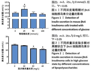
2.1 βtc6细胞胰岛素分泌量 细胞接种48 h后,镜下可见细胞呈片状、岛状等不规则形状生长,极少个别细胞独立生长。葡萄糖浓度为50 mmol/L时,βtc6细胞胰岛素分泌量与对照组相比差异无显著性意义(P > 0.05),在后续实验中选择葡萄糖浓度为50 mmol/L,见图1。当脂多糖浓度为20 mg/L时,βtc6细胞胰岛素分泌量(11.109 5±0.181 2) mIU/L显著下降,差异有显著性意义(P < 0.05),见图2。因此在后续实验中选择脂多糖质量浓度为20 mg/L。"

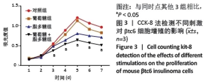
2.2 βtc6细胞增殖能力 相较于对照组,高浓度葡萄糖对βtc6细胞增殖总体呈现抑制趋势,脂多糖抑制βtc6细胞增殖的能力较强。当脂多糖联合50 mmol/L 葡萄糖作用于βtc6细胞时,抑制增殖的能力最强,除了第2天外,其余5 d增殖能力均低于另外3组(P < 0.05),见图3。"

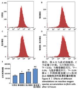
2.3 βtc6细胞中活性氧聚集量 与对照组相比,葡萄糖组和脂多糖组生成的活性氧水平有所增加(P < 0.05),脂多糖+葡萄糖组的活性氧生成量显著增加(P < 0.05);且脂多糖+葡萄糖组水平高于葡萄糖组和脂多糖组(P < 0.05);脂多糖组生成的活性氧水平高于葡萄糖组,但二者之间差异无显著性意义(P > 0.05),见图4。"

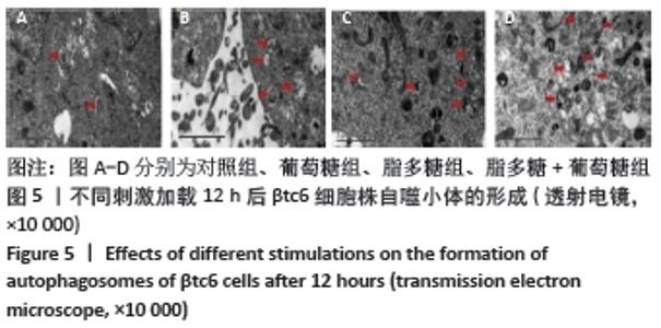
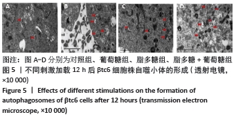
2.4 自噬小体变化 电镜下,对照组细胞自噬小体数量少,体积小,见图5A;脂多糖组略多于葡萄糖组,自噬小体数量较对照组增加,见图5B,C;脂多糖+葡萄糖组自噬小体增加更明显,体积较大,部分细胞器被包裹其中,见图5D。"
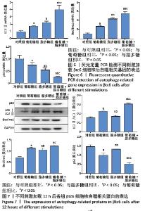
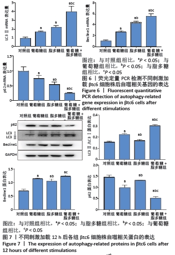
2.5 自噬相关基因相对表达 与对照组相比,葡萄糖组、脂多糖组及脂多糖+葡萄糖组的LC3Ⅱ基因相对表达量依次升高(P < 0.05),脂多糖组LC3Ⅱ虽略高于葡萄糖组,但两者相比差异无显著性意义(P > 0.05)。Becline1基因相对表达量依次升高,特别是脂多糖+葡萄糖组升高更剧烈(P < 0.05);另外,p62基因相对表达量在葡萄糖组、脂多糖组和脂多糖+葡萄糖组中均低于对照组,且脂多糖+葡萄糖组低于葡萄糖组和脂多糖组(P < 0.05),见图6。LC3Ⅱ/LC3Ⅰ、Becline1、p62蛋白表达与荧光定量PCR结果一致(P < 0.05),见图7。"
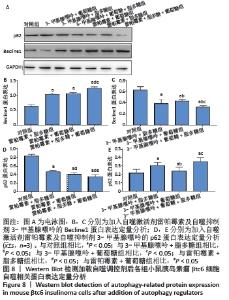
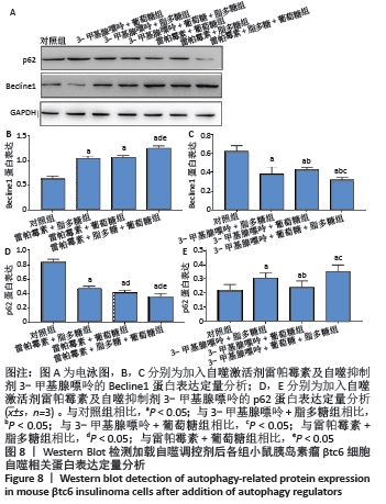
2.6 PI3K/AKT/mTOR信号通路的调控作用 加入3-甲基腺嘌呤后,与对照组相比,3-甲基腺嘌呤+葡萄糖组、3-甲基腺嘌呤+脂多糖组、3-甲基腺嘌呤+葡萄糖+脂多糖组Becline1蛋白表达显著降低(P < 0.05);与3-甲基腺嘌呤+葡萄糖组、3-甲基腺嘌呤+脂多糖组相比,3-甲基腺嘌呤+葡萄糖+脂多糖组Becline1蛋白表达显著降低(P < 0.05);p62蛋白表达则呈相反趋势(P < 0.05);加入雷帕霉素后, Becline1蛋白表达与加入3-甲基腺嘌呤后相反(P < 0.05);p62蛋白表达趋势与Becline1相反(P < 0.05),见图8。"
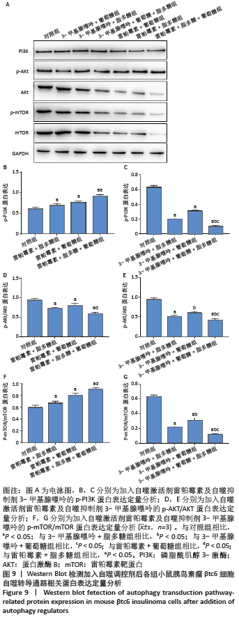
加入3-甲基腺嘌呤后,与对照组相比,3-甲基腺嘌呤+葡萄糖组、3-甲基腺嘌呤+脂多糖组、3-甲基腺嘌呤+脂多糖+葡萄糖组中p-PI3K显著降低(P < 0.05);与3-甲基腺嘌呤+葡萄糖组和3-甲基腺嘌呤+脂多糖组相比,3-甲基腺嘌呤+葡萄糖+脂多糖组p-PI3K显著降低(P < 0.05);与对照组相比,雷帕霉素+葡萄糖组、雷帕霉素+脂多糖组、雷帕霉素+葡萄糖+脂多糖组中p-AKT/AKT比值显著降低(P < 0.05),p-mTOR/mTOR比值趋势与p-PI3K一致(P < 0.05)。自噬激活组中p-PI3K、p-mTOR/mTOR 比值与自噬抑制组相反(P < 0.05),p-AKT/AKT比值趋势与自噬抑制组一致(P < 0.05),见图9。"
|
[1] 刘尔黎,钟雯怡,刘增一,等. 2型糖尿病伴牙周炎牙周膜干细胞成牙骨质分化能力的研究[J]. 遵义医科大学学报,2020,43(5):51-57. |
| [1] | Gao Lei, Qin Xinyuan, Nie Xin, Wang Lei, Wang Jiangning. Extracorporeal circulation compression perfusion in the reconstruction of limb microcirculation from the mechanism of mechanical and chemical signal transduction [J]. Chinese Journal of Tissue Engineering Research, 2022, 26(9): 1334-1340. |
| [2] | Lü Yiyan, Li Hanbing, Ma Xiaoqing, Zhang Han, Zhang Yuhang, Li Genlin. Establishment and characteristic analysis of interior heat and diabetes mouse model using compound factors [J]. Chinese Journal of Tissue Engineering Research, 2022, 26(8): 1187-1193. |
| [3] | Chen Xianghe, Liu Bo, Yang Kang, Lu Pengcheng, Yu Huilin. Treadmill exercise improves the myocardial fibrosis of spontaneous type 2 diabetic mice: an exploration on the functional pathway [J]. Chinese Journal of Tissue Engineering Research, 2022, 26(8): 1210-1215. |
| [4] | Cui Xing, Sun Xiaoqi, Zheng Wei, Ma Dexin. Huangqin Decoction regulates autophagy to intervene with intestinal acute graft-versus-host disease in mice [J]. Chinese Journal of Tissue Engineering Research, 2022, 26(7): 1057-1062. |
| [5] | Shui Xiaoping, Li Chunying, Li Shunchang, Sun Junzhi, Su Quansheng . Effects of aerobic and resistance exercises on brain-derived neurotrophic factor, nuclear factor-kappa B and inflammatory cytokines in skeletal muscle of type II diabetic rats [J]. Chinese Journal of Tissue Engineering Research, 2022, 26(5): 669-675. |
| [6] | Mi Jianguo, Qiao Rongqin, Liu Shaojin. Bushen Jianpi Huoxue Recipe improves bone metabolism, oxidative stress, and autophagy in osteoporotic rats [J]. Chinese Journal of Tissue Engineering Research, 2022, 26(26): 4147-4152. |
| [7] | Zeng Chunrong, Liu Menglan, Xie Yang, Li Zuoxiao. Preventive and therapeutic effects of naringin on experimental autoimmune encephalomyelitis via regulating microglial polarization [J]. Chinese Journal of Tissue Engineering Research, 2022, 26(26): 4127-4135. |
| [8] | Gong Yuqing, Yao Wei, Li Ran. Osteoimmunological effect of tumor necrosis factor alpha in alveolar bone remodeling [J]. Chinese Journal of Tissue Engineering Research, 2022, 26(26): 4242-4251. |
| [9] | Zhang Gaofei, Wang Di, Li Jiamei, Lou Hanxiao, Zeng Yueqin, Liu Wenjun. Mesenchymal stem cells promote wound healing by regulating the autophagy [J]. Chinese Journal of Tissue Engineering Research, 2022, 26(25): 4058-4063. |
| [10] | Jiang Jie, Zhao Baixiao, Chen Libin, Wen Li, Zhang Shanshan, Ma Jie, Zhao Hua. Effect of moxibustion pretreatment on autophagy and NLRP3 inflammasome expression in cerebral ischemia-reperfusion model rats [J]. Chinese Journal of Tissue Engineering Research, 2022, 26(23): 3615-3619. |
| [11] | Huang Jie, Ren Jing, Peng Pairan, Mu Yandong. Treatment of periodontitis in rats with a novel temperature-sensitive gel with immunomodulatory peptide [J]. Chinese Journal of Tissue Engineering Research, 2022, 26(22): 3514-3520. |
| [12] | Lyu Yuhu, Cheng Lin, Lin Wentao, Peng Fenglin. Role and regulatory mechanism of glycophagy in glycogen metabolism [J]. Chinese Journal of Tissue Engineering Research, 2022, 26(20): 3243-3249. |
| [13] | Liu Qiqi, Liu Min, Yang Jian, Yu Ke. Lipopolysaccharide stimulates the expression of interleukin-6 and nuclear factor kappa B receptor activator ligand in mouse MLO-Y4 osteoblasts [J]. Chinese Journal of Tissue Engineering Research, 2022, 26(20): 3121-3126. |
| [14] | Fan Shuai, Wu Chunfei, Liang Zujian, Xu Zhaohui, Xie Pingjin, Zhu Genfu. Bushen Tiaogan Prescription containing serum in the treatment of osteoarthritis by promoting chondrocyte autophagy in rats [J]. Chinese Journal of Tissue Engineering Research, 2022, 26(20): 3178-3183. |
| [15] | Wang Sihan, Chen Junji, Mo Weibin. Effects of Dendrobium officinale flavonoid on oxidative stress and autophagy in the liver of an exhaustive exercise rat model [J]. Chinese Journal of Tissue Engineering Research, 2022, 26(20): 3212-3219. |
| Viewed | ||||||
|
Full text |
|
|||||
|
Abstract |
|
|||||