Chinese Journal of Tissue Engineering Research ›› 2022, Vol. 26 ›› Issue (12): 1931-1936.doi: 10.12307/2022.519
Previous Articles Next Articles
Advantages of micro CT in three-dimensional reconstruction of specimens and its application in animal models of osteoarthritis
Du Longlong1, Yuan Puwei1, Yang Wei2, Li Xuefeng1, Gao Qimeng1
- 1Shaanxi University of Chinese Medicine, Xianyang 712046, Shaanxi Province, China; 2Guangzhou University of Chinese Medicine, Guangzhou 510006, Guangdong Province, China
-
Received:2021-06-26Revised:2021-07-08Accepted:2021-09-04Online:2022-04-28Published:2021-12-15 -
Contact:Yuan Puwei, MD, Professor, Chief physician, Shaanxi University of Chinese Medicine, Xianyang 712046, Shaanxi Province, China -
About author:Du Longlong, Master candidate, Shaanxi University of Chinese Medicine, Xianyang 712046, Shaanxi Province, China -
Supported by:the Li’s Orthopedic Inheritance Studio Construction Project of Chang’an Medicine, No. [2018] 40 (to YPW); Shaanxi Provincial Key Laboratory of Bone Degenerative Disease Prevention and Treatment of Integrated Traditional Chinese and Western Medicine Construction Project, No. [2018] 32 (to YPW); Xianyang Key Laboratory Construction Project, No. 2019k01-53 (to YPW)
CLC Number:
Cite this article
Du Longlong, Yuan Puwei, Yang Wei, Li Xuefeng, Gao Qimeng. Advantages of micro CT in three-dimensional reconstruction of specimens and its application in animal models of osteoarthritis[J]. Chinese Journal of Tissue Engineering Research, 2022, 26(12): 1931-1936.
share this article
Add to citation manager EndNote|Reference Manager|ProCite|BibTeX|RefWorks
"
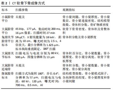
2.1 骨关节炎全关节micro CT表现 在骨关节炎病程进展中,全关节的X射线片为临床最重要的诊断标准[4],可清楚地反映关节间隙的变化、骨赘的位置及大小等,甚至可以通过X射线片的情况对骨关节炎进行病程划分,更好地指导治疗。但X射线片在动物实验中的应用较少,更多应用的是micro CT。通过micro CT扫描的图像可以对全关节的整体形态进行评估,对于关节局部影像表现,如关节间隙、关节边缘骨赘大小、骨硬化程度等表现予以研判。Micro CT可以提供高分辨率的图像,包括关节的断层扫描、骨的2D形态及3D重建,以定量评估骨特征,如骨体积分数(BV/TV)、骨小梁厚度和骨组织矿物密度(TMD),能够根据实验动物的骨关节炎进展的时间节点准确进行研究。 有研究对不同月龄豚鼠膝关节进行全关节micro CT扫描和组织学评分评估,结果显示随着月龄增加,全关节micro CT和组织学评分均增加,具有统计学意义;还能提示骨关节病理变化,通过影像分级结合定量micro CT测量,可以提高豚鼠动物模型在人类骨关节炎中的转化价值[5]。张永亮等[6]用木瓜蛋白酶注射制备膝关节炎大鼠模型,在造模成功后取大鼠完整膝关节标本进行micro CT扫描,观察其二维重建图像下的髁间嵴、膑股关节间隙和髌骨侧缘相关变化,并设立正常组进行对照,观察不同组别间存在的变化。 Micro CT扫描相关标本后,可以通过软件重建标本的二维及三维图像,可以直观、清楚地观察到病变关节的大致状况,还可以了解整体关节的结构变化,打破了传统病理切片对关节炎病理的唯一性,为骨关节炎的关节病理提供了一种简单、便捷的新方式。 2.2 骨关节炎软骨micro CT表现 软骨的丢失是骨关节炎的重要病理改变,早期的骨关节炎治疗也提倡对关节软骨的保护[7]。 在骨关节炎的相关机制研究中,关节软骨也是研究的热点。虽然关节软骨在micro CT下不能直接显影,但在多种增强对比剂的辅助下可以帮助软骨显影。在软骨对比造影成像技术主要是通过软骨局部关节软骨糖胺聚糖和对比剂局部浓度的反比关系,来实现软骨显影[8]。CT软骨成像方法的总结,见表1。"
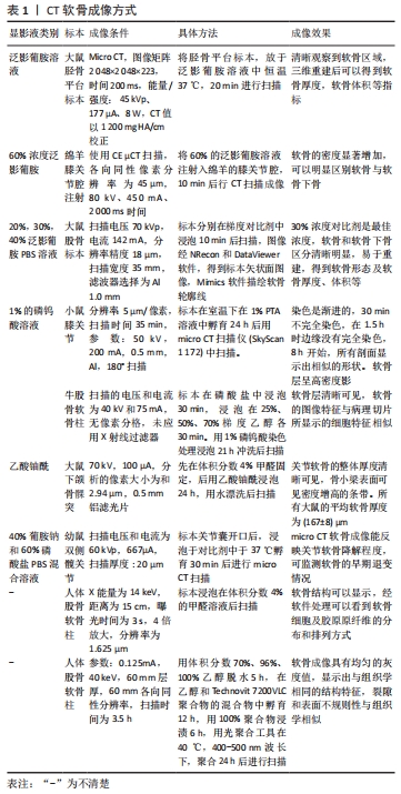
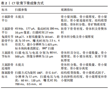
巫旭娜等[9]将切除卵巢及关节腔注射碘乙酸钠的大鼠模型的胫骨平台标本,放于泛影葡胺溶液中后进行micro CT扫描,选取感兴趣区域对其进行3D重建,最终可以得到关于关节软骨的定量参数:软骨厚度,软骨体积。FLYNN等[10]采用关节腔注射的方式,将6 mL 60%泛影葡胺溶液注射在正常组和膝骨关节炎模型组绵羊的膝关节腔行micro CT扫描成像,使得软骨在CT图像中得以显影。还有研究表明30%浓度的泛影葡胺溶液浸泡关节后显影,软骨及软骨下骨的分界更加清楚,对软骨的相关分析也具有更高的精度[11]。增强软骨显影的溶液也并非只有泛影葡胺。有研究表明小鼠膝关节炎模型的胫骨标本在1%磷钨酸溶液室温下浸泡24 h后,进行micro CT扫描并对其进行3D重建,对重建模型进行矢状位、冠状位平面分析,关节软骨呈现高密度影,可以对软骨进行定量分析[12]。关于磷钨酸溶液对增强关节软骨成像的方案还有别的方式,如CLARK等[13]在幼牛膝关节股骨髁取圆柱形骨软骨标本,先用梯度乙醇逐级脱水固定后用含1%磷钨酸的乙醇溶液浸泡行micro CT扫描,软骨有较好的成像表现。还有别的一些增强对比剂对软骨成像有帮助作用,如有研究将大鼠下颌骨浸渍在乙酸铀酰溶液后进行micro CT成像,关节软骨的全部厚度都清晰可见,而在浸渍之前软骨是不可见的[14]。刘进等[15]在发育性髋关节发育不良模型大鼠的幼鼠出生后不同时期分别取双侧髋关节标本,放置于40%葡胺钠溶液和60%磷酸盐PBS混合溶液后进行micro CT扫描,结果显示对比剂在软骨层的浓聚度随着时间的推移而升高,不同组别的股骨头软骨层的浓聚度具有统计学意义,提示软骨存在不同程度的改变。刘成磊等[16]对不同程度损伤的人体离体软骨标本,采用同步辐射显微CT(SR-μCT)进行扫描,对扫描图像进行二维重建及三维处理,结果显示软骨陷窝及软骨细胞的形态、排列方式等与病理切片的表现相似,软骨基质胶原原纤维网也可以清晰显示。Micro CT对于人体软骨的成像还有其他的方式,如有研究对人体软骨柱标本通过梯度乙醇脱水和Technovit 7200 VLC聚合物包埋后,使用micro CT扫描,结果显示软骨成像具有均匀的灰度值,裂隙和表面不规则与组织学相似[17]。 近年来,关节软骨micro CT成像的研究越来越多,随着micro CT的图像分辨率越来越高,可以成为一种非破坏性病理形态学技术,这样可以更加清楚地了解软骨病理形态在骨关节炎病程进展中的确切变化,有利于骨关节炎的软骨机制研究方面的深层次突破。 2.3 骨关节炎软骨下骨micro CT表现 软骨下骨有着较为丰富的血供,为关节软骨提供营养供应和缓冲应力[18],因为关节软骨位于软骨下骨表面,一旦软骨下骨出现微损伤、硬化及囊变时,表层的软骨所承受的应力会无法被缓冲,久而久之软骨就会发生退变。软骨下骨显微结构还帮助维持关节形态,对骨关节炎病程的进展具有重要意义[19]。 近年来,软骨下骨对于骨关节炎病程的影响越来越受到重视[20],有研究表明在骨关节炎病理变化中,软骨下骨的病理变化应早于关节软骨的改变[21],甚至有研究认为软骨下骨的病理改变是骨关节炎的始发因素[22]。软骨下骨在骨关节炎早期,主要以软骨下骨骨吸收和骨重塑为主要特征,而骨关节炎晚期主要病理改变为骨形成,表现为软骨下骨骨硬化[23]。软骨下骨在micro CT成像中可以完全显影,并通过micro CT的重建软件得以形成三维形态,更加直观地体现了软骨下骨形态方面的改变,同时对软骨下骨的感兴趣区域进行圈定,对骨体积、骨体积分数、骨小梁数目等数据整理,从而对软骨下骨的微观病理改变进行绝对定量分析。CT软骨下骨成像的总结,见表2。"

有研究将膝关节炎模型小鼠和正常小鼠胫骨平台的软骨下骨进行micro CT扫描后,对骨小梁间隙、骨小梁厚度、骨小梁数目、骨小梁连接密度、结构模型指数、骨体积分数等数据进行统计学分析,差异均有统计学意义[24]。PUCHA等[25]对正常大鼠和手术模型大鼠,用micro CT定量评估骨赘体积和软骨下骨结构等,最终结果提示手术组的软骨下骨密度增加,骨赘体积更大。还有研究应用改良Hulth法构建骨关节炎新西兰大白兔模型,将骨关节炎组和正常组兔的胫骨平台内侧圆柱形标本用SR-μCT扫描,三维重建后可以得到清晰的软骨下骨三维立体结构,结果显示相较于对照组,骨关节炎组的骨小梁网结构紊乱,骨小梁厚度降低,骨体积分数下降,差异有统计学意义[26]。谭启钊等[27]通过对不同骨关节炎模型大鼠的胫骨平台进行micro CT扫描和病理切片分析,探究前交叉韧带切除、木瓜蛋白酶关节腔注射及卵巢切除建立骨关节炎模型大鼠的软骨及软骨下骨的病理改变特征,结果表明3组的软骨下松质骨有显著改变,卵巢切除组软骨下骨显微结构变化最为明显,骨小梁数量、骨小梁分离度均有显著降低,3组相比于对照组,软骨下骨板厚度均变薄,差异有统计学意义。孙光华等[28]对膝骨关节炎-骨质疏松症大鼠模型的胫骨平台近端标本进行micro CT扫描,得出骨体积分数、骨小梁数目、骨小梁厚度、骨小梁分离度等相关数据,结果显示以上数据模型组较空白组均有显著改变,具有统计学意义。陶剑锋等[29]研究不同月龄的豚鼠软骨及软骨下骨的变化情况,在达到要求月龄后分别取实验豚鼠左右两侧完整膝关节,观察其关节及软骨损伤大体观后,对胫骨平台进行micro CT扫描,后通过相关软件三维重建、图像二值化等流程,得出范围内结构模型指数、各向异性的程度、骨小梁厚度、间隙及骨小梁模式因子等数据,以上数据组间均具有差异。 软骨下骨微结构的变化是由力学变化引起的骨关节炎的继发过程,防止软骨下骨的显微改变可以防止或减缓骨关节炎的进展[30]。在早期骨关节炎过程中,软骨下骨的显微结构会有明显的病理改变,micro CT对关节软骨下骨的损伤较为敏感,可以通过micro CT对早期骨关节炎病程中软骨下骨的病理改变做出更明确的研究。 2.4 骨关节炎滑膜micro CT表现 关节滑膜可以分泌关节滑液,为关节软骨提供营养物质,主要成分包括滑膜成纤维细胞和滑膜巨噬细胞。滑膜成纤维样细胞可以参与免疫调节维持关节局部稳态,滑膜巨噬细胞主要作用是吞噬关节局部细菌和凋亡细胞[31]。如果滑膜炎症持续发生时,两者会进一步诱发炎症反应,造成局部关节的进一步损害,加剧骨关节炎的程度。 近年来,滑膜被认为与骨关节炎的发生具有密切联系[32]。研究表明90%以上的骨关节炎患者存在不同程度的滑膜病变[33],滑膜细胞通过炎症因子的作用,也可直接或间接导致多种关节组织细胞的代谢紊乱,在骨关节炎的病程进展中起到重要的作用[34]。在分子和基因水平上,也证明滑膜相关的多种基因和通路对骨关节炎的发生与发展有重要的作用[35]。在针对滑膜的检查中主要以超声和核磁为主,但新的micro CT技术能完成对小型动物全身脂肪的扫描,还可以得到如脂肪分布、形态、体积的数据,同样可以通过micro CT对骨关节炎动物模型进行滑膜的相关检测。CT不同软组织成像的总结,见表3。"
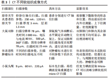
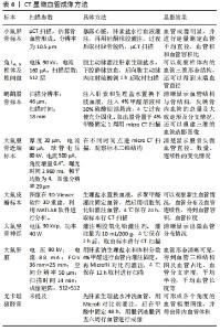
王琳等[36]在膝骨关节炎患者的双膝关节治疗前后分别进行CT轴位扫描,得出髌下脂肪垫的前后径、上下径、左右径、密度值等相关数据,经统计学分析,治疗组髌下脂肪垫相关数据治疗前后具有明显差异。目前micro CT在门控技术的使用下,可以实现实验动物活体成像。安国亮等[37]采用micro CT活体扫描正常大鼠和矽肺模型大鼠,扫描完成后用相关软件进行三维重建,并观察肺组织变化,结果显示micro CT可以清楚地观察到肺纹理及支气管和血管所形成的纹理影,双肺清晰可见,不同组别大鼠的肺组织图像存在差别。姜伟乾等[38]对患者体表恶性肿瘤进行手术切除后,将肿瘤标本进行micro CT扫描,通过对扫描时间和扫描电压的不断调整,结果发现不同的恶性肿瘤标本随着扫描电压和时间的变化,成像质量也在不断变化,且micro CT的扫描图像和病理切片的结果接近,micro-CT对体表软组织标本皮肤结构成像清楚。杨榕等[39]对正常小鼠进行颅底-颞下区注射癌细胞,造模3周后取头颅标本进行micro CT扫描,用3.75%的复方碘剂浸泡4 d再次以相同扫描条件进行micro CT扫描,结果显示没有用碘液浸泡的标本可以看到颅骨的整体形态及颞骨的局部破坏,软组织无法显影;经碘液浸泡的头颅标本可以清晰呈现出局部肌肉的分布情况。 脂肪和肌肉组织之间碳氧比的巨大差异解释了X射线能量下吸收的差异,其中光电吸收是主要的相互作用机制。因此在动物软组织中,脂肪组织与其他软组织相比具有不同的X射线衰减度,在micro CT图像中具有不同的密度,也可以利用不用组织对增强对比剂的吸收分布不同,再次增加不同软组织的密度,可以对髌下脂肪垫等组织病理变化在骨关节炎的进展中进行研究。滑膜位于关节腔内壁,可以产生关节液润滑关节,保证软骨的正常代谢环境及营养供应,一旦某些因素导致滑膜发生炎症,大量的炎症递质会改变关节局部基质环境,造成软骨基质的降解和软骨的退变[40]。骨关节炎有关于micro CT检测滑膜病变鲜有文献报道,但micro CT的技术的进步,活体成像技术愈发成熟,它的图像分辨率更高,这会让micro CT在滑膜方面的检测成为一个新的研究方向。 2.5 骨关节炎血管micro CT表现 血管病理在骨关节炎中的作用在近年来被人们不断所探索,被认为是促进骨关节炎病程发展的重要因素。血管病理状况主要包括各种原因所致的局部缺血、血液高黏滞、高血压、静脉淤血等,都会导致骨关节炎软骨、软骨下骨的营养物质的丢失,造成软骨及软骨下骨的缺损,加速骨关节炎病程的进展[41]。如研究表明在膝骨关节炎小鼠模型的软骨下骨H-型血管比正常组明显增多,还会导致实验组动物软骨下骨重塑、应力改变等,且模型组的软骨退变情况严重[42]。CT血管造影在临床应用中已经是很成熟的技术,动物实验中,micro CT主要针对肺、肾、肝脏等脏器、较大血管及肿瘤的血管成像,对于骨的显微血管成像较少。 血管病理在骨关节炎的进展中起到不可忽视的作用,在骨关节炎实验动物模型中也开始关注局部血管的改变。Micro CT可以对结合血管造影剂对末梢血管进行血管成像,得到高分辨率和三维的血管结构图像。CT显微血管成像方法的总结,见表4。"
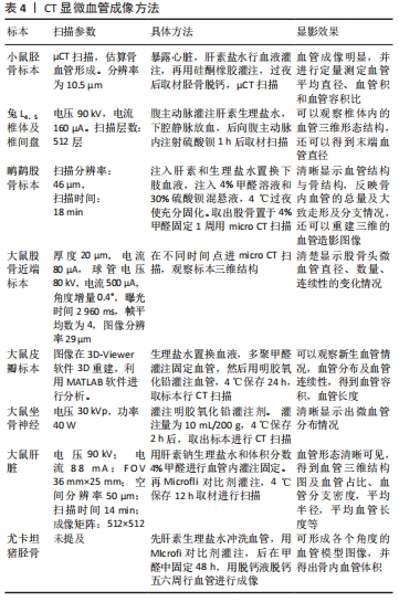

丁文鸽等[43]对卵巢切除组和正常组小鼠进行硅酮橡胶血液灌注后,取出胫骨标本脱钙后进行CT扫描以获得清晰的三维血管结构图像,还能得到血管容积、容积比、平均直径的定量参数。徐鸿明等[44]对大白兔麻醉后从腹主动脉进行硫酸钡对比剂血管灌注,L4、5椎体取材用micro CT进行扫描,可以得到完整的椎体终板内血管三维图像及末梢血管平均直径。范猛等[45]对成年鸸鹋麻醉行血液置换后,注射硫酸钡溶液做血管造影操作,之后取实验动物股骨标本脱钙,再用micro CT扫描,通过得到的图像进行软件处理获得血管造影的二维和三维图像,可以得到清晰的骨内显微血管图像。朱觉新等[46]对正常大鼠和股骨头坏死模型大鼠进行micro CT扫描,骨内微血管显影清楚。李国栋等[47]制备大鼠背部皮瓣,用明胶氧化铅行血管灌注,取下皮瓣标本进行micro CT扫描,图像经三维重建处理后,可以看到治疗组的皮瓣远端有大量新生血管长入,皮瓣局部血管的连续性、血管分布优于对照组,还可以得出扫描皮瓣血管容积和血管长度等数据。朱昭炜等[48]对正常大鼠颈总动脉血管灌注明胶氧化铅对比剂,通过micro CT扫描去观察神经内显微血管,可以显现血管形态和血管分布情况并得到神经内显微血管的可视化模型。刘巧遇等[49]对正常组和肝癌模型小鼠行Microfli对比剂灌注后,取出肝脏进行micro CT扫描,借助最大密度投影和容积再现技术重建血管图像,形成肝脏血管可视化三维模型,还可以得到血管占比、血管分支密度、血管长度、平均半径等数据。对Microfli对比剂还有别的应用方式,有研究对尤卡坦猪使用Microfli进行血管造影,脱钙后行扫描,可以得出血管体积的定量数据[50]。 血管病因学说是促进骨关节炎病程进展的重要因素,包括软骨下骨缺血、骨髓水肿、静脉淤血、血液高凝和低纤溶状态等都会促进骨关节炎病程的继续发展。血管病理在骨关节炎中的重要性被人们越来越重视,一种针对动物血管的检查方法尤为关键,从而去验证临床发现的血管基础病理研究。同时micro CT对微血管成像测量的准确性是肯定的,微血管系统树结构的呈现可以通过micro CT完成,这种分析的时间比传统方法要快,而且不易出错[51]。Micro CT可以通过对比剂对实验动物骨内微血管进行成像,得到血管三维图像及容积、直径等定量数据,对探索血管病理在骨关节炎病程中的作用有重要意义。"

| [1] KOLASINSKI SL, NEOGI T, HOCHBERG MC, et al. 2019 American College of Rheumatology/Arthritis Foundation Guideline for the Management of Osteoarthritis of the Hand, Hip, and Knee. Arthritis Care Res (Hoboken). 2020;72(2):149-162. [2] 张莹莹,李旭东,杨佳娟,等.中国40岁及以上人群骨关节炎患病率的Meta分析[J].中国循证医学杂志,2021,21(4):407-414. [3] 奚阳,石银朋,宋志伟,等.骨关节炎影像学研究进展[J].中国实用内科杂志, 2020,40(2):170-173. [4] YOON YJ, CHANG S, KIM OY, et al. Three-dimensional imaging of hepatic sinusoids in mice using synchrotron radiation micro-computed tomography. PLoS One. 2013; 8(7):e68600. [5] RADAKOVICH LB, MAROLF AJ, SHANNON JP, et al. Development of a microcomputed tomography scoring system to characterize disease progression in the Hartley guinea pig model of spontaneous osteoarthritis. Connect Tissue Res. 2018;59(6):523-533. [6] 张永亮,宓轶群,刚嘉鸿,等.温针灸对膝骨关节炎大鼠关节软骨及形态的影响[J].中国针灸,2016,36(2):175-179. [7] 杨波,周明旺,吉星,等.中药有效成分调节线粒体保护骨关节炎软骨的研究进展[J].中草药,2021,52(7):2117-2133. [8] 贾艳辉,卢世璧,汪爱媛,等.CT增强软骨成像技术的研究进展[J].中国医药生物技术,2013,8(2):143-146. [9] 巫旭娜,许碧莲,田佳,等.用Micro-CT评价大鼠骨质疏松合并骨性关节炎模型[J].中国老年学杂志,2020,40(5):1044-1047. [10] FLYNN C, HURTIG M, LINDEN AZ. Anionic Contrast-Enhanced MicroCT Imaging Correlates with Biochemical and Histological Evaluations of Osteoarthritic Articular Cartilage. Cartilage. 2020:1947603520924748. [11] 王江雪,高玉,李辉,等.离子造影剂增强Micro-CT扫描对大鼠关节软骨形态的定量分析[J].生物医学工程研究,2014,33(3):157-161. [12] DAS NEVES BORGES P, FORTE AE, VINCENT TL, et al. Rapid, automated imaging of mouse articular cartilage by microCT for early detection of osteoarthritis and finite element modelling of joint mechanics. Osteoarthritis Cartilage. 2014; 22(10):1419-1428. [13] CLARK JN, GARBOUT A, FERREIRA SA, et al. Propagation phase-contrast micro-computed tomography allows laboratory-based three-dimensional imaging of articular cartilage down to the cellular level. Osteoarthritis Cartilage. 2020;28(1):102-111. [14] KÜN-DARBOIS JD, MANERO F, RONY L, et al. Contrast enhancement with uranyl acetate allows quantitative analysis of the articular cartilage by microCT: Application to mandibular condyles in the BTX rat model of disuse. Micron.2017;97:35-40. [15] 刘进,傅明,黄广鑫,等.Micro-CT软骨成像监测发育性髋关节发育不良大鼠髋关节软骨早期退变的应用价值[J].中华关节外科杂志(电子版),2013,7(2):213-219. [16] 刘成磊,郗艳,左后东,等.同步辐射显微CT的人关节软骨三维成像研究[J].CT理论与应用研究,2015,24(6):793-799. [17] MOHR A, HEISS C, BERGMANN I, et al. Value of micro-CT as an investigative tool for osteochondritis dissecans: A preliminary study with comparison to histology. Acta Radiologica. 2003;44:5. doi:10.1080/j.1600-0455.2003.00113. [18] 王前源,刘水涛,卫小春,等.软骨下骨在骨性关节炎进程中的作用[J].中国医学前沿杂志(电子版),2017,9(2):53-57. [19] MOUNTCASTLE SE, ALLEN P, MELLORS BOL, et al. Dynamic viscoelastic characterisation of human osteochondral tissue: understanding the effect of the cartilage-bone interface. BMC Musculoskelet Disord. 2019; 20(1):575. [20] 常亮,秦江辉,史冬泉,等.骨关节炎与软骨下骨研究进展[J].中华骨与关节外科杂志,2019,12(10):827-832. [21] POURAN B, ARBABI V, BLEYS RL, et al. Solute transport at the interface of cartilage and subchondral bone plate: Effect of micro-architecture. J Biomech. 2017;52:148-154. [22] 郭洁梅,陈鹏,肖艳,等.软骨下骨重塑与骨关节炎综述[J].福建中医药,2021, 52(1):54-57. [23] 陈文杰,李益军,郑小飞,等.软骨下骨的病理改变及其在骨关节炎发病机制中的作用[J].中国骨科临床与基础研究杂志,2020, 12(4):234-241. [24] 郇松玮,査振刚,王华军,等.miR-214在小鼠骨关节炎软骨及软骨下骨中的表达分析[J].中国矫形外科杂志,2019,27(7):641-645. [25] PUCHA KA, MCKINNEY JM, FULLER JM, et al. Characterization of OA development between sexes in the rat medial meniscal transection model. Osteoarthr Cartilage Open. 2020;(3):100066. [26] 耿佳,星月,胡扬帆,等.同步辐射X线显微断层成像在兔膝骨关节炎软骨及软骨下骨三维成像中的应用研究[J].诊断学理论与实践,2020,19(3):238-242. [27] 谭启钊,牛国栋,赵振达,等.关节软骨与软骨下骨改变在不同骨关节炎动物模型中的特点[J].中国实验动物学报,2019,27(4):450-455. [28] 孙光华,廖源,彭婷,等.依降钙素对骨质疏松-骨关节炎大鼠的影响[J].中国矫形外科杂志,2020,28(18):1685-1689. [29] 陶剑锋,王莹,王超,等.豚鼠不同年龄自发性骨关节炎显微变化的观察[J].骨科临床与研究杂志,2019,4(3):167-172. [30] HOLZER LA ,KRAIGER M, TALAKIC E, et al. Microstructural analysis of subchondral bone in knee osteoarthritis. Osteoporos Int. 2020;31(10):2037-2045. . [31] 韩广弢,李皓桓,张宇标,等.滑膜在炎性关节病中的作用[J].广西医学,2019, 41(12):1545-1548. [32] 刘金富,曾平,农焦,等.整合多组微阵列芯片分析骨关节炎患者滑膜中生物标志物和治疗靶点[J].中国组织工程研究,2021,25(23): 3690-3696. [33] SCANZELLO CR, GOLDRING SR. The role of synovitis in osteoarthritis pathogenesis. Bone. 2012;51(2):249-257. [34] 郑洁,袁普卫.骨性关节炎的代谢机制研究进展[J].中国骨质疏松杂志,2018, 24(3):406-410. [35] 宋珊,胡方媛,乔军,等.基于生物信息学途径认识骨关节炎滑膜的生物学标志物[J].中国组织工程研究,2021,25(5):785-790. [36] 王琳,张洁,刘太运,等.膝关节骨性关节炎髌下脂肪垫在中药治疗前后的CT变化研究[J].中国中西医结合影像学杂志,2014,12(4): 351-354. [37] 安国亮,李小丽,王炎,等.Micro CT对骨髓间充质干细胞拮抗矽肺大鼠肺纤维化的效果评价[J].首都医科大学学报,2017,38(2):238-243. [38] 姜伟乾,陈犹白,陶然,等.显微计算机断层扫描对体表恶性肿瘤成像的研究[J].中华整形外科杂志,2020(3):242-243-244-245-246-247-248-249-250. [39] 杨榕,李庆祥,王逸飞,等.碘液浸染在Micro-CT下识别小鼠颅底-颞下区肿瘤组织中的应用[J].北京大学学报(医学版),2021,53(3): 598-601. [40] 江攀,李大鹏,毛良浩,等.滑膜在骨关节炎发病机制及治疗中的作用[J].中国矫形外科杂志,2020,28(5):430-434. [41] 李晗,陈百成,邵德成,等.血管病理与骨关节炎发病关系的研究进展[J].中国矫形外科杂志,2010,18(1):46-48. [42] 卢键森,柳鑫,曾春,等.H-型血管在骨关节炎软骨下骨中的表达及作用[J].中国组织工程研究,2017,21(20):3135-3140. [43] 丁文鸽,戴力扬,蒋雷生.卵巢切除小鼠胫骨干骺端血管的显微CT观察[J].上海交通大学学报(医学版),2008(10):1238-1241. [44] 徐鸿明,胡斐,王雍立,等.兔椎体软骨终板内血管三维影像结构研究[J].中国脊柱脊髓杂志,2015,25(11):1013-1017. [45] 范猛,汪爱媛,王玉,等.基于Micro-CT的骨内微血管显影和三维重建[J].南开大学学报(自然科学版),2011,44(1):78-84. [46] 朱觉新,黄连芳.骨内微血管显影及三维重建在激素致大鼠股骨头缺血性坏死模型中的应用[J].广东医科大学学报,2020,38(5):562-565. [47] 李国栋,徐永清,何晓清,等.Tempol对大鼠任意皮瓣成活影响的实验研究[J].中国修复重建外科杂志,2016,30(10):1264-1269. [48] 朱昭炜,毛以华,何波,等.SD大鼠坐骨神经微血管三维可视化研究初探[J].中国修复重建外科杂志,2013,27(2):189-192. [49] 刘巧遇,李若坤.基于Micro-CT成像对肝癌原位移植瘤血管三维结构的定量研究[J].肝脏,2020,25(12):1290-1293. [50] KOTSOUGIANI D, HUNDEPOOL CA, BULSTRA LF, et al. Bone vascularized composite allotransplantation model in swine tibial defect: Evaluation of surgical angiogenesis and transplant viability. Microsurgery. 2019; 39(2):160-166. [51] KLINE TL, ZAMIR M, RITMAN EL. Accuracy of microvascular measurements obtained from micro-CT images. Ann Biomed Eng. 2010;38(9):2851-64. [52] 许云腾,许丽梅,李慧,等.从筋骨的力学特性探讨膝关节软骨-软骨下骨稳态失衡的生物力学机制[J].风湿病与关节炎,2019,8(12): 43-45+57. [53] 赵泽明,张柳.NF-κB信号通路与骨关节炎的关系研究进展[J].华北理工大学学报(医学版),2021,23(3):232-238. |
| [1] | Tan Xinfang, Guo Yanxing, Qin Xiaofei, Zhang Binqing, Zhao Dongliang, Pan Kunkun, Li Yuzhuo, Chen Haoyu. Effect of uniaxial fatigue exercise on patellofemoral cartilage injury in a rabbit [J]. Chinese Journal of Tissue Engineering Research, 2022, 26(在线): 1-6. |
| [2] | Zhu Chan, Han Xuke, Yao Chengjiao, Zhou Qian, Zhang Qiang, Chen Qiu. Human salivary components and osteoporosis/osteopenia [J]. Chinese Journal of Tissue Engineering Research, 2022, 26(9): 1439-1444. |
| [3] | Jin Tao, Liu Lin, Zhu Xiaoyan, Shi Yucong, Niu Jianxiong, Zhang Tongtong, Wu Shujin, Yang Qingshan. Osteoarthritis and mitochondrial abnormalities [J]. Chinese Journal of Tissue Engineering Research, 2022, 26(9): 1452-1458. |
| [4] | Zhang Lichuang, Xu Hao, Ma Yinghui, Xiong Mengting, Han Haihui, Bao Jiamin, Zhai Weitao, Liang Qianqian. Mechanism and prospects of regulating lymphatic reflux function in the treatment of rheumatoid arthritis [J]. Chinese Journal of Tissue Engineering Research, 2022, 26(9): 1459-1466. |
| [5] | Li Huo, Wang Peng, Gao Jianming, Jiang Haoran, Lu Xiaobo, Peng Jiang. Relationship between revascularization and internal microstructure changes in osteonecrosis of the femoral head [J]. Chinese Journal of Tissue Engineering Research, 2022, 26(9): 1323-1328. |
| [6] | Zhang Jichao, Dong Yuefu, Mou Zhifang, Zhang Zhen, Li Bingyan, Xu Xiangjun, Li Jiayi, Ren Meng, Dong Wanpeng. Finite element analysis of biomechanical changes in the osteoarthritis knee joint in different gait flexion angles [J]. Chinese Journal of Tissue Engineering Research, 2022, 26(9): 1357-1361. |
| [7] | Wu Cong, Jia Quanzhong, Liu Lun. Relationship between transforming growth factor beta1 expression and chondrocyte migration in adult articular cartilage after fragmentation [J]. Chinese Journal of Tissue Engineering Research, 2022, 26(8): 1167-1172. |
| [8] | Wang Baojuan, Zheng Shuguang, Zhang Qi, Li Tianyang. Miao medicine fumigation can delay extracellular matrix destruction in a rabbit model of knee osteoarthritis [J]. Chinese Journal of Tissue Engineering Research, 2022, 26(8): 1180-1186. |
| [9] | Lü Yiyan, Li Hanbing, Ma Xiaoqing, Zhang Han, Zhang Yuhang, Li Genlin. Establishment and characteristic analysis of interior heat and diabetes mouse model using compound factors [J]. Chinese Journal of Tissue Engineering Research, 2022, 26(8): 1187-1193. |
| [10] | Wang Jing, Xiong Shan, Cao Jin, Feng Linwei, Wang Xin. Role and mechanism of interleukin-3 in bone metabolism [J]. Chinese Journal of Tissue Engineering Research, 2022, 26(8): 1260-1265. |
| [11] | Zhu Chan, Han Xuke, Yao Chengjiao, Zhang Qiang, Liu Jing, Shao Ming. Acupuncture for Parkinson’s disease: an insight into the action mechanism in animal experiments [J]. Chinese Journal of Tissue Engineering Research, 2022, 26(8): 1272-1277. |
| [12] | Hui Xiaoshan, Bai Jing, Zhou Siyuan, Wang Jie, Zhang Jinsheng, He Qingyong, Meng Peipei. Theoretical mechanism of traditional Chinese medicine theory on stem cell induced differentiation [J]. Chinese Journal of Tissue Engineering Research, 2022, 26(7): 1125-1129. |
| [13] | An Weizheng, He Xiao, Ren Shuai, Liu Jianyu. Potential of muscle-derived stem cells in peripheral nerve regeneration [J]. Chinese Journal of Tissue Engineering Research, 2022, 26(7): 1130-1136. |
| [14] | Fan Yiming, Liu Fangyu, Zhang Hongyu, Li Shuai, Wang Yansong. Serial questions about endogenous neural stem cell response in the ependymal zone after spinal cord injury [J]. Chinese Journal of Tissue Engineering Research, 2022, 26(7): 1137-1142. |
| [15] | Wang Xinmin, Liu Fei, Xu Jie, Bai Yuxi, Lü Jian. Core decompression combined with dental pulp stem cells in the treatment of steroid-associated femoral head necrosis in rabbits [J]. Chinese Journal of Tissue Engineering Research, 2022, 26(7): 1074-1079. |
| Viewed | ||||||
|
Full text |
|
|||||
|
Abstract |
|
|||||