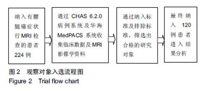
| [1]Du CF, Yang N, Guo JC, et al. Biomechanical response of lumbar facet joints under follower preload: a finite element study. BMC Musculoskelet Disord. 2016; 17(1):126.[2]王洪岗.小关节方向不规则变化与腰椎间盘突出症的临床相关性研究[D].重庆:解放军陆军军医大学;第三军医大学,2017.[3]刘玉成. 腰椎管狭窄临床症状用影像检查验证的临床意义[J]. 中国医药指南, 2017,15(30):82-83.[4]Sun D , Liu P , Cheng J , et al. Correlation between intervertebral disc degeneration, paraspinal muscle atrophy, and lumbar facet joints degeneration in patients with lumbar disc herniation. BMC Musculoskelet Disord. 2017;18(1):167.[5]Pathria M, Sartoris DJ, Resnick D. Osteoarthritis of the facet joints: accuracy of oblique radiographic assessment. Radiology. 1987;164:227–230.[6]Clarençon F, Lawye B, Bienvenot P, et al. The degenerative spine. Magn Reson Imaging Clin N Am. 2016;24(3):495-513. [7]曹勇. 单排螺旋CT对腰椎间盘突出症并发椎管狭窄的诊断价值分析[J]. 中外医学研究, 2016, 14(18):42-43.[8]张春,刘红梅. 下腰椎小关节退行性病变的X线、CT、MRI影像学比较[J]. 包头医学院学报, 2016, 32(3):84-85.[9]乔聚义. 下腰椎小关节退变的磁共振成像及CT表现特征分析[J]. 实用医学影像杂志,2016,17(3):265-266.[10]Weishaupt D, Zanetti M, Boos N, et al. MR imaging and CT in osteoarthritis of the lumbar facet joints. Skeletal Radiol. 1999;28(4):215-219.[11]Abbas J, Slon V, Stein D, et al. In the quest for degenerative lumbar spinal stenosis etiology: The Schmorl's nodes model. BMC Musculoskelet Disord. 2017;18(1):164.[12]Lurie J, Tomkinslane C. Management of lumbar spinal stenosis. BMJ. 2016;352(1):6234.[13]史少岩,黄研生,郝定均. 腰椎管狭窄的治疗进展[J]. 中国骨伤,2017,30(5): 484-488.[14]Kalichman L, Li L, Kim DH, et al. Facet joint osteoarthritis and low back pain in the community-based population. Spine. 2008; 33(23):2560-2565.[15]Yabuki S, Fukumori N, Takegami M, et al. Prevalence of lumbar spinal stenosis, using the diagnostic support tool, and correlated factors in Japan: a population-based study. J Orthop Sci. 2013;18(6):893-900.[16]殷刚,邱勇. 腰椎小关节骨性关节炎危险因素的研究进展[J]. 中国矫形外科杂志,2009,17(13):995-997.[17]Abbas J, Hamoud K, Peleg S, et al. Facet joints arthrosis in normal and stenotic lumbar spines. Spine. 2011;36(24):1541-1546.[18]郭增峰,于滨生. 腰椎小关节骨性关节炎的研究进展[J]. 中国矫形外科杂志, 2017,25(17):1587-1591.[19]祝恺,谭军,张紫豪,等. 腰椎小关节骨关节炎宏观及微观改变的研究进展[J]. 中国骨与关节杂志, 2017, 6(10):764-767.[20]Simon P, Espinoza Orías AA, Andersson GB, et al. In vivo topographic analysis of lumbar facet joint space width distribution in healthy and symptomatic subjects. Spine. 2012;37(12):1058-1064.[21]Grobler LJ, Robertson PA, Novotny JE, et al. Etiology of spondylolisthesis. Assessment of the role played by lumbar facet joint morphology. Spine. 1993; 18(1):80-91.[22]Jentzsch T, Geiger J, Zimmermann SM, et al. Lumbar facet joint arthritis is associated with more coronal orientation of the facet joints at the upper lumbar spine. Radiol Res Pract. 2013;2013:693971. [23]Linov L, Klindukhov A, Li L, et al. Lumbar facet joint orientation and osteoarthritis: a cross-sectional study. J Back Musculoskelet Rehabil. 2013;26(4):421-426.[24]顾树明,张彦东,肖京,等. 腰椎小关节方向和骨性关节炎分级与侧隐窝狭窄症的关系[J]. 中国医刊,2015,50(1):43-45.[25]闫广辉,高春光,李华,等. 腰椎小关节精确测量及其与腰椎管狭窄症的相关性研究[J]. 中国现代医学杂志, 2015, 25(35):88-91.[26]黄宗文,黄创新,郭远清,等. 下腰椎形态与椎管退行性狭窄的关系及其临床意义[J]. 中国脊柱脊髓杂志, 2004,14(11):663-665.[27]肖伟平,杨凤云.腰椎小关节矢状化与年龄、体重指数之间的相关性研究[C].//第二十四届全国中西医结合骨伤科学术年会论文集, 2017:233-233.[28]李志赏, 闫广辉, 张庆胜, 等. 年龄相关的腰椎小关节三维角度的变化规律[J]. 中国现代医学杂志, 2016, 26(20):78-81.[29]覃柳生. 腰椎关节突关节退变的影像学表现及意义[J]. 实用医学影像杂志, 2014,15(5):357-359.[30]Boden SD, Riew KD, Yamaguchi K, et al. Orientation of the lumbar facet joints: association with degenerative disc disease. J Bone Joint Surg Am. 1996;78(3):403-411.[31]程若勤,程小杰. 腰椎小关节骨性关节炎与小关节角度及不对称性的关系[J]. 现代仪器与医疗, 2013, 19(2):28-31.[32]闫广辉,徐宝山,夏群,等. 腰椎小关节方向性与腰椎间盘突出症[J]. 中国组织工程研究, 2011,15(35):6563-6566.[33]荆慧田,冯世庆,班德翔. 关节突关节角度与退行性腰椎滑脱的关系[J]. 中国脊柱脊髓杂志, 2011,21(4):299-302. [34]Fujiwara A, Tamai K, An HS, et al. Orientation and osteoarthritis of the lumbar facet joint. Clin Orthop Relat Res. 2001; 385(385):88-94. |