Chinese Journal of Tissue Engineering Research ›› 2020, Vol. 24 ›› Issue (36): 5741-5748.doi: 10.3969/j.issn.2095-4344.2916
Changes in cervical sagittal balance after three-dimensional printing ACT titanium cage in anterior cervical discectomy with fusion
Yang Xu, Zhao Xiaofeng, Qi Detai, Wang Xiaonan, Jin Yuanzhang, Zhou Runtian, Zhao Bin
Department of Orthopedics, Second Hospital of Shanxi Medical University, Taiyuan 030001, Shanxi Province, China
-
Received:2020-01-18Revised:2020-01-20Accepted:2020-03-09Online:2020-12-28Published:2020-10-27 -
Contact:Zhao Bin, Chief physician, Professor, Master’s supervisor, Department of Orthopedics, Second Hospital of Shanxi Medical University, Taiyuan 030001, Shanxi Province, China -
About author:Yang Xu, Master candidate, Department of Orthopedics, Second Hospital of Shanxi Medical University, Taiyuan 030001, Shanxi Province, China
CLC Number:
Cite this article
Yang Xu, Zhao Xiaofeng, Qi Detai, Wang Xiaonan, Jin Yuanzhang, Zhou Runtian, Zhao Bin. Changes in cervical sagittal balance after three-dimensional printing ACT titanium cage in anterior cervical discectomy with fusion[J]. Chinese Journal of Tissue Engineering Research, 2020, 24(36): 5741-5748.
share this article
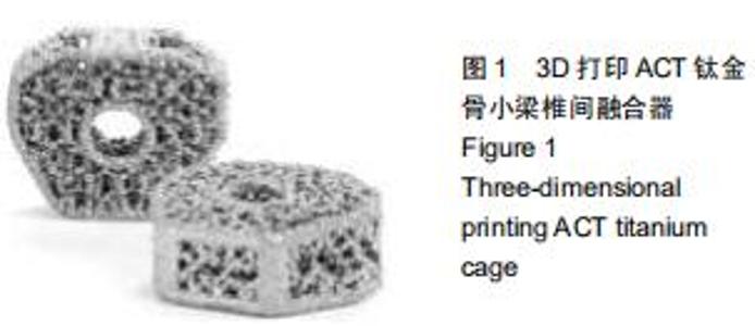
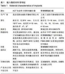
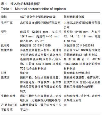
1.4.1 ACT钛金骨小梁椎间融合器 北京爱康宜诚医疗器械产品,医疗器械注册证编号:国械注准20163461289;产品技术要求编号:国械注准20163461289;生产许可证号:京食药监械生产证20040045号。融合器为多边棱柱形,上下表面为弧形,前后径12.5/14 mm,左右径15/17 mm,高度有4,5,6,7,8,9,10 mm共7种规格,具有0°,4°,8° 3种前凸角型号,具有多孔类似骨小梁结构,见图1及表1。 "
1.4.1 ACT钛金骨小梁椎间融合器 北京爱康宜诚医疗器械产品,医疗器械注册证编号:国械注准20163461289;产品技术要求编号:国械注准20163461289;生产许可证号:京食药监械生产证20040045号。融合器为多边棱柱形,上下表面为弧形,前后径12.5/14 mm,左右径15/17 mm,高度有4,5,6,7,8,9,10 mm共7种规格,具有0°,4°,8° 3种前凸角型号,具有多孔类似骨小梁结构,见图1及表1。 "
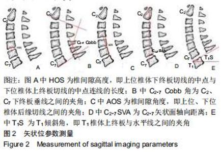
(1)完善术前、术后3 d、术后3,6个月及末次随访时颈椎正侧位及动力位X射线片(拍摄侧位X射线片时要求下颚角与枕骨下角保持在一个水平面上)。应用医院影像归档和通信系统测量相关颈椎矢状面参数,包括:①椎间隙高度:上位椎体下终板切线的中点与下位椎体上终板切线的中点连线的长度;②C2-7 Cobb角:C2、C7椎体下终板平面垂线之间的夹角;③椎间隙角度:手术节段上位、下位椎体后缘切线之间的夹角;④C2-7矢状面轴向距离:经过C2椎体几何中心作铅垂线,该线与C7椎体后上角的水平距离;⑤T1倾斜角:T1椎体上终板与水平线之间的夹角。各影像学参数示意图见图2。 "
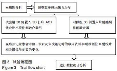
2.2 试验流程图 见图3。 "
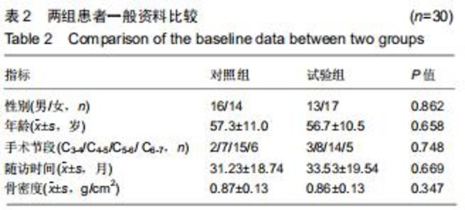
2.3 一般资料比较 2组一般资料具有可比性,差异无显著性意义,见表2。 "
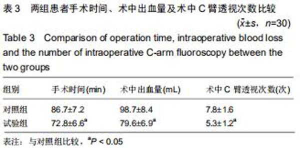
2.4 围术期指标比较 试验组手术时间、术中出血量及术中C臂透视次数均小于对照组,差异有显著性意义(P < 0.05),见表3。 "
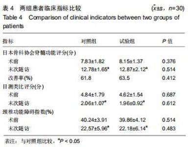
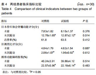
2.5 临床疗效评估 随访时间15-49个月,所有患者均未出现脊髓、神经血管损伤、血肿压迫、脑脊液漏以及感染和内固定松动等并发症。日本骨科协会脊髓功能评分、目测类比评分和颈椎功能障碍指数结果见表4。术后2组日本骨科协会脊髓功能评分均较术前显著升高,目测类比评分以及颈椎功能障碍指数显著降低,术前、术后差异有显著性意义(P < 0.05);2组间对比差异无显著性意义。 "
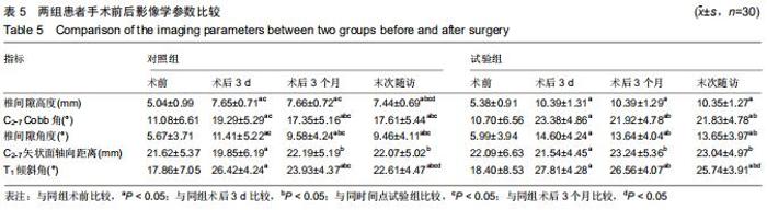
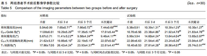
2.6 影像学参数评价 影像学参数测量见表5。术前2组各参数对比差异无显著性意义(P > 0.05)。术后3 d、3个月及末次随访时,2组手术节段椎间隙高度、椎间隙角度、C2-7 Cobb角及T1倾斜角均较术前增大(P < 0.05),C2-7矢状面轴向距离无明显变化;术后2组C2-7 Cobb角、椎间隙角度及T1倾斜角均呈下降趋势,在术后3 d、3个月及末次随访时,2组间手术节段椎间隙高度、C2-7 Cobb角、椎间隙角度及T1倾斜角差异有显著性意义(P < 0.05),即对照组各参数值减小更为显著,但2组间C2-7矢状面轴向距离对比差异无显著性意义(P > 0.05)。 "
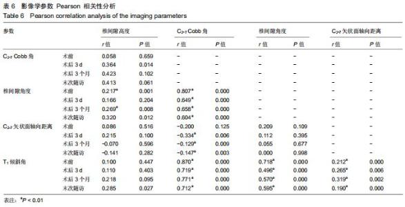
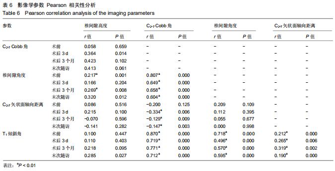
2组影像学参数Pearson相关性分析结果见表6,术前、术后3 d、术后3个月及末次随访时 C2-7 Cobb角与椎间隙角度及T1倾斜角、椎间隙角度与T1倾斜角、T1倾斜角与C2-7 矢状面轴向距离均呈正相关(P < 0.01),C2-7矢状面轴向距离与C2-7 Cobb角呈负相关(P < 0.01,术前时除外)。 "
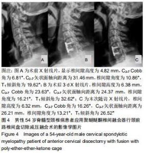
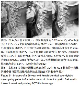
2.7 典型病例 见图4,5。 "
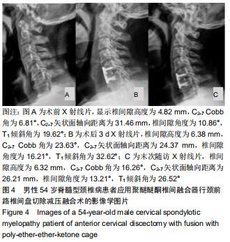
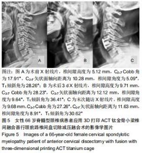
2.7 典型病例 见图4,5。 "
|
[1] MUZEVIĆ D, SPLAVSKI B, BOOP FA, et al. Anterior cervical discectomy with instrumented allograft fusion: lordosis restoration and comparison of functional outcomes among patients of different age groups. World Neurosurg. 2018;109: e233-e243.
[2] OLIVER JD, GONCALVES S, KEREZOUDIS P, et al. Comparison of outcomes for anterior cervical discectomy and fusion with and without anterior plate fixation: a systematic review and meta-analysis. Spine. 2018;43(7): E413-E422.
[3] 陈丹华,葛许锋,成红兵,等.颈椎桥型锁定融合器在颈椎前路减压融合术中的应用[J].实用临床医药杂志,2020,24(1):32-35.
[4] HAWS BE, KHECHEN B, NARAIN AS, et al. Iliac crest bone graft for minimally invasive transforaminal lumbar interbody fusion: a prospective analysis of inpatient pain, narcotics consumption, and costs. Spine (Phila Pa 1976).2018;43(18): 1307-1312.
[5] SCHMITZ P, NEUMANN CC, NEUMANN C, et al. Biomechanical analysis of iliac crest loading following cortico-cancellous bone harvesting. J Orthop Surg Res. 2018;13(1):108.
[6] LEE YS, KIM YB, PARK SW. Risk factors for postoperative subsidence of single-level anterior cervical discectomy and fusion: the significance of the preoperative cervical alignment. Spine. 2014;39(16):1280-1287.
[7] 程文俊,王俊文,焦竞, 等.3D打印钛合金骨小梁金属臼杯在初次全髋关节置换术应用的临床和影像学评估:5年临床随访[J].中华创伤骨科杂志,2018,20(12):1066-1071.
[8] PATWARDHAN AG, KHAYATZADEH S, HAVEY RM, et al. Cervical Sagittal Balance: A Biomechanical Perspective Can Help Clinical Practice. Eur Spine J. 2018; 27(Suppl1):25-38.
[9] 杨洋,黎庆初,朱召银,等. 双节段前路颈椎自锁式融合器融合术后矢状位影像学参数的变化[J].中国脊柱脊髓杂志,2016,26(2):116-123.
[10] 刘涛,邱水强,徐志刚,等.颈椎前路椎间盘切除减压不同融合节段对脊柱-骨盆矢状位平衡的影响[J].中国修复重建外科杂志,2019,33(3): 265-272.
[11] 许艺荠,张雪松,孙太存,等.新型Zero-P与cage钛板椎间融合器修复颈椎病:早期稳定性对比[J].中国组织工程研究,2016, 20(22): 3227-3234.
[12] 刘涛,李浩曦,黄宇峰,等.下颈椎前路减压融合术后颈椎矢状位平衡的变化[J].中国脊柱脊髓杂志,2018,28(6):496-502.
[13] FURLAN JC, CRAVEN BC. Psychometric analysis and critical appraisal of the original, revised, and modified versions of the Japanese Orthopaedic Association score in the assessment of patients with cervical spondylotic myelopathy. Neurosurg Focus. 2016;40(6): E6.
[14] BRIDGES KJ, SIMPSON LN, BULLIS CL, et al. Combined laminoplasty and posterior fusion for cervical spondylotic myelopathy treatment: A literature review. Asian Spine J. 2018; 12(3):446-458.
[15] KAUR M, SINGH K. Review on titanium and titanium based alloys as biomaterials for orthopaedic applications. Mater Sci Eng C Mater Biol Appl. 2019;102:844-862. [16] MCGILVRAY KC, EASLEY J, SEIM HB, et al. Bony Ingrowth Potential of 3D-printed Porous Titanium Alloy: A Direct Comparison of Interbody Cage Materials in an in Vivo Ovine Lumbar Fusion Model. Spine J. 2018;18(7):1250-1260.
[17] VAZ G, ROUSSOULY P, BERTHONNAUD E, et al. Sagittal morphology and equilibrium of pelvis and spine. Eur Spine J. 2002; 11(1):80-87.
[18] RAO H, HUANG Y, LAN Z, et al. Does Preoperative T1 Slope and Cervical Lordosis Mismatching Affect Surgical Outcomes After Laminoplasty in Patients with Cervical Spondylotic Myelopathy? World Neurosurg. 2019;130:e687-e693.
[19] YOU J, TANG X, GAO W, et al. Factors predicting adjacent segment disease after anterior cervical discectomy and fusion treating cervical spondylotic myelopathy: A retrospective study with 5-year follow-up. Medicine. 2018;97(43): e12893.
[20] LING FP, CHEVILLOTTE T, LEGLISE A, et al. Which parameters are relevant in sagittal balance analysis of the cervical spine? A literature review. Eur Spine J. 2018; 27 (Suppl 1):8-15.
[21] HUANG Y, LAN Z, XU W. Analysis of sagittal alignment parameters following anterior cervical hybrid decompression and fusion of multilevel cervical Spondylotic myelopathy. BMC Musculoskelet Disord. 2019;20(1):1.
[22] PAVEL B, PETR S. Factors affecting sagittal malalignment due to cage subsidence in standalone cage assisted anterior cervical fusion. Eur Spine J.2007;16(9): 1395-1400.
[23] TOMÉ-BERMEJO F, MORALES-VALENCIA JA, MORENO-PÉREZ J, et al. Degenerative cervical disc disease: long-term changes in sagittal alignment and their clinical implications after cervical interbody fusion cage subsidence: a prospective study with standalone lordotic tantalum cages. Clin Spine Surg. 2017;30(5):E648-E655.
[24] 李斌,夏卿,华永新,等.多孔钽金属的骨外科应用:关节置换假体及软骨重建支架[J].中国组织工程研究,2015,9(12):1943-1947.
[25] LEE SH, KIM KT, SEO EM, et al. The influence of thoracic inlet alignment on the craniocervical sagittal balance in asymptomatic adults. J Spinal Disord Tech. 2011;25(2): E41-47.
[26] 赵文奎,于淼,韦峰,等.无症状成人颈椎矢状位曲度分析及其与全脊柱矢状位参数的关系[J].中国脊柱脊髓杂志,2015,25(3):231-238.
[27] WEI W, LIAO S, SHI S, et al. Straightened cervical lordosis causes stress concentration:a finite element model study. Australas Phys Eng Sci Med. 2013;36(1):27-33.
[28] TANG JA, SCHEER JK, SMITH JS, et al. The impact of standing regional cervical sagittal alignment on outcomes in posterior cervical fusion surgery. Neurosurgery (Baltimore). 2012;71(3): 662-669.
[29] PATWARDHAN AG, KHAYATZADEH S, HAVEY RM, et al. Cervical sagittal balance: a biomechanical perspective can help clinical practice. Eur spine J,2018;27(Suppl 1):25-38.
[30] ZHANG Y, LIU H, YANG H, et al. Between sagittal balance and axial symptoms in patients with cervical spondylotic myelopathy treated with anterior cervical discectomy and fusion. J Invest Surg. 2020;33(5):404-411.
[31] 余文超,袁文,陈华江,等.脊髓型颈椎病颈前路手术对术后颈椎矢状位平衡参数的影响[J].中华骨科杂志, 2018,38(21):1285-1292.
[32] WENG C, WANG J, TUCHMAN A, et al. Influence of T1 Slope on the Cervical Sagittal Balance in Degenerative Cervical Spine: An Analysis Using Kinematic MRI. Spine. 2016;41(3): 185-190.
[33] LIU W, FAN J, BAI J, et al. Magnetic resonance imaging: A possible alternative to a standing lateral radiograph for evaluating cervical sagittal alignment in patients with cervical disc herniation? Medicine (Baltimore). 2017;96(39):e8194. [34] 缪健荣,周志平,田守进,等.传统钛板加Cage与ROI-C治疗颈椎病术后矢状位参数的变化[J].南京医科大学学报(自然科学版), 2018, 38(11):1572-1575. |
| [1] | . Changes in Lumbosacral Sagittal Plane Parameters of L5/S1 Disc Herniation [J]. Chinese Journal of Tissue Engineering Research, 2023, 27(在线): 1-6. |
| [2] | Zhong Yizheng, Huang Peizhen, Cai Qunbin, Zheng Liqin, He Xingpeng, Dong Hang. Microstructural indexes that determine the trabecular bone maximum stress of micro-finite element models [J]. Chinese Journal of Tissue Engineering Research, 2023, 27(9): 1313-1318. |
| [3] | Peng Zhixin, Yan Wengang, Wang Kun, Zhang Zhenjiang. Finite element analysis and structural optimization design of 3D printed forearm braces [J]. Chinese Journal of Tissue Engineering Research, 2023, 27(9): 1340-1345. |
| [4] | Wu Tianliang, Tao Xiuxia, Xu Hongguang. Influence of different bone mineral densities on cage subsidence after stand-alone oblique lateral interbody fusion: three-dimensional finite element analysis [J]. Chinese Journal of Tissue Engineering Research, 2023, 27(9): 1352-1358. |
| [5] | Wen Xinghua, Ding Huanwen, Cheng Kai, Yan Xiaonan, Peng Yuanhao, Wang Yuning, Liu Kang, Zhang Huiwu. Three-dimensional finite element model analysis of intramedullary nailing fixation design for large femoral defects in Beagle dogs [J]. Chinese Journal of Tissue Engineering Research, 2023, 27(9): 1371-1376. |
| [6] | He Yinhao, Li Xiaosheng, Chen Hongwen, Chen Tiezhu. 3D printed porous tantalum metal in the treatment of developmental dysplasia of the hip: current status and application prospect [J]. Chinese Journal of Tissue Engineering Research, 2023, 27(9): 1455-1461. |
| [7] | Dang Yi, Du Chengyan, Yao Honglin, Yuan Nenghua, Cao Jin, Xiong Shan, Zhang Dingmei, Wang Xin. Hormonal osteonecrosis and oxidative stress [J]. Chinese Journal of Tissue Engineering Research, 2023, 27(9): 1469-1476. |
| [8] | Wang Yanjin, Zhou Yingjie, Chai Xubin, Zhuo Hanjie. Meta-analysis of the efficacy and safety of 3D printed porous titanium alloy fusion cage in anterior cervical discectomy and fusion [J]. Chinese Journal of Tissue Engineering Research, 2023, 27(9): 1434-1440. |
| [9] | Jiang Xiaocheng, Shi Lu, Wang Yinbin, Li Qiujiang, Xi Chuangzhen, Ma Zefeng, Cai Lijun. Systematical evaluation of bone fusion rate after interbody fusion in patients with osteoporosis and lumbar degenerative disease treated with teriparatide [J]. Chinese Journal of Tissue Engineering Research, 2023, 27(9): 1427-1433. |
| [10] | Yang Zhishan, Tang Zhenglong. YAP/TAZ, a core factor of the Hippo signaling pathway, is involved in bone formation [J]. Chinese Journal of Tissue Engineering Research, 2023, 27(8): 1264-1271. |
| [11] | Nie Chenchen, Su Kaiqi, Gao Jing, Fan Yongfu, Ruan Xiaodi, Yuan Jie, Duan Zhaoyuan, Feng Xiaodong. The regulatory role of circular RNAs in cerebral ischemia-reperfusion injury [J]. Chinese Journal of Tissue Engineering Research, 2023, 27(8): 1286-1291. |
| [12] | Gao Yu, Han Jiahui, Ge Xin. Immunoinflammatory microenvironment after spinal cord ischemia-reperfusion injury [J]. Chinese Journal of Tissue Engineering Research, 2023, 27(8): 1300-1305. |
| [13] | Bai Yulong, Li Zhonghai, Zhao Yantao, Xia Cencan, Shi Lei. History, current situation and prospect of tissue banks in China [J]. Chinese Journal of Tissue Engineering Research, 2023, 27(8): 1306-1312. |
| [14] | Sun Jiajia, Zhu Haidi, Lu Yun, Zhang Kai. Comparison of bone metabolism markers between type 2 diabetes mellitus and non-type 2 diabetes mellitus patients with hip fracture [J]. Chinese Journal of Tissue Engineering Research, 2023, 27(8): 1156-1160. |
| [15] | Huang Linke, Wei Linhua, Jiang Jie, Liu Qian, Chen Weiwei. Effects of estrogen combined with treadmill exercise on bone mass and articular cartilage in ovariectomized mice [J]. Chinese Journal of Tissue Engineering Research, 2023, 27(8): 1166-1171. |
| Viewed | ||||||
|
Full text |
|
|||||
|
Abstract |
|
|||||