Chinese Journal of Tissue Engineering Research ›› 2020, Vol. 24 ›› Issue (9): 1400-1404.doi: 10.3969/j.issn.2095-4344.2512

Previous Articles     Next Articles

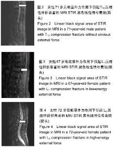
Relationship between a linear black signal area of STIR image in MRI of osteoporotic thoracolumbar fracture and the size of external force

Zhong Yuanming1, Luo Man2, Tang Fubo1, Tang Cheng3   

  1. 1The First Affiliated Hospital of Guangxi University of Chinese Medicine, Nanning 530001, Guangxi Zhuang Autonomous Region, China; 2Guangxi International Zhuang Medical Hospital, Nanning 530001, Guangxi Zhuang Autonomous Region, China; 3Liuzhou Traditional Chinese Medicine Hospital, Liuzhou 545001, Guangxi Zhuang Autonomous Region, China
  • Received:2017-11-20 Revised:2017-11-24 Accepted:2019-02-06 Online:2020-03-28 Published:2020-02-12
  • About author:Zhong Yuanming, Master, Doctoral supervisor, Chief physician, The First Affiliated Hospital of Guangxi University of Chinese Medicine, Nanning 530001, Guangxi Zhuang Autonomous Region, China

Abstract:

BACKGROUND: With the aging of the society, the number of patients with osteoporotic vertebral fracture is increasing, mainly manifesting compression fracture of thoracolumbar body, which seriously affects the daily life of the elderly. Therefore, to study the relationship between the degree of external force and the performance of osteoporotic thoracolumbar body fracture on MRI STIR is to provide a better basis for clinical diagnosis and treatment.

OBJECTIVE: To explore the relationship between the size of external force and a linear black signal area of STIR image in MRI of thoracic and lumbar osteoporosis vertebral compression fractures.

METHODS: The hospitalized patients, who were diagnosed as thoracic and lumbar osteoporosis vertebral compression fractures, were retrospectively analyzed from September 2013 to September 2016 at the Department of Spine Surgery of The First Affiliated Hospital of Guangxi University of Chinese Medicine. All cases in the three groups were diagnosed as osteoporosis by quantitative CT (bone mineral density ≤ 80 mg/cm3). All patients signed the informed consent. This study was approved by the Hospital Ethics Committee. The patients were divided into three groups according to the different trauma history: Non-obvious external force group (without apparent cause or external force), low energy group (sprains, bent down to lift heavy objects, and carrying heavy items), high energy group (flat road down hips touchdown, falls, and bruise). Gender, age, fracture site (thoracic lumbar segment and non-thoracic lumbar segment), the number of the vertebrae and the position where would they occur with a linear black signal area of STIR image in MRI were analyzed in each group. Age was analyzed by analysis of variance. Gender, fracture site and the number of the vertebrae and the position were analyzed by Pearson chi-square test.

RESULTS AND CONCLUSION: (1) All the 782 cases were included in the three groups. There were 334 in the non-obvious external force group, which a linear black signal area of STIR image in MRI existed in 114 cases. There were 186 cases in low energy group, which a linear black signal area of STIR image in MRI existed in 124 cases. There were 262 cases in high energy group, which a linear black signal area of STIR image in MRI existed in 87 cases. (2) The age, gender, fracture site and the number of the vertebrae and the position in three groups were not statistically significantly different among the three groups (P > 0.05). (3) There were significant differences in a linear black signal area of STIR image in MRI among the three groups (P < 0.05) (α´=0.05/3=0.017). Significant differences were found as compared the low energy group with non-obvious external force group and high energy group (P < 0.017). No significant difference was determined between non-obvious external force group and high energy group (P > 0.017). (4) The occurrence rate of linear black signal area of STIR image in MRI was 66.7% and higher than other groups (43.1% and 33.2%). (5) In the history of trauma, low energy in external force has more opportunity to cause a linear black signal area of STIR image in MRI than non-obvious external force and high energy; and they often occur in thoracic and lumbar osteoporosis vertebrae.

Key words: osteoporosis, thoracolumbar compression fracture, linear black signals of MRI STIR, external force

CLC Number: