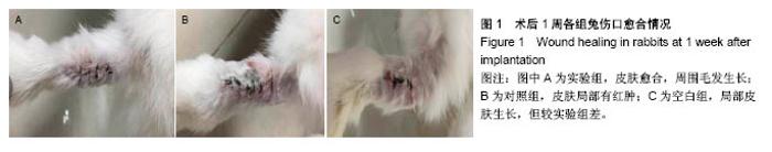
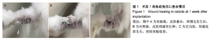
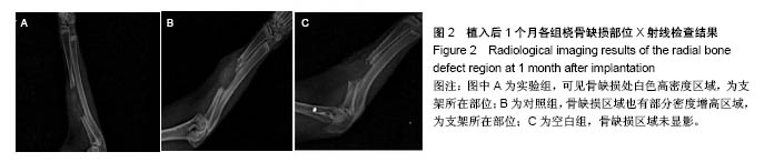
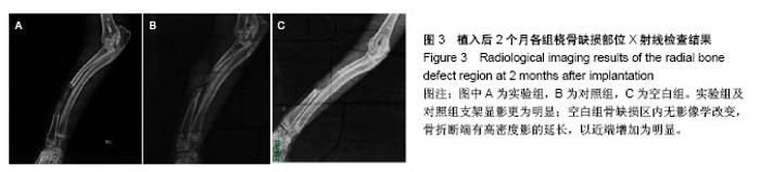
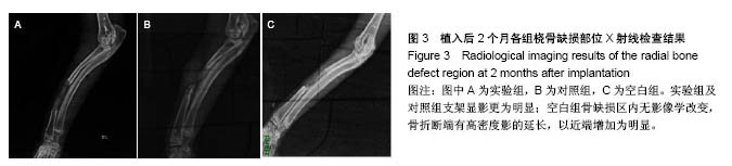
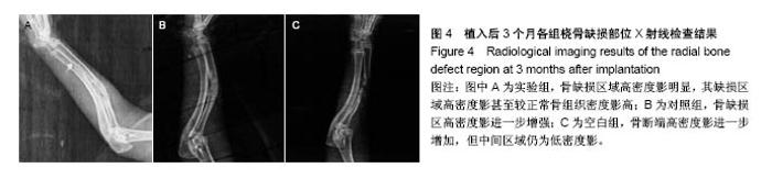
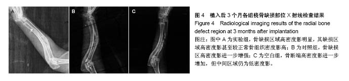
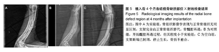
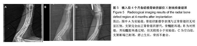
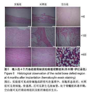
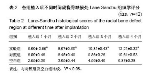
| [1] Jing ZW,Ma ZW,Li C,et al.Chitosan cross-linked with poly (ethylene glycol)dialdehyde via reductive amination as effective controlled release carriers for oral protein drug delivery.Bioorg Med Chem Lett. 2017;27(4): 1003-1006. [2] Braz L,Grenha A,Ferreira D,et al.Chitosan/sulfated locust bean gum nanoparticles: In vitro and in vivo evaluation towards an application in oral immunization.Int J Biol Macromol.2017;96:786-797. [3] Banerjee SL,Khamrai M,Sarkar K,et al. Modified chitosan encapsulated core-shell Ag Nps for superior antimicrobial and anticancer activity.Int J Biol Macromol.2016;85:157-167.[4] Song K,Li L,Li W,et al.Three-dimensional dynamic fabrication of engineered cartilage based on chitosan/gelatin hybrid hydrogel scaffold in a spinner flask with a special designed steel frame.Mater Sci Eng C Mater Biol Appl. 2015;55:384-392.[5] Aznar-Cervantes S,Martinez JG,Bernabeu-Esclapez A,et al.Fabrication of electrospun silk fibroin scaffolds coated with graphene oxide and reduced graphene for applications in biomedicine. Bioelectrochemistry. 2016;108:36-45.[6] Choudhury AJ,Gogoi D,Kandimalla R,et al.Penicillin impregnation on oxygen plasma surface functionalized chitosan/Antheraea assama silk fibroin: Studies of antibacterial activity and antithrombogenic property.Mater Sci Eng C Mater Biol Appl.2016;60:475-484.[7] Srisawasdi T,Petcharoen K,Tetteh G,et al. Electromechanical response of silk fibroin hydrogel and conductive polycarbazole/silk fibroin hydrogel composites as actuator material.Mater Sci Eng C Mater Biol Appl. 2015;56:1-8.[8] Kim SM,Jo JH,Lee SM,et al.Hydroxyapatite-coated magnesium implants with improved in vitro and in vivo biocorrosion, biocompatibility, and bone response.J Biomed Mater Res A.2014;102(2):429-441.[9] Mallik PK,Basu B.Better early osteogenesis of electroconductive hydroxyapatite-calcium titanate composites in a rabbit animal model.J Biomed Mater Res A. 2014;102(3):842-851. [10] Rabiee SM,Ravarian R,Mehmanchi M,et al.Effect of alumina on microstructure and compressive strength of a porous silicated hydroxyapatite.J Appl Biomater Funct Mater. 2014;12(2):102-106. [11] Yin P,Feng FF,Lei T,et al.Osteoblastic cell response on biphasic fluorhydroxyapatite/strontium-substituted hydroxyapatite coatings.J Biomed Mater Res A. 2014;102(3):621-627. [12] Deplaine H,Lebourg M,Ripalda P,et al.Biomimetic hydroxyapatite coating on pore walls improves osteointegration of poly(L-lactic acid) scaffolds.J Biomed Mater Res B Appl Biomater. 2013;101(1):173-186. [13] 蒋科,熊雁,余江,等.负载转化生长因子β3微球的壳聚糖三维支架的制备[J].第三军医大学学报,2013,35(10):988-991.[14] 赵建忠,张广程,狄东华,等.壳聚糖支架与BGC-823细胞的体外生物相容性研究[J].临床检验杂志,2013,31(11):856-858.[15] 康献刚,赵智远,吴旭芝,等.壳聚糖-同种异体骨粉复合多孔支架修复大鼠骨缺损的实验研究[J].中国修复重建外科杂志, 2016,30(3):298-302.[16] Inci I,Kirsebom H,Galaev IY,et al.Gelatin cryogels crosslinked with oxidized dextran and containing freshly formed hydroxyapatite as potential bone tissue-engineering scaffolds.J Tissue Eng Regen Med. 2013;7(7):584-588.[17] 王恺,马绍扬,张钧.利用装载内皮细胞生长因子的丝素纳米球制备具有生物活性的电纺支架材料[J].南开大学学报(自然科学版), 2016,49(6):80-85.[18] 张家庆.等离子体磺酸化丝素蛋白膜膨体聚四氟乙烯复合小口径人工血管的体内实验研究[D].南方医科大学,2016.[19] 曾超,朱美峰,徐宝山,等.丝素蛋白多孔支架与兔髓核细胞生物相容性研究[J].中国矫形外科杂志,2013,21(23):2402-2407.[20] Xing ZC,Han SJ,Shin YS,et al.Enhanced osteoblast responses to poly(methyl methacrylate)/hydroxyapatite electrospun nanocomposites for bone tissue engineering.J Biomater Sci Polym Ed.2013;24(1):61-76.[21] 李斯,胡晓文.纳米羟基磷灰石/明胶仿生复合材料的制备及其细胞相容性[J].中南大学学报(医学版),2014,39(9):949-958.[22] Lambert F,Lecloux G,Léonard A,et al.Bone regeneration using porous titanium particles versus bovine hydroxyapatite: a sinus lift study in rabbits.Clin Implant Dent Relat Res. 2013;15(3):412-426.[23] 王大平,熊建义,朱伟民,等.纳米羟基磷灰石人工骨在骨缺损修复中的临床应用研究[J].中国临床解剖学杂志, 2014,32(2):206-209.[24] 荣子杰,杨联军,张赞杰,等.载ADM-PLGA微球的纳米羟基磷灰石/胶原支架修复兔骨缺损的实验研究[J]. 实用医学杂志, 2014,30(22):3559-3562.[25] Khajuria DK,Zahra SF,Razdan R.Effect of locally administered novel biodegradable chitosan based risedronate/ zinc-hydroxyapatite intra-pocket dental film on alveolar bone density in rat model of periodontitis.J Biomater Sci Polym Ed. 2018;29(1):74-91. [26] 李海建,龙志成,张峥,等. HA/β-TCA复合材料与同种异体骨体外兔脂肪干细胞生物相容性的对比实验[J]. 新疆医科大学学报, 2015,38(8):949-953.[27] 金光辉,张馨雯,孙晓飞,等.组织工程化纳米羟基磷灰石/聚己内酯人工骨支架修复兔桡骨大段骨缺损的实验研究[J].中华损伤与修复杂志(电子版), 2015,10(1):43-49.[28] 朱文赫,钟秀宏,张巍,等.鹿茸多肽-胶原蛋白/壳聚糖复合材料对家兔下颌骨缺损愈合的促进作用及其机制[J].吉林大学学报(医学版), 2017,43(3): 527-531.[29] Nazeer MA,Yilgör E,Yilgör I.Intercalated chitosan/ hydroxyapatite nanocomposites: Promising materials for bone tissue engineering applications.Carbohydr Polym. 2017;175:38-46. [30] Przekora A,Ginalska G.Chitosan/β-1,3-glucan/hydroxyapatite bone scaffold enhances osteogenic differentiation through TNF-α-mediated mechanism.Mater Sci Eng C Mater Biol Appl.2017;73:225-233. |