Chinese Journal of Tissue Engineering Research ›› 2017, Vol. 21 ›› Issue (16): 2600-2605.doi: 10.3969/j.issn.2095-4344.2017.16.024
Previous Articles Next Articles
Tissue-engineered skin substitutes: a prospect evaluation from the aspects of morphology and function
Wang Xiao-jing1, Wang Guo-wei2, Hui Guang-yan1, Zhao Yi-min3
- 1Department of Stomatology, Qingdao First Sanatorium of PLA Navy, Qingdao 266071, Shandong Province, China; 2Department of Stomatology, Laoshan Branch of No. 401 Hospital of PLA Navy, Qingdao 266061, Shandong Province, China; 3Stomatological Hospital of the Fourth Military Medical University, Xi’an 710032 , Shaanxi Province, China
-
Revised:2017-05-08Online:2017-06-08Published:2017-07-06 -
Contact:Wang Xiao-jing, Department of Stomatology, Qingdao First Sanatorium of PLA Navy, Qingdao 266071, Shandong Province, China -
About author:Wang Xiao-jing, M.D., Attending physician, Department of Stomatology, Qingdao First Sanatorium of PLA Navy, Qingdao 266071, Shandong Province, China -
Supported by:the National Natural Science Foundation of China, No. 81271104
CLC Number:
Cite this article
Wang Xiao-jing, Wang Guo-wei, Hui Guang-yan, Zhao Yi-min. Tissue-engineered skin substitutes: a prospect evaluation from the aspects of morphology and function[J]. Chinese Journal of Tissue Engineering Research, 2017, 21(16): 2600-2605.
share this article
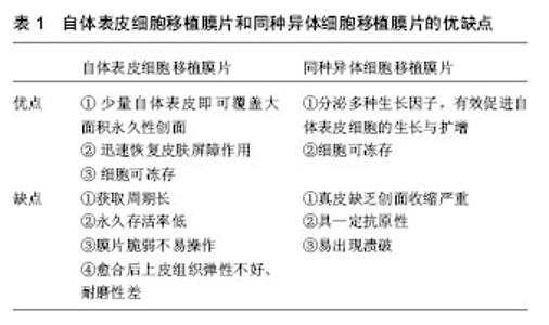
2.1 表皮替代物 主要分为:自体表皮细胞移植膜片、同种异体细胞移植膜片以及其他表皮细胞移植膜片。培养的自体表皮移植物首先从供者获取皮肤,培养角化细胞,而后在实验室操作来生成大片的可移植表皮,此过程约需3周来获取大片可用于永久覆盖受区的移植皮片。然而,这种表皮移植物十分脆弱,由于缺乏真皮部分而容易起泡。培养异体表皮移植物对于即刻使用很有帮助。由于是来源为同种异体的,不存在延迟稳定性,也不存在移植排斥反应。供者细胞会逐渐被患者自身的细胞所替代。因此,这些移植物作为临时的覆盖和刺激因素作用在急慢性伤口区。应当注意这些移植物较薄且受限于其内在的脆弱性。 1975年,Rheinwald等将人表皮角质形成细胞体外培养在经放射处理的鼠成纤维细胞滋养层上获得成功。1979年,Green等首次培养出自体表皮膜片(cultured epithelial autograft,CEA)并将此项技术应用于大面积烧伤患者的创面治疗。自此,美国Genzyme Tissue Repair公司于1981年成功生产出自体表皮膜片(Epicel)。 表1中将自体表皮细胞移植膜片和同种异体细胞移植膜片的优缺点进行了比较。其他表皮细胞移植膜片的研究工作中包含自体与异体表皮细胞混合移植,表皮干细胞移植及骨髓间充质干细胞移植等。 目前,自体表皮细胞移植临床上多用于治疗大面积烧伤患者,国外有学者报道还可用于白癜风,慢性乳突炎及巨大型痣切除的皮肤创面。同种异体表皮细胞移植膜片临床多用于大面积烧伤,大疱性表皮松解症以及慢性静脉溃疡等的治疗[11-12]。"

| [1] Pickerill HP. On the possibility of establishing skin banks. Br J Phst Surg. 1951;(4):157-165.[2] Reinwald JG, Green H. Serial cultivation of strains of human epidermal keratinocytes: the formation of keratinizing colonies from single cells. Cell. 1975;6(3):331-343.[3] Vig K, Chaudhari A, Tripathi S, et al. Advances in Skin Regeneration Using Tissue Engineering. Int J Mol Sci. 2017. doi: 10.3390/ijms18040789.[4] Marino D, Reichmann E, Meuli M. Skingineering. Eur J Pediatr Surg. 2014;24(3):205-213. [5] Chua AW, Khoo YC, Tan BK, et al. Skin tissue engineering advances in severe burns: review and therapeutic applications. Burns Trauma. 2016; 4:3. [6] Bello YM, Falabella AF. Use of skin substitutes in dermatology. Dermatol Clin. 2001;19:555-561.[7] Nyame TT, Chiang HA, Leavitt T, et al. Tissue-Engineered Skin Substitutes. Plast Reconstr Surg. 2015;136(6): 1379-1388.[8] Varkey M, Ding J, Tredget EE. Advances in Skin Substitutes-Potential of Tissue Engineered Skin for Facilitating Anti-Fibrotic Healing. J Funct Biomater. 2015; 6(3):547-563. [9] Markeson D, Pleat JM, Sharpe JR, et al. Scarring, stem cells, scaffolds and skin repair. J Tissue Eng Regen Med. 2015; 9(6):649-668. [10] Catalano E, Cochis A, Varoni E, et al. Tissue-engineered skin substitutes: an overview. J Artif Organs. 2013;16(4):397-403. [11] Kitala D, Kawecki M, Klama-Bary?a A, et al. Autologous skin grafts in the therapy of patients with burn injuries: a restrospective, open-label clinical study with pair matching. Adv Clin Exp Med. 2016;25(5):923-929. [12] Nyame TT, Chiang HA, Orgill DP. Clinical applications of skin substitutes. Surg Clin North Am. 2014;94(4):839-850. [13] Límová M. Active wound coverings: bioengineered skin and dermal substitutes. Surg Clin North Am. 2010;90(6): 1237-1255.[14] Van Der Veen VC, van der Wal MBA, van Leuwen MCE, et al. Biological background of dermal substitutes. Burns. 2010;36: 305-321.[15] Murray RC, Gordin EA, Saigal K, et al. Reconstruction of the radial forearm free flap donor site using Integra artificial dermis. Microsurgery. 2011;31:104-108.[16] Nguyen DQ, Potokar TS, Price P. An objective long-term evaluation of Integra (a dermal skin substitute) and split thickness skin grafts, in acute burns and reconstructive surgery. Burns. 2010;36:23-28.[17] Rnjak J, Wise SG, Mithieux SM, et al. Severe burn injuries and the role of elastin in the design of dermal substitutes. Tissue Eng. 2010;17(2):81-93.[18] Haslik W, Kamolz LP, Manna F et al. Management of full-thickness skin defects in the hand and wrist region: first long-term experiences with the dermal matrix matriderm. J Plast Reconstr Aesthet Surg. 2010;63:360-364.[19] Halim AS, Khoo TL, Mohd Yussof SJ. Biologic and synthetic skin substitutes: an overview. Indian J Plast Surg. 2010;43: S23-S28. [20] Varkey M, Ding J, Tredget EE. Advances in Skin Substitutes-Potential of Tissue Engineered Skin for Facilitating Anti-Fibrotic Healing. J Funct Biomater. 2015:6(3): 547-563. [21] Watt FM,Hogan BL. Out of Eden:Stem cells and their niches. Science. 2000:25;287(5457):1427-1430.[22] Nishimura E, Siobhan A, Oshima JH, et al. Dominant role of the niche in melanoeyte stem-cell fate determination. Nature. 2002;25;416(6883):854-860.[23] Xie T, Spradling AC. A niche maintaining germ line stem cells in the drosophila ovary. Science. 2000;13;290(5490): 328-330.[24] Ideta R, Soma T, Tsunenaga M, et al. Cultured human dermal papilla cells secrete a chemotactic factor for melanoeytes. J Dermatol Sci. 2002;28(1):48-59.[25] Gragnani A, Morgan JR, Ferreira LM. Differentiation and barrier formation of a cultured composite skin graft. J Burn Care Rehabil. 2002;23(2):126-131.[26] Wencel A, Zakrzewska KE, Samluk A, et al. Dried human skin fibroblasts as a new substratum for functional culture of hepatic cells. Acta Biochim Pol. 2017;64(2):357-363.[27] Koch M, Ehrenreich T, Koehl G, et al. Do cell based Tissue Engineering products for meniscus regeneration influence vascularization? Clin Hemorheol Microcirc. 2017. doi: 10.3233/CH-17085. [28] Wang JP, Zhou XL, Yan JP, et al. Nanobubbles as ultrasound contrast agent for facilitating small cell lung cancer imaging. Oncotarget. 2017. doi: 10.18632/oncotarget.18155.[29] Abaci HE, Guo Z, Doucet Y, et al. Next generation human skin constructs as advanced tools for drug development. Exp Biol Med (Maywood). 2017. doi: 10.1177/1535370217712690.[30] Pielesz A, Binia? D, Bobiński R, et al. The role of topically applied l-ascorbic acid in ex-vivo examination of burn-injured human skin. Spectrochim Acta A Mol Biomol Spectrosc. 2017. doi: 10.1016/j.saa.2017.05.055.[31] Modena DAO, da Silva CN Pt, Grecco C, et al. Extracorporeal shockwave: mechanisms of action and physiological aspects for cellulite, body shaping and localized fat - systematic review. J Cosmet Laser Ther. 2017. doi: 10.1080/14764172.2017. 1334928. [32] Kühbacher A, Burger-Kentischer A, Rupp S. Interaction of Candida Species with the Skin.Microorganisms. 2017. doi: 10.3390/microorganisms5020032. [33] Khan TK, Wender PA, Alkon DL. Bryostatin and its synthetic analog, picolog rescue dermal fibroblasts from prolonged stress and contribute to survival and rejuvenation of human skin equivalents. J Cell Physiol. 2017. doi: 10.1002/jcp.26043. [34] Szlavicz E, Szabo K, Groma G, et al. Splicing factors differentially expressed in psoriasis alter mRNA maturation of disease-associated EDA+ fibronectin. Mol Cell Biochem. 2017. doi: 10.1007/s11010-017-3090-1.[35] Kim BS, Lee JS, Gao G, et al. Direct 3D cell-printing of human skin with functional transwell system. Biofabrication. 2017; 9(2):025034. [36] Desmet E, Ramadhas A, Lambert J, et al. In vitro psoriasis models with focus on reconstructed skin models as promising tools in psoriasis research. Exp Biol Med (Maywood). 2017. doi: 10.1177/1535370217710637.[37] Henderson F, Alho R, Riches P, et al. Assessment of knee alignment with varus and valgus force through the range of flexion with non-invasive navigation. J Med Eng Technol. 2017. doi: 10.1080/03091902.2017.1333164.[38] Hu B, Leow WR, Amini S, et al. Orientational Coupling Locally Orchestrates a Cell Migration Pattern for Re-Epithelialization. Adv Mater. 2017. doi: 10.1002/adma.201700145. [39] Cai M, Shen S, Li H. The effect of facial expressions on respirators contact pressures. Comput Methods Biomech Biomed Engin. 2017. doi: 10.1080/10255842.2017.1336549. [40] Wang XR, Gao SQ, Niu XQ, et al. Capsaicin-loaded nanolipoidal carriers for topical application: design, characterization, and in vitro/in vivo evaluation. Int J Nanomedicine. 2017. doi: 10.2147/IJN.S131901.[41] Ma J, Chen B, Zhang Y, et al. Multiple laser pulses in conjunction with an optical clearing agent to improve the curative effect of cutaneous vascular lesions. Lasers Med Sci. 2017. doi: 10.1007/s10103-017-2244-4. [42] Strong AL, Neumeister MW, Levi B. Stem Cells and Tissue Engineering: regeneration of the Skin and Its Contents. Clin Plast Surg. 2017. doi: 10.1016/j.cps.2017.02.020. [43] Carvalho IC, Mansur HS. Engineered 3D-scaffolds of photocrosslinked chitosan-gelatin hydrogel hybrids for chronic wound dressings and regeneration. Mater Sci Eng C Mater Biol Appl. 2017. doi: 10.1016/j.msec.2017.04.126.[44] Beiki B, Zeynali B, Seyedjafari E. Fabrication of a three dimensional spongy scaffold using human Wharton's jelly derived extra cellular matrix for wound healing. Mater Sci Eng C Mater Biol Appl. 2017. doi: 10.1016/j.msec.2017.04.074. [45] Tan J, Zhao C, Zhou J, et al. Co-culturing epidermal keratinocytes and dermal fibroblasts on nano-structured titanium surfaces. Mater Sci Eng C Mater Biol Appl. 2017. doi: 10.1016/j.msec.2017.04.036. [46] Mostmans Y, Cutolo M, Giddelo C, et al. The role of endothelial cells in the vasculopathy of systemic sclerosis: A systematic review: SSc vasculopathy: The role of endothelial cells. Autoimmun Rev. 2017. doi: 10.1016/j.autrev.2017.05.024.[47] Chen X, Zhang M, Wang X, et al. Peptide-modified chitosan hydrogels promote skin wound healing by enhancing wound angiogenesis and inhibiting inflammation. Am J Transl Res. 2017;9(5):2352-2362. [48] Lee MJ, Hung SH, Huang MC, et al. Doxycycline potentiates antitumor effect of 5-aminolevulinic acid-mediated photodynamic therapy in malignant peripheral nerve sheath tumor cells. PLoS One. 2017. doi: 10.1371/journal.pone.0178493.[49] Cortese FAB, Aguiar S, Santostasi G. Induced Cell Turnover: a novel therapeutic modality for in situ tissue regeneration. Hum Gene Ther. 2017. doi: 10.1089/hum.2016.167. [50] Lotz C, Schmid FF, Oechsle E, et al. Cross-linked Collagen Hydrogel Matrix Resisting Contraction To Facilitate Full-Thickness Skin Equivalents. ACS Appl Mater Interfaces. 2017. doi: 10.1021/acsami.7b04017. [51] Mohafez H, Ahmad SA, Hadizadeh M, et al. Quantitative assessment of wound healing using high-frequency ultrasound image analysis. Skin Res Technol. 2017. doi: 10.1111/srt.12388.[52] Bhardwaj N, Chouhan D, Mandal BB. Tissue engineered skin and wound healing: current strategies and future directions. Curr Pharm Des. 2017. doi: 10.2174/1381612823666170526094606. [53] von Byern J, Mebs D, Heiss E, et al. Salamanders on the bench - A biocompatibility study of salamander skin secretions in cell cultures. Toxicon. 2017. doi: 10.1016/j.toxicon.2017.05.021. [54] Bonnet V, Richard V, Camomilla V, et al. Joint kinematics estimation using a multi-body kinematics optimisation and an extended Kalman filter, and embedding a soft tissue artefact model. J Biomech. 2017. doi: 10.1016/j.jbiomech.2017.04.033. [55] Aurand AM, Dufour JS, Marras WS. Accuracy map of an optical motion capture system with 42 or 21 cameras in a large measurement volume. J Biomech. 2017. doi: 10.1016/j.jbiomech.2017.05.006. [56] Fong CJ, Garzon MC, Hoi JW, et al. Assessment of Infantile Hemangiomas Using a Handheld Wireless Diffuse Optical Spectroscopic Device. Pediatr Dermatol. 2017. doi: 10.1111/pde.13150. [57] van den Broek LJ, Bergers LIJC, Reijnders CMA, et al. Progress and Future Prospectives in Skin-on-Chip Development with Emphasis on the use of Different Cell Types and Technical Challenges. Stem Cell Rev. 2017. doi: 10.1007/s12015-017-9737-1.[58] Sasaki Y, Sathi GA, Yamamoto O. Wound healing effect of bioactive ion released from Mg-smectite. Mater Sci Eng C Mater Biol Appl. 2017. doi: 10.1016/j.msec.2017.03.236.[59] Levengood SL, Erickson AE, Chang FC, et al. Chitosan-Poly(caprolactone) Nanofibers for Skin Repair. J Mater Chem B Mater Biol Med. 2017. doi: 10.1039/C6TB03223K. [60] Wang H, Dong XX, Yang JC, et al. Finite element method simulating temperature distribution in skin induced by 980-nm pulsed laser based on pain stimulation. Lasers Med Sci. 2017. doi: 10.1007/s10103-017-2223-9. [61] Scalise A, Torresetti M, Verdini F, et al. Acellular dermal matrix and heel reconstruction: a new prospective. J Appl Biomater Funct Mater. 2017. doi: 10.5301/jabfm.5000357. [62] Yu K, Lu F, Li Q, et al. In situ assembly of Ag nanoparticles (AgNPs) on porous silkworm cocoon-based would film: enhanced antimicrobial and wound healing activity. Sci Rep. 2017 May 18;7(1):2107. doi: 10.1038/s41598-017-02270-6.[63] Motamed S, Taghiabadi E, Molaei H, et al. Cell-based skin substitutes accelerate regeneration of extensive burn wounds in rats. Am J Surg. 2017. doi: 10.1016/j.amjsurg.2017.04.010. [64] Chang CJ, Yu DY, Hsiao YC, et al. Noninvasive imaging analysis of biological tissue associated with laser thermal injury. Biomed J. 2017. doi: 10.1016/j.bj.2016.10.004.[65] Chen S, Liu B, Carlson MA, et al. Recent advances in electrospun nanofibers for wound healing. Nanomedicine (Lond). 2017. doi: 10.2217/nnm-2017-0017. |
| [1] | Yao Xiaoling, Peng Jiancheng, Xu Yuerong, Yang Zhidong, Zhang Shuncong. Variable-angle zero-notch anterior interbody fusion system in the treatment of cervical spondylotic myelopathy: 30-month follow-up [J]. Chinese Journal of Tissue Engineering Research, 2022, 26(9): 1377-1382. |
| [2] | Zhou Ying, Zhang Huan, Liao Song, Hu Fanqi, Yi Jing, Liu Yubin, Jin Jide. Immunomodulatory effects of deferoxamine and interferon gamma on human dental pulp stem cells [J]. Chinese Journal of Tissue Engineering Research, 2022, 26(7): 1012-1019. |
| [3] | Zhang Jinglin, Leng Min, Zhu Boheng, Wang Hong. Mechanism and application of stem cell-derived exosomes in promoting diabetic wound healing [J]. Chinese Journal of Tissue Engineering Research, 2022, 26(7): 1113-1118. |
| [4] | An Weizheng, He Xiao, Ren Shuai, Liu Jianyu. Potential of muscle-derived stem cells in peripheral nerve regeneration [J]. Chinese Journal of Tissue Engineering Research, 2022, 26(7): 1130-1136. |
| [5] | He Yunying, Li Lingjie, Zhang Shuqi, Li Yuzhou, Yang Sheng, Ji Ping. Method of constructing cell spheroids based on agarose and polyacrylic molds [J]. Chinese Journal of Tissue Engineering Research, 2022, 26(4): 553-559. |
| [6] | He Guanyu, Xu Baoshan, Du Lilong, Zhang Tongxing, Huo Zhenxin, Shen Li. Biomimetic orientated microchannel annulus fibrosus scaffold constructed by silk fibroin [J]. Chinese Journal of Tissue Engineering Research, 2022, 26(4): 560-566. |
| [7] | Chen Xiaoxu, Luo Yaxin, Bi Haoran, Yang Kun. Preparation and application of acellular scaffold in tissue engineering and regenerative medicine [J]. Chinese Journal of Tissue Engineering Research, 2022, 26(4): 591-596. |
| [8] | Kang Kunlong, Wang Xintao. Research hotspot of biological scaffold materials promoting osteogenic differentiation of bone marrow mesenchymal stem cells [J]. Chinese Journal of Tissue Engineering Research, 2022, 26(4): 597-603. |
| [9] | Shen Jiahua, Fu Yong. Application of graphene-based nanomaterials in stem cells [J]. Chinese Journal of Tissue Engineering Research, 2022, 26(4): 604-609. |
| [10] | Zhang Tong, Cai Jinchi, Yuan Zhifa, Zhao Haiyan, Han Xingwen, Wang Wenji. Hyaluronic acid-based composite hydrogel in cartilage injury caused by osteoarthritis: application and mechanism [J]. Chinese Journal of Tissue Engineering Research, 2022, 26(4): 617-625. |
| [11] | Li Hui, Chen Lianglong. Application and characteristics of bone graft materials in the treatment of spinal tuberculosis [J]. Chinese Journal of Tissue Engineering Research, 2022, 26(4): 626-630. |
| [12] | Gao Cangjian, Yang Zhen, Liu Shuyun, Li Hao, Fu Liwei, Zhao Tianyuan, Chen Wei, Liao Zhiyao, Li Pinxue, Sui Xiang, Guo Quanyi. Electrospinning for rotator cuff repair [J]. Chinese Journal of Tissue Engineering Research, 2022, 26(4): 637-642. |
| [13] | Guan Jian, Jia Yanfei, Zhang Baoxin , Zhao Guozhong. Application of 4D bioprinting in tissue engineering [J]. Chinese Journal of Tissue Engineering Research, 2022, 26(3): 446-455. |
| [14] | Maihemuti•Yakufu, Sun Qinqin, Chen Hongtao, Liu Xu, Yiliyaer•Abudusimu, Abudushalamu•Abudukelimu, Liu Jianjiang. Tension of 3D printed controllable tension band and skin regeneration of skin defect model rats [J]. Chinese Journal of Tissue Engineering Research, 2022, 26(3): 371-375. |
| [15] | Liu Jiali, Suo Hairui, Yang Han, Wang Ling, Xu Mingen. Influence of lay-down angles on mechanical properties of three-dimensional printed polycaprolactone scaffolds [J]. Chinese Journal of Tissue Engineering Research, 2022, 10(16): 2612-2617. |
| Viewed | ||||||
|
Full text |
|
|||||
|
Abstract |
|
|||||