Chinese Journal of Tissue Engineering Research ›› 2017, Vol. 21 ›› Issue (14): 2163-2169.doi: 10.3969/j.issn.2095-4344.2017.14.006
Previous Articles Next Articles
Titanium intramedullary nail coated with vancomycin-hydroxyapatite in a model of open long bone fracture with wound infection
Wang Yong, Wan Yong-xian, Zhang Xi-hai, Ye Jun-wu, Zhuo Nai-qiang
- Department of Bone and Joint Surgery, Affiliated Hospital of Southwest Medical University, Luzhou 646000, Sichuan Province, China
-
Received:2016-12-22Online:2017-05-18Published:2017-06-10 -
Contact:Zhuo Nai-qiang, Professor, Department of Bone and Joint Surgery, Affiliated Hospital of Southwest Medical University, Luzhou 646000, Sichuan Province, China -
About author:Wang Yong, Master, Physician, Department of Bone and Joint Surgery, Affiliated Hospital of Southwest Medical University, Luzhou 646000, Sichuan Province, China -
Supported by:the Project of Luzhou Science and Technology Bureau, No. 2011-S-39(3/6)
CLC Number:
Cite this article
Wang Yong, Wan Yong-xian, Zhang Xi-hai, Ye Jun-wu, Zhuo Nai-qiang. Titanium intramedullary nail coated with vancomycin-hydroxyapatite in a model of open long bone fracture with wound infection[J]. Chinese Journal of Tissue Engineering Research, 2017, 21(14): 2163-2169.
share this article
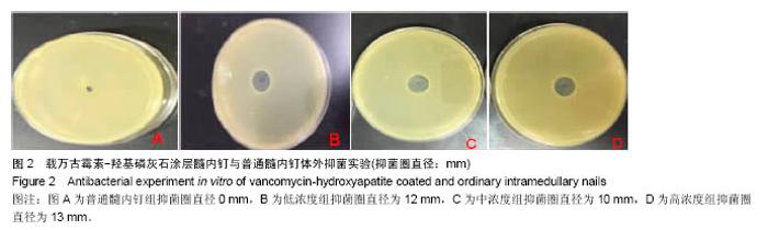
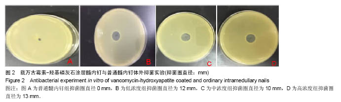
2.1 实验动物数量分析 实验选用新西兰大白兔40只,分为4组,实验过程中无脱失,全部进入结果分析。 2.2 载万古霉素-羟基磷灰石涂层髓内钉体外抑菌实验 将实验制备的髓内钉与普通髓内钉插入细菌培养皿中, 24 h后测量抑菌圈大小(图2)。其中普通髓内钉周围24 h内无抑菌圈的产生,低浓度组抑菌圈直径为12 mm,中浓度组抑菌圈直径为10 mm,高浓度组抑菌圈直径为13 mm。由此表明,实验所制备的载万古霉素-羟基磷灰石涂层髓内钉与普通髓内钉相比具有明显的抑菌性能,而3种不同抗生素浓度之间的体外抑菌性能无明显差异。"
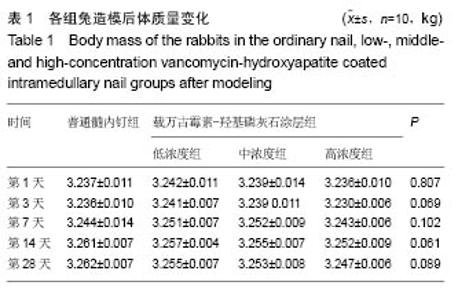
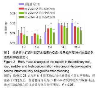
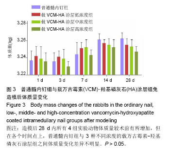
2.3 载万古霉素-羟基磷灰石涂层髓内钉对实验家兔造模后体质量的影响 造模后28 d内所有实验兔体质量较术前有所增加,但普通髓内钉组及低、中、高浓度抗生素涂层组在造模后第1,3,7,14,28天内各时间点上4组间体质量差异无显著性意义(P > 0.05),见表1,图3。"

"
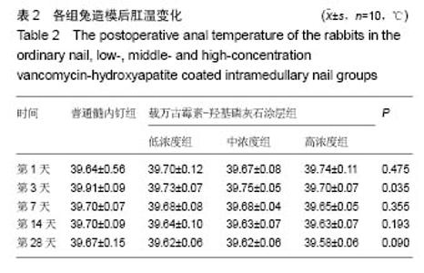
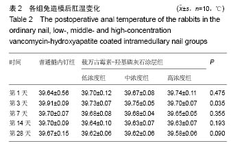
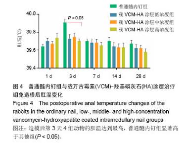
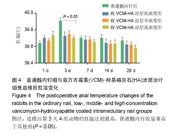
2.4 载万古霉素-羟基磷灰石涂层髓内钉对实验家兔造模后肛温的影响 普通髓内钉组及低、中、高浓度抗生素涂层组家兔造模后第1天肛温开始升高,其中造模后第3天4组动物肛温达到最高,且普通髓内钉组肛温升高比低、中、高、浓度组明显,差异有显著性意义(P < 0.05)。而后体温开始降低,其余各个时间点上普通髓内钉组肛温较低、中、高浓度组高,但其组间差异无显著性意义(P > 0.05),见表2,图4)。 "
"
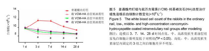
2.5 载万古霉素-羟基磷灰石涂层髓内钉对实验家兔造模后白细胞的影响 使用普通髓内钉固定的实验动物造模后第1,3,7,14,28天其体内白细胞计数均升高,低、中、高浓度的载万古霉素-羟基磷灰石涂层髓内钉固定组兔于造模后第1天内升高,第3,7,14,28天后逐渐降低。其中第1天普通髓内钉组和低、中、高浓度抗生素涂层组白细胞升高)其差异无显著性意义(P > 0.05),造模后3,7,14,28 d时间点低、中、高浓度抗生素涂层组家兔白细胞计数明显低于对照组,差异有显著性意义(P < 0.05)。说明载万古霉素-羟基磷灰石涂层髓内钉在开放性骨折治疗早期能有效降低血液中白细胞计数,有利于早期控制炎症的发生(图5)。"

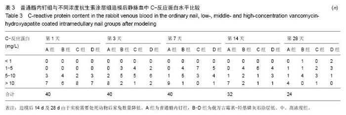
2.6 载万古霉素-羟基磷灰石涂层髓内钉对实验家兔造模后C-反应蛋白的影响 普通髓内钉固定组造模后第3,7,14,28天静脉血中C-反应蛋白含量大于10 mg/L的家兔数量明显高于低、中、高不同浓度抗生素涂层组,造模后第1天兔静脉血中C-反应蛋白含量大于10 mg/L的家兔数量4组间无明显区别。而低、中、高不同浓度抗生素涂层组在造模后第1,3,7,14,28天其兔静脉血中C-反应蛋白含量大于10 mg/L的家兔数量无明显差异。见表3。"
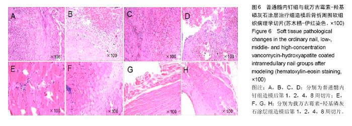
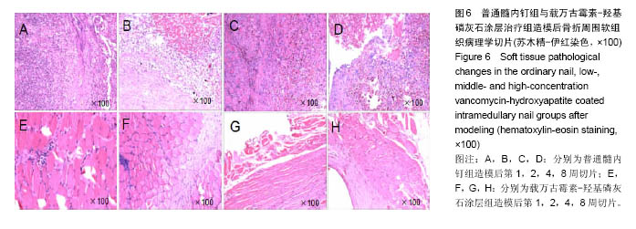
2.7 骨折周围软组织病理学检查结果 普通髓内钉组感染兔于第1周病理检查可见局部大量纤维组织渗出、淋巴细胞及中性粒细胞浸润、血管壁充血扩张明显。第2周时可见局部坏死,组织结构轮廓消失。第4周时可见巨噬细胞浸润,局部炎性反应性增生,呈慢性炎症表现。第8周可见局部炎性增生,纤维瘢痕。而载万古霉素-羟基磷灰石涂层组在各个时间点与普通髓内钉组相比,可见少量中性粒细胞、淋巴细胞浸润,周围组织结构清晰,未见明显坏死及脓腔等形成。表明载万古霉素-羟基磷灰石涂层髓内钉组骨折周围软组织早期炎症反应明显低于普通髓内钉组。见图6。"
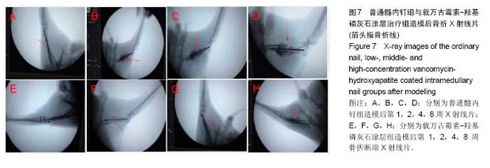
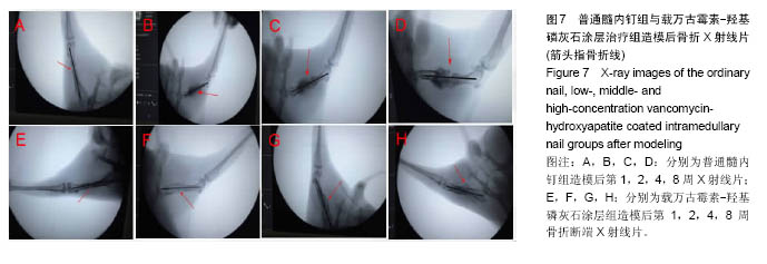
2.8 普通髓内钉组与载万古霉素-羟基磷灰石涂层髓内钉组造模后伤肢X射线片检查 造模后第1周股骨X射线片可见普通髓内钉组骨折断端稳定,周围软组织影,肿胀明显,骨折线清晰,未见明显骨痂生长;载万古霉素-羟基磷灰石涂层组可见骨折对位对线良好,周围软组织肿胀不明显,骨折线清晰,周围未见明显骨痂。造模后第2周可见普通髓内钉组骨折线清晰,周围可见团状组织显影,骨折周围未见明显骨痂生长;载万古霉素-羟基磷灰石涂层组骨折线稍模糊,局部可见骨痂生长。造模后第4周可见普通髓内钉组骨折线稍模糊,而骨痂生长缓慢;载万古霉素-羟基磷灰石涂层组骨折线模糊,骨痂生长明显好于普通髓内钉组。造模后第8周X射线片可见普通髓内钉组骨折线模糊,可见少量骨痂生长,周围组织团块影明显,而载万古霉素-羟基磷灰石涂层组骨折线消失,周围见大量骨痂生成,骨折骨性愈合。见图7。"
| [1]Grote S, Polzer H, Prall WC, et al. [Prevention of infection in the current treatment of open fractures: an evidence-based systematic analysis. Orthopade. 2012;41(1): 32-42.[2]Court-Brown CM, Rimmer S, Prakash U, et al.The epidemiology of open long bonefractures.Injury.1998;29(7): 529-534. [3]Pavoni GL, Giannella M, Falcone M, et al. Conservative medical therapy of prosthetic joint infections: retrospective analysis of an 8-year experience. Clin Microbiol Infect. 2004; 10(9): 831-837.[4]Lew DP, Waldvogel FA. Osteomyelitis. Lancet. 2004; 364 (9431): 369-379.[5]Patzakis MJ, Wilkins J. Factors influencing infection rate in open fracture wounds. Clin Orthop Relat Res.1989; 246:36Y40.[6]Smirnov AY, Savel'ev SE, Nori F.Diffusion-controlled generation of a proton-motive force across a biomembrane. Phys Rev E Stat Nonlin Soft Matter Phys. 2009;80(1 Pt 1): 011916.[7]Jorge LS, Chueire AG, Rossit AR. Osteomyelitis: a current hallenge.Braz J Infect Dis.2010;14(3):310-315.[8]Calhoun JH, Manring MM, Shirtliff M. Osteomyelitis of the long bones.Semin Plast Surg.2009;23(2):59-72.[9]Saveli CC, Morgan SJ, Belknap RW, et al.Prophylactic antibiotics in open fractures: a pilot randomized clinical safety study. J Orthop Trauma. 2013;27:552-557.[10]唐良华.载万古霉素-PDLLA钢板与-HA钢板的研制及其机制探讨[D].第三军医大学,2009.[11]Zelle BA, Boni G. Safe surgical technique: intramedullary nail fixation of tibial shaft fractures.Patient Saf Surg.2015’9: 40.[12]Elamurugan TP, Jagdish S, Kate V. Chandra Parija S. Role of bone biopsy spec-imen culture in the management of diabetic foot osteomyelitis.Int J Surg 2011;9:214-216. [13]Hofmann A, Dietz SO, Pairon P, et al. The role of intramedullary nailing in treatment of open fractures.Eur J Trauma Emerg Surg.2015;41(1):39-47.[14]Singh D, Garg R, Bassi JL, Tripathi SK Open grade III fractures of tibia: outcome after early unreamed intramedullary nailing. Eur J Orthop Surg Traumatol 2011; 21:321-325[15]Craig J, Fuchs T, Jenks M, et al. Systematic review and meta-analysis of the additional benefit of local prophylactic antibiotic therapy for infection rates in open tibia fractures treated with intramedullary nailing.Int Orthop.2014;;38(5): 1025-1030.[16]Phaff M, Aird J, Rollinson PD. Delayed implants sepsis in HIV-positive patients following open fractures treated with orthopaedic implants.Injury 2015;46:590-594.[17]Mérens A, Rapp C, Delaune D,et al. Prevention of com-bat-related infections: Antimicrobial therapy in battlefield and barrier mea-sures in French military medical treatment facilities. Travel Med Infect Dis.2014;12:318-329.[18]Otchwemah R,Grams V,Tjardes T,et al.Bacterial contamination of open fractures- pathogens, antibiotic resistances and therapeuti -c regimes in four hospitals of the trauma network Cologne, Germany.Injury. 2015. 46 Suppl 4: S104-108.[19]张春,张俊,吕战虎,等.股骨骨折患者术后医院感染病原菌耐药特征与相关危险因素研究[J].中华医院感染学杂志,2016,26(2): 379-381. [20]王彤,王立新,白雪,等. 2014年骨科伤口感染患者病原菌分布耐药性分析[J].国际检验医学杂志,2015,36 (20):2989-2991.[21]Dunkel N, Pittet D, Tovmirzaeva L, et al. Short duration of antibiotic prophylaxis in open fractures does notenhance risk of subsequent infection. Bone Joint J.2013;95(6):831Y837.[22]Von EC, Proctor RA, Peters G. Coagulase-negative staphylococci. Pathogens have major role in nosocomial infections.Postgrad Med. 2001;110(4): 63-64, 69-70, 73-76.[23]Nanchahal J. Reply:Soft-tissue reconstruction of open fractures of the lower limb: muscle versus fasciocutaneous flaps. Plast Reconstr Surg. 2013; 131(3): 448e-9e.[24]Mounasamy V,Fulco P,Desai P,et al.The successful use of vancomycin-impregnated cement beads in a patient with vancomycin systemic toxicity: a case report with review of literature.Eur J Orthop Surg Traumatol. 2013;23 Suppl 2: S299-302.[25]林岚云,刘榕芳,肖秀峰.羟基磷灰石涂层的生物仿生法研究进展[J].硅酸盐通报,2005,24(1):70-75.[26]Hoiby N, Bjarnsholt T, Givskov M, et al. Antibiotic resistance of bacterial biofilms.Int J Antimicrob Agents.2010;35(4): 322-332.[27]Jensen T,Jakobsen T,Baas J,et al.Hydroxyapatite nanoparticles in poly-D,L-lactic acid coatings on porous titanium implants conducts bone formation.Biomed Mater Res A.2010;95(3): 665-672. |
| [1] | Yao Xiaoling, Peng Jiancheng, Xu Yuerong, Yang Zhidong, Zhang Shuncong. Variable-angle zero-notch anterior interbody fusion system in the treatment of cervical spondylotic myelopathy: 30-month follow-up [J]. Chinese Journal of Tissue Engineering Research, 2022, 26(9): 1377-1382. |
| [2] | Zhang Jinglin, Leng Min, Zhu Boheng, Wang Hong. Mechanism and application of stem cell-derived exosomes in promoting diabetic wound healing [J]. Chinese Journal of Tissue Engineering Research, 2022, 26(7): 1113-1118. |
| [3] | An Weizheng, He Xiao, Ren Shuai, Liu Jianyu. Potential of muscle-derived stem cells in peripheral nerve regeneration [J]. Chinese Journal of Tissue Engineering Research, 2022, 26(7): 1130-1136. |
| [4] | Chen Xiaoxu, Luo Yaxin, Bi Haoran, Yang Kun. Preparation and application of acellular scaffold in tissue engineering and regenerative medicine [J]. Chinese Journal of Tissue Engineering Research, 2022, 26(4): 591-596. |
| [5] | Kang Kunlong, Wang Xintao. Research hotspot of biological scaffold materials promoting osteogenic differentiation of bone marrow mesenchymal stem cells [J]. Chinese Journal of Tissue Engineering Research, 2022, 26(4): 597-603. |
| [6] | Shen Jiahua, Fu Yong. Application of graphene-based nanomaterials in stem cells [J]. Chinese Journal of Tissue Engineering Research, 2022, 26(4): 604-609. |
| [7] | Zhang Tong, Cai Jinchi, Yuan Zhifa, Zhao Haiyan, Han Xingwen, Wang Wenji. Hyaluronic acid-based composite hydrogel in cartilage injury caused by osteoarthritis: application and mechanism [J]. Chinese Journal of Tissue Engineering Research, 2022, 26(4): 617-625. |
| [8] | Li Hui, Chen Lianglong. Application and characteristics of bone graft materials in the treatment of spinal tuberculosis [J]. Chinese Journal of Tissue Engineering Research, 2022, 26(4): 626-630. |
| [9] | Gao Cangjian, Yang Zhen, Liu Shuyun, Li Hao, Fu Liwei, Zhao Tianyuan, Chen Wei, Liao Zhiyao, Li Pinxue, Sui Xiang, Guo Quanyi. Electrospinning for rotator cuff repair [J]. Chinese Journal of Tissue Engineering Research, 2022, 26(4): 637-642. |
| [10] | Le Guoping, Zhang Ming, Xi Licheng, Luo Hanwen. Preparation and in vitro evaluation of vancomycin hydrochloride@polylactic acid-glycolic acid copolymer-chitosan-hyaluronic acid composite sustained-release microspheres [J]. Chinese Journal of Tissue Engineering Research, 2022, 26(4): 528-534. |
| [11] | He Yunying, Li Lingjie, Zhang Shuqi, Li Yuzhou, Yang Sheng, Ji Ping. Method of constructing cell spheroids based on agarose and polyacrylic molds [J]. Chinese Journal of Tissue Engineering Research, 2022, 26(4): 553-559. |
| [12] | He Guanyu, Xu Baoshan, Du Lilong, Zhang Tongxing, Huo Zhenxin, Shen Li. Biomimetic orientated microchannel annulus fibrosus scaffold constructed by silk fibroin [J]. Chinese Journal of Tissue Engineering Research, 2022, 26(4): 560-566. |
| [13] | Guan Jian, Jia Yanfei, Zhang Baoxin , Zhao Guozhong. Application of 4D bioprinting in tissue engineering [J]. Chinese Journal of Tissue Engineering Research, 2022, 26(3): 446-455. |
| [14] | Liu Jiali, Suo Hairui, Yang Han, Wang Ling, Xu Mingen. Influence of lay-down angles on mechanical properties of three-dimensional printed polycaprolactone scaffolds [J]. Chinese Journal of Tissue Engineering Research, 2022, 10(16): 2612-2617. |
| [15] | Huang Bo, Chen Mingxue, Peng Liqing, Luo Xujiang, Li Huo, Wang Hao, Tian Qinyu, Lu Xiaobo, Liu Shuyun, Guo Quanyi . Fabrication and biocompatibility of injectable gelatin-methacryloyl/cartilage-derived matrix particles composite hydrogel scaffold [J]. Chinese Journal of Tissue Engineering Research, 2022, 10(16): 2600-2606. |
| Viewed | ||||||
|
Full text |
|
|||||
|
Abstract |
|
|||||