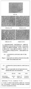
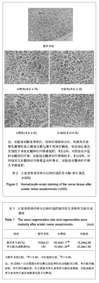
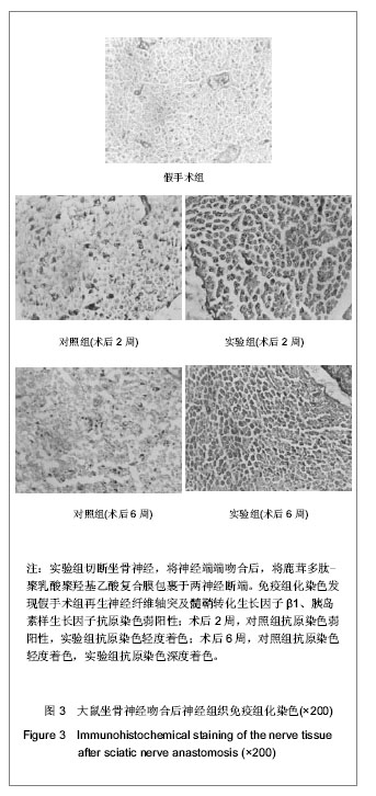
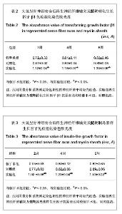
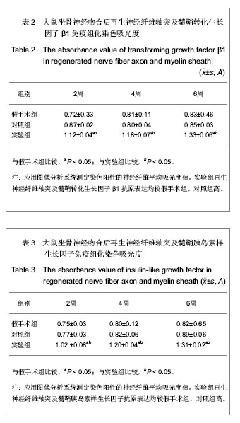
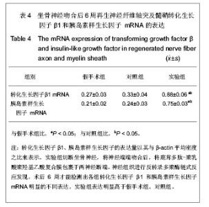
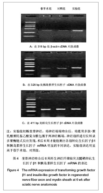
| [1] Lee Sk, Wolfe SW. Peripheral nerve injury and repair. J Am Acad Orthop surg. 2000;4:243-252.[2] Gu LQ, Pei GX. Beijing: Renmin Junyi Chubanshe. 2001: 70-100.顾立强,裴国献.周围神经损伤基础与临床[M].北京:人民军医出版社,2001:70-100.[3] Seckel BR. Enhancement of peripheral nerve regeneration. Mucscle Nerve. 1990;13(9):785-791.[4] Mendell LM. Neurotrophins and sensary neurons: role in development, maintance and injury: a thematic Summary. Philos Trans R Socland B Biol Sci. 1996;351(1338):463-367.[5] Brushart TM, Mesulam MM. Alteration in connections between muscle and anterior horn motoneurons after peripheral nerve repair. Science. 1980;208(4444):603-605.[6] Zhang ZH, Ruan HZ. Chuangshang Waike Zazhi. 2002;4(6): 377-380.张泽华,阮怀珍.基因治疗在周围神经损伤修复中的应用[J].创伤外科杂志,2002,4(6):377-380.[7] Zhao LG, Duan KD. Shengwu Yixue Gongcheng yu Linchuang. 2003;7(2):120-123.赵立国,端康德.神经导管修复周围神经损伤的研究进展[J].生物医学工程与临床.2003,7(2):120-123.[8] Wang TG, Wang SK, Hong GX, et al. Zhonghua Xianwei Wailke Zazhi. 2001;24:198-200.王同光,王栓科,洪光祥,等.细胞外ATP对外周神经再生作用的实验研究[J].中华显微外科杂志,2001,24:198-200.[9] Zhong SZ. Zhonghua Chuangshang Zazhi. 2000;16(2):69-70.钟世镇.创伤骨科基础研究展望[J].中华创伤杂志,2000,16(2): 69-70.[10] Sobue G. Neurotrophic factors and their receptor expressions in human neuropathies. Rinsho Shinkeigaku. 1997;37(12): 1107-1108.[11] Rutkowski GE, Heath CA. Development of a bioartificiaI nerve graft II Nerve regeneration in vitro. Biotechnol Prog. 2002; 18(2):373-379.[12] Matsumoto K, Ohnishi K, Kiyotani T, et al. Peripheral nerve regeneration across an 80-mm gap bridged by a polyglycolic acid (PGA)-collagen tube filled with laminin-coated collagen fibers: a histological and electrophysiological evaluation of regenerated nerves. Brain Res. 2000;868(2):315-328.[13] Agnew SP, Dumanian GA. Technical use of synthetic conduits for nerve repair. J Hand Surg Am. 2010;35(5):838-841.[14] Lundborg G, Dahlin LB, Danielsen N, et al. Tissue specificity in nerve regeneration. Scand J Hast Recoustr surg. 1986; 20(3):279-283.[15] Hung V, Dellon AL. Reconstruction of a 4-cm human median nerve gap by including an autogenous nerve slice in a bioabsorbable nerve conduit: case report. J Hand Surg Am. 2008;33(3):313-315.[16] Sun M, Kingham PJ, Reid AJ, et al. In vitro and in vivo testing of novel ultrathin PCL and PCL/PLA blend films as peripheral nerve conduit. J Biomed Mater Res A. 2010;93(4):1470-1481.[17] Zhang ZJ, Liu Q, Wang SJ. Yixue Zongshu. 2010;16(5): 647-650.张志军,刘强,王世杰.神经导管的基础与临床研究[J].医学综述, 2010,16(5):647-650.[18] Huang JF, Zheng HY, Li SP, et al. Zhonghua Shou Waike Zazhi. 2009;25(3):154-157.黄继锋,征华勇,李世普,等.RGD多肽接枝聚复合导管桥接神经缺损的实验研究[J].中华手外科杂志,2009,25(3):154-157.[19] Ding T, Luo ZJ, Zheng Y, et al. Rapid repair and regeneration of damaged rabbit sciatic nerves by tissue-engineered scaffold made from nano-silver and collagen type I. Injury. 2010;41(5):522-527.[20] Bushnell BD, McWilliams AD, Whitener GB, et al. Early clinical experience with collagen nerve tubes in digital nerve repair. J Hand Surg Am. 2008;33(7):1081-1087.[21] Yuan Q, Shah J, Hein S, et al. Controlled and extended drug release behavior of chitosan-based nanoparticle carrier. Acta Biomater. 2010;6(3):1140-1148.[22] Johansson F, Wallman L, Danielsen N, et al. Porous silicon as a potential electrode material in a nerve repair setting: Tissue reactions. Acta Biomater. 2009;5(6):2230-2237.[23] Oh SH, Kim JH, Song KS, et al. Peripheral nerve regeneration within an asymmetrically porous PLGA/Pluronic F127 nerve guide conduit. Biomaterials. 2008;29(11): 1601-1609.[24] Pabari A, Yang SY, Seifalian AM, et al. Modern surgical management of peripheral nerve gap. J Plast Reconstr Aesthet Surg. 2010;63(12):1941-1948.[25] Jiang X, Lim SH, Mao HQ, et al. Current applications and future perspectives of artificial nerve conduits. Exp Neurol. 2010;223(1):86-101.[26] Ji Y, Xu GP, Yan JL, et al. Transplanted bone morphogenetic protein/poly(lactic-co-glycolic acid) delayed-release microcysts combined with rat micromorselized bone and collagen for bone tissue engineering. J Int Med Res. 2009; 37(4):1075-1087.[27] Ling Y, Huang YS, Liang CY. Zhongguo Zuzhi Gongcheng Yanjiu yu Linchuang Kangfu. 2008;12(10):1899-1902.凌友,黄岳山,梁常艳.乳酸-羟基乙酸共聚物纳米粒的研究进展[J].中国组织工程研究与临床康复,2008,12(10):1899-1902.[28] Lee ES, Park KH, Park IS, et al. Glycol chitosan as a stabilizer for protein encapsulated into poly(lactide-co-glycolide) microparticle. Int J Pharm. 2007;338(1-2):310-316.[29] Yi X, Jin GH, Tian ML, et al. Shenjing Jiepouxue Zazhi. 2007; 23(5):506-510. 衣昕,金国华,田美玲,等.壳聚糖支架与神经神干细胞生物相容性的研究[J].神经解剖学杂志,2007,23(5):506-510.[30] Rochkind S, Astachov L, el-Ani D, et al. Further development of reconstructive and cell tissue-engineering technology fortreatment of complete peripheral nerve injury in rats. Neurol Res. 2004;26(2):161-166.[31] Wang KL, Lu LJ, Zhang JL. Zhongguo Zuzhi Gongcheng Yanjiu yu Lingchuang Kangfu Zazhi. 2012;34(16):6307-6311.王克利,路来金,张静玲.聚乳酸和聚羟基乙酸共聚物复合膜修复坐骨神经损伤[J].中国组织工程研究与临床康复杂志,2012, 34(16):6307-6311.[32] Wei YY, Wang JG, Huang L, et al. Zhonghua Chuangshang Guke Zazhi. 2009;11(1):51-55.魏延云,王建广,黄磊,等.复合神经生长因子的纳米纤维导管促神经再生的初步研究[J].中华创伤骨科杂志,2009,11(1):51-55.[33] Chen D, Meng XT, Liu JM, et al. Jiepou Xuebao. 2004;35: 240-243.陈东,孟晓婷,刘佳梅,等.鹿茸多肽对胎大鼠脑神经干细胞体外诱导分化的实验研究[J].解剖学报,2004,35:240-243.[34] Li LJ, Lu LJ, Chen L, et al. Liaoning Zhongyi Zazhi. 2004; 31(4): 343-344.李立军,路来金,陈雷,等.鹿茸多肽促进大鼠坐骨神经再生的实验研究[J].辽宁中医杂志,2004,31(4):343-344.[35] Sadighi M, Haines SR, Skottner A, et al. Effects of insulin-like growth factor-I (IGF-I) and IGF-II on the growth of antler cells in vitro. J Endocrinol. 1994;143(3):461-469.[36] Luetteke NC, Lee DC. Transforming growth factor alpha: expression, regulation and biological action of its integral membrane precursor. Semin Cancer Biol. 1990;1(4):265-275.[37] Liu LL, Cong B, Jiang HM, et al. Techan Yanjiu. 2009;31(2): 60-70.刘琳玲,丛波,姜洪梅,等.鹿茸多肽功能的研究进展[J].特产研究, 2009,31(2):60-70.[38] Pan FG, Sun W, Zhou Y. Zhongguo Shengwu Zhipin Xue Zazhi. 2007;20(9):669-671.潘风光,孙威,周玉,等.梅花鹿鹿茸多肽的提取及免疫功效的初步研究[J].中国生物制品学杂志,2007,20(9):669-671.[39] Gu YD. Zhonghua Shou Waike Zazhi. 2000;16(1):7-9.顾玉东. 21世纪臂丛损伤诊治的研究方向与任务[J].中华手外科杂志,2000,16(1):7-9. |