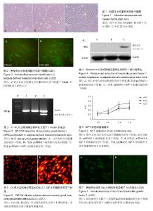
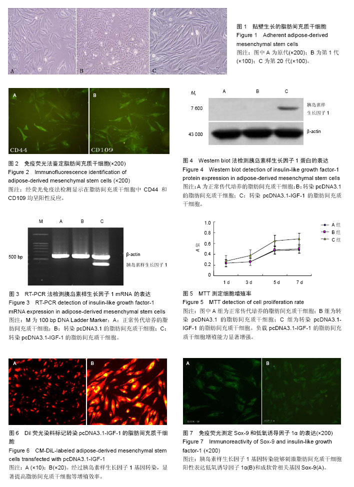
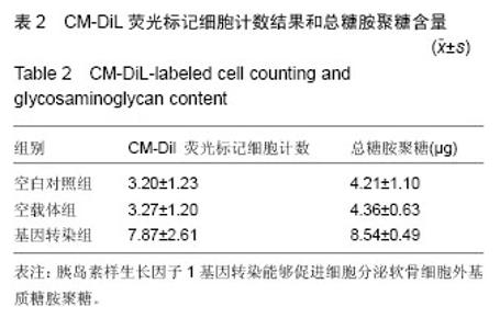
| [1] Mandelbaum B, Browne JE, Fu F,et al.Treatment outcomes of autologous chondrocyte implantation for full-thickness articular cartilage defects of the trochlea.Am J Sports Med.2007;35(6):915-921.
[2] 戴兵,徐海艇,金海东,等.低氧对脂肪干细胞和关节软骨细胞三维共培养成软骨能力的影响[J].中国组织工程研究,2014,18(29):4630-4635.
[3] Zuk PA,Zhu M,Mizuno H, et al.Multilineage cells from human adipose tissue: implications for cell-based therapies. Tissue Eng.2001;7(2):211-228.
[4] Strem BM, Hicok KC, Zhu M,et al.Multipotential differentiation of adipose tissue-derived stem cells. Keio J Med.2005;54(3):132-141.
[5] Zuk PA, Zhu M, Ashjian P, et al. Human adipose tissue is a source of multipotent stem cells. Mol Biol Cell. 2002;13(12): 4279-4295.
[6] Ogawa R. The importance of adipose-derived stem cells and vascularized tissue regeneration in the field of tissue transplantation. Curr Stem Cell Res Ther. 2006; 1(1):13-20.
[7] Fraser JK, Wulur I, Alfonso Z,et al.Fat tissue: an underappreciated source of stem cells for biotechnology. Trends Biotechnol. 2006;24(4): 150-154.
[8] Schäffler A,Büchler C.Concise review: adipose tissue-derived stromal cells-basic and clinical implications for novel cell-based therapies. Stem Cells.2008;25(4):818-827.
[9] 宋克东,杨延飞,李文芳,等.ADSCs-壳聚糖/明胶水凝胶工程化软骨的三维动态构建[J].功能材料2015;15(46): 15021-15029.
[10] 张勇,陈晓菊,王燕杰,等.兔脂肪干细胞直接修复陈旧性全层兔膝关节软骨缺损[J].临床骨科杂志,2015,18(4): 492-497.
[11] 赵明璨,刘畅.脂肪干细胞在软骨组织工程中的研究进展[J].中国生物医学工程学报,2014,33(4):475-481.
[12] 杨强,丁晓明,徐宝山,等.取向性丝素蛋白支架复合脂肪干细胞体外构建组织工程软骨[J],中华骨科杂志,2015, 35(12):1235-1242.
[13] Wang DW, Fermor B, Gimble JM,et al.Influence of oxygen on the proliferation and metabolism of adipose derived adult stem cells.J Cell Physiol. 2005;204(1): 184-191.
[14] Bunnell BA, Flaat M, Gagliardi C,et al. Adipose-derived stem cells: isolation, expansion and differentiation. Methods.2008;45(2)115-120.
[15] Shu W, Shu YT, Shi HB, et al. An easy method to discover cell membrane antigen with atomic force microscopy. Mol Biol Rep.2008;35(4):557-561.
[16] Lin Y, Tang W, Wu L, et al. Bone regeneration by BMP-2 enhanced adipose stem cells loading on alginate gel. Histochem Cell Biol. 2008; 129(2): 203-210.
[17] 张云松,高建华,鲁峰,等.荧光活性染料DiI标记人脂肪干细胞[J].中国组织工程研究与临床康复,2007,11(15): 2897-2899.
[18] de Villiers JA, Houreld N, Abrahamse H. Adipose derived stem cells and smooth muscle cells: implications for regenerative medicine. Stem Cell Rev and Rep.2009;5:256-265.
[19] 於明明,黄立渠,邓永继.脂肪干细胞体外诱导分化为平滑肌细胞的研究进展[J/CD].中华临床医师杂志:电子版, 2016,10(3):429-432.
[20] Guilak F,Awad HA,Fermor B,et al .Adipose-derived adult stem cells for cartilage tissue engineering. Biorheology.2004;41(3-4):389-399.
[21] Yaeger PC, Masi TL, de Ortiz JL, et al. Synergistic action of transforming growth factor-β and insulin-like growth factor-1 induces expression of type II collagen and aggrecan genes in adult human articular chondrocytes. Exp Cell Res.1997;237:318-25.
[22] Wright E,Hargrave MR,Christiansen J,et al.The Sry-related gene SOX-9 is expressed during chondrogenesis in mouse embryos.NatGenet.1995;9(1): 15-20.
[23] Yin W,Park JI,Loeser RF.oxidative stress inhibits insulin-like growth factor-I induction of chondrocyte proteoglycan synthesis through differential regulation of phosphatidylinositol 3-Kinase-Akt and MEK-ERK MAPK signaling pathways. J Biol Chem 2009;284(46): 31972-31981.
[24] Gelse K, Mühle C, Knaup K, et al.Chondrogenic differentiation of growth factor-stimulated precursor cells in cartilage repair tissue is associated with increased HIF-1a activity. Osteoarthritis Cartilage. 2008;16:1457-1465.
[25] Treins C, Giorgetti-Peraldi S, Murdaca J, et al. Regulation of hypoxia-inducible factor (HIF)-1 activity and expression of HIF hydroxylases in response to insulin-like growth factor I. Mol Endocrinol.2005;19: 1304-1317.
[26] Treins C, Giorgetti-Peraldi S, Murdaca J, et al.Insulin stimulates hypoxia-inducible factor 1 through a phosphatidylinositol 3-kinase/target of rapamycin-dependent signaling pathway. J Biol Chem.2002;277: 27975-27981.
[27] Treins C, Murdaca J, Van Obberghen E, et al.AMPK activation inhibits the expression of HIF-1a induced by insulin and IGF-1. Biochem Biophys Res Commun. 2006;342:1197-1202.
[28] Robins JC, Akeno N, Mukherjee A ,et al. Hypoxia induces chondrocyte -specific gene expression in mesenchymal cells in association with transcriptional activation of Sox9. Bone.2005;37:313-22.
[29] 陈少坚,郭风劲,王春,等. SiRNA沉默Sox-9基因调控骨骺干细胞的增殖及凋亡[J].中国组织工程研究,2013,17(40): 7068-7075.
[30] 李锋,聂喜增,刘金辉,等 胰岛素样生长因子-1及转化生长因子-β2对幼年及成年兔软骨细胞合成糖胺多糖的影响[J]. 中国老年学杂志,2015,35(16):4458-4460. |