Chinese Journal of Tissue Engineering Research ›› 2016, Vol. 20 ›› Issue (38): 5657-5663.doi: 10.3969/j.issn.2095-4344.2016.38.005
Previous Articles Next Articles
Effects of nano porous beta-tricalcium phosphate/collagen scaffold modified with human bone morphogenetic protein 2 gene on differentiation of MC3T3-E1 cell lines
- School of Stomatology, Jiamusi University, Jiamusi 154007, Heilongjiang Province, China
-
Received:2016-07-10Online:2016-09-16Published:2016-09-16 -
Contact:Zhao Gang, Master’s supervisor, Associate professor, School of Stomatology, Jiamusi University, Jiamusi 154007, Heilongjiang Province, China -
About author:Ruan Qiang, Studying for master’s degree, School of Stomatology, Jiamusi University, Jiamusi 154007, Heilongjiang Province, China -
Supported by:the Natural Science Foundation of Heilongjiang Province, No. H201487; the Postgraduate Innovation Project of Jiamusi University, No. LZR2015_015
CLC Number:
Cite this article
Ruan Qiang, Zhao Gang, Guo Rui, Xiao Yue, Li Chao.
share this article

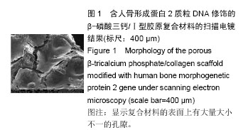
2.1 形态学观察结果 2.1.1 支架电镜观察结果 含人骨形成蛋白2质粒DNA修饰的纳米级β-磷酸三钙/Ⅰ型胶原复合材料的扫描电镜观察结果见图1,显示复合材料的表面上有大量大小不一的孔隙。"
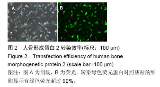
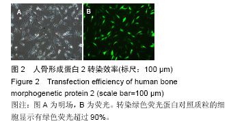
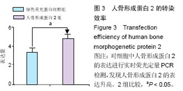
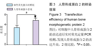
2.1.2 人骨形成蛋白2转染效率 转染绿色荧光蛋白对照质粒的细胞显示绿色荧光超过90%(见图2A,B),说明成功将外源基因转染至细胞中。同时对转染了人骨形成蛋白2的细胞中人骨形成蛋白2的表达进行实时荧光定量PCR检测,发现人骨形成蛋白2的表达升高(见图3)。"
"
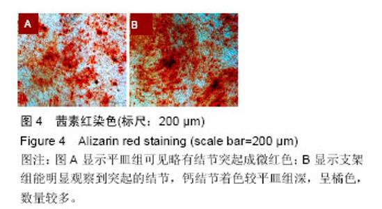
2.2 茜素红染色检测钙结节 小鼠前成骨细胞在成骨诱导第1,3,7,14天分别进行茜素红染色,在第1,3天对照结果不是很显著,第7,14天后,茜素红结果显示平皿组可见略有结节突起成微红色(见图4A),支架组能明显观察到突起的结节,钙结节着色较平皿组深,呈橘色,数量较多(见图4B)。"

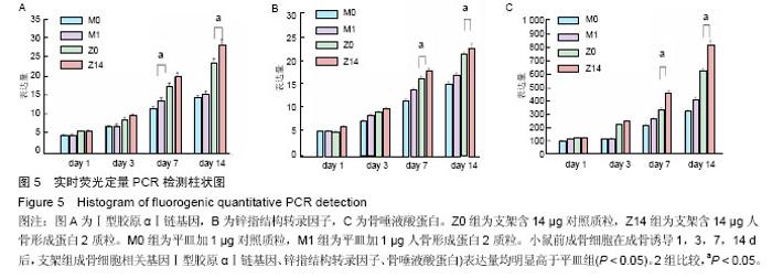
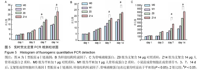
2.3 实时荧光定量PCR检测细胞成骨相关基因表达结果 实验使用实时荧光定量 PCR 法测定成骨细胞相关基因Ⅰ型胶原αⅠ链基因、锌指结构转录因子、骨唾液酸蛋白的表达,从而证明细胞的成骨活性。小鼠前成骨细胞在成骨诱导1,3,7,14 d后,支架组成骨细胞相关基因Ⅰ型胶原αⅠ链基因、锌指结构转录因子、骨唾液酸蛋白表达量均明显高于平皿组(P < 0.05)。图5结果表明,复合支架材料由于掺入了具有生物活性的质粒,在一定时期内提高了成骨细胞的相关基因表达水平。据此,推测该类材料的成骨活性主要通过影响和调控成骨基因的表达,从而促进成骨细胞的生长、分化和骨基质形成。"
| [1] Robert F. Tissue engineers build new bone. Science. 2000;289: 1498-1500.
[2] Kim HM, Uenoyama M, Kobubo T, et al. Biomimetic apatite formation on polyethylene photografted with vinyltrimethxysilane and hydrolyzed. Biomaterials. 2001;22(18): 2489-2494.
[3] Wei J, Li YB. Tissue engineering scaffold material of nano-apatite crystals and polyamide composite. Eur Polymer J. 2004;40:509-515.
[4] Kondo N, Ogose A, Tokunaga K, et al. Osteoinduction with highly purified b-tricalcium phosphate in dog dorsal muscles and the proliferation of osteoclasts before heterotopic bone formation. Biomaterials. 2006;27:4419-4427.
[5] Bauer TW, Muschler GF. Bone graft material: an overview of the basic science. Clin Orthop. 2000;(371): 10-27.
[6] Hosseinkhani H,Hosseinkhani M, Gabrielson NP, et al. DNA nanoparticles encapsulated in 3D tissue-engineered scaffolds enhance osteogenic differentiation of mesenchymal stem cells .Biomed Mater Res. 2007;85A(1):47-60.
[7] Hosseinkhani H, Inatsugu Y, Hiraoka Y, et al. Impregnation of plasmid DNA into three-dimensional scaffolds and medium perfusion enhance in vitro DNA expression of mesenchymal stem cells. Tissue Eng. 2005,11(9-10):1459-1475.
[8] Gugala Z, Davis AR, Fouletier-Dilling CM, et al. Adenovirus BMP2-induced osteogenesis in combination with collagen carriers. Biomaterials. 2012;28(30):4469-4479.
[9] Park J, Lutz R, Felszeghy E, et al. The effect on bone regeneration of a liposomal v ector to deliver BMP2 gene to bone grafts in peri-implant bone defects. Biomaterials. 2007;28(17):2772-2782.
[10] Parsons B, Strauss E. Surgical management of chronic osteomyelitis. Am J Surg. 2004;188: 57-64.
[11] 范丽,卢岩,林慧平,等.骨形成蛋白-2转染MG-63细胞增加其对壳聚糖膜的粘附[J].材料科学与工程学报,2011,29(6):911-915.
[12] Zou C, Weng W, Deng X, et al. Preparation and characterization of porous b-tricalcium phosphaye/ collagen composites with an integrated structure. Biomaterials. 2005;26:5276-5284.
[13] Silva GA,Ducheyne P,Reis RL.Materials in particulate form for tissue engineering.1.Basic concepts. J Tissue Eng Regen Med.2007;1(1):4-24.
[14] Lu K, Zeng D, Zhang Y,et al.BMP-2 Gene Modified Canine bMSCs Promote Ectopic Bone Formation Mediated by a Nonviral PEI Derivative. Ann Biomed Eng. 2011;39(6):1829-1839.
[15] Zeng D,Xia L,Zhang W,et al. Maxillary Sinus Floor Elevation Using a Tissue-Engineered Bone with Calcium-Magnesium Phosphate Cement and Bone Marrow Stromal Cells in Rabbits. Tissue Eng Part A. 2012;18(7-8):870-881.
[16] 王晓娜,赵静辉,储顺礼,等.骨替代材料在口腔种植领域中的成骨效果[J]. 国际口腔医学杂志,2016,43(1):113-117.
[17] Chen H, Zhao G, Ye J. Autosolidifying calcium phosphate cement in the repair of bone defects due to different etiology among 94 cases. J Clin Rehabil Tissue Eng Res. 2008;12(19): 3779-3781.
[18] Joosten U, Joist A, Gosheger G, et al. Burkhard Brandt.Christof von Eiff. Effectiveness of hydroxyapatite- vancomycin bone cement in thetreatment of Staphylococcus aureus induced chronic osteomyelitis. Biomaterials. 2005;26: 5251-5258.
[19] Hartman GA, Arnold RM,mills MP, et al. Clinical and histologic evaluation of anorganic bovine bone collagen with or without a collagen barrier. Int J Periodontics Restorative Dent. 2004;24(2):127-135.
[20] Chen D, Zhao M, Mundy GR. Bone morphogenetic Proteins. Growth Factors. 2004;22(4):233-241.
[21] Urist MR.Bone formation by autoinduction. Science. 1965;150(3698):893-899.
[22] Shimer AL, Oner FC, Vaccaro AR. Spinal reconstruction and bone morphogenetic proteins: open questions. Injury. 2009;40(3):32-38.
[23] Wegman F, Bijenhof A, Schuijll L, et al. Osteogenic differentiation as a result of BMP-2 plasmid DNA based gene therapy in vitro and in vitro and in vivo. Eur Cell Mater. 2011;15(21):230-242
[24] Lee JH, Kim CS. The induction of bone form ation in rat calvarial defects and subcutaneous tissues by recombinant human BMP-2, produced in Escherichia coli. Biomaterials. 2010;31(13):3512-3519.
[25] Bozidar M,Hari S,Michael D,et al. β-tricalcium phosphate-type I collagen cones with or without a barrier membrane in hume extraction socket healing-clinical, histologic, histomorph- ometric, and immunohistochemical evaluation. Clin Oral Invest. 2012;16(10):581-590.
[26] Zou C, Weng WJ, Deng XL, et al. Preparation and characterization of porous β-tricalcium phosphaye/ collagen composites with an integrated structure. Biomaterials. 2005;26:5276-5284.
[27] Cao X, Deng W, Wei Y, et al. Encapsulation of plasmid DNA in ralrium phosphate nanoparticles:stemcell uptake and gene transfer efficiency.Int J Nanomedicine. 2011;6: 333-3349.
[28] Liu T, Tang A, Zhang G, et al. Calcium phosphate nanoparticles as a novel nonviral vector for efficient transfection of DNA in cancer gene therapy. Cancer Biother Radiopharm. 2005;20:141-149.
[29] Keeney M, van den Beucken LL,van der Kraan PM, et al. The ability of a collagen/calcium phosphate scaffold to act as its own vector for gene delivery and to promote bone formation via transfection with VECF(165). Biomaterials. 2010; 31(10):2893-902.
[30] Glassman SD,Carreon LY,Campbell MJ,et al.The perioperative cost of Infuse bone graft in posterolateral lumbar spine fusion.Spine J. 2008;8:443-448.
[31] Chamberlain JR,Schwarze U,Wang PR,et al.Gene targeting instem cells from individuals with osteogenesis imperfecta.Science.2004;303(5661): 1198-1201.
[32] Zhang JY,Doll BA,Beckman EJ,et al. Three-dimensional biocompatible ascorbic acid-containing scaffold for bone tissue engineering. Tissue Eng. 2003.9(6):1143-1157.
[33] Petite H, Viateau V, Bensaid W,et al.Tissue-engineered bone regeneration. Nature Bioteehnology. 2000;18(9): 959-963.
[34] Orban JM,Marra KG,Hollinger JO.Composition options for tissue-engineered bone.Tissue Eng. 2002;8(4):529-539.
[35] Bauer TW,Muschler GF.Bone graft matcrials.An overview of the basic science.Clin Orthop. 2000;371:10-27.
[36] Hosseinkhani H,Hosseinkhani M,Gabrielson NP,et al. DNA nanoparticles encapsulated in 3D tissue-engineered scaffolds enhance osteogenic differentiation of mesenchymal stem cells.Biomed Mater Res A. 2007; 85A(1):47-60.
[37] Hosseinkhani H,Inatsugu Y,Hiraoka Y,et al. Impregnation of plasmid DNA into three-dimensional scaffolds and medium perfusion enhance in vitro DNA expression of mesenchymal stem cells. Tissue Eng. 2005;11(9-10): 1459-1475.
[38] Gugala Z,Davis AR,Fouletier-Dilling CM,et al. Adenovirus BMP2-induced osteogenesis in combination with collagen carriers. Biomaterials. 2012;28(30):4469-4479.
[39] Hinz P,Wolf E,Sehwesinger G,et al.A new resorbable bone void filler intrauma:early clinical experience and histologic evaluation.Orthopedics. 2002;25(S5):597-600.
[40] Sikavitsas VI,Temenoff JS,Mikos AG.Biomaterials and bone mechanotrans Duction. Biomaterials. 2001; 22(19):2581-2593.
[41] Hinz P, Wolf E, Schwesinger G, et al. A new resorbable bone void filler in trauma: early clinical experience and histologic evaluation. Orthopedics. 2002;25(S5):597-600.
[42] Yuan H, Li Y, de Bruijn JD, et al. Tissue responses of calcium phosphate cement: a study in dogs. Biomaterials. 2000;21(12): 1283-1290. |
| [1] | Zhang Tongtong, Wang Zhonghua, Wen Jie, Song Yuxin, Liu Lin. Application of three-dimensional printing model in surgical resection and reconstruction of cervical tumor [J]. Chinese Journal of Tissue Engineering Research, 2021, 25(9): 1335-1339. |
| [2] | Shen Jinbo, Zhang Lin. Micro-injury of the Achilles tendon caused by acute exhaustive exercise in rats: ultrastructural changes and mechanism [J]. Chinese Journal of Tissue Engineering Research, 2021, 25(8): 1190-1195. |
| [3] | Liang Xueqi, Guo Lijiao, Chen Hejie, Wu Jie, Sun Yaqi, Xing Zhikun, Zou Hailiang, Chen Xueling, Wu Xiangwei. Alveolar echinococcosis protoscolices inhibits the differentiation of bone marrow mesenchymal stem cells into fibroblasts [J]. Chinese Journal of Tissue Engineering Research, 2021, 25(7): 996-1001. |
| [4] | Duan Liyun, Cao Xiaocang. Human placenta mesenchymal stem cells-derived extracellular vesicles regulate collagen deposition in intestinal mucosa of mice with colitis [J]. Chinese Journal of Tissue Engineering Research, 2021, 25(7): 1026-1031. |
| [5] | Li Cai, Zhao Ting, Tan Ge, Zheng Yulin, Zhang Ruonan, Wu Yan, Tang Junming. Platelet-derived growth factor-BB promotes proliferation, differentiation and migration of skeletal muscle myoblast [J]. Chinese Journal of Tissue Engineering Research, 2021, 25(7): 1050-1055. |
| [6] | Zeng Yanhua, Hao Yanlei. In vitro culture and purification of Schwann cells: a systematic review [J]. Chinese Journal of Tissue Engineering Research, 2021, 25(7): 1135-1141. |
| [7] | Xu Dongzi, Zhang Ting, Ouyang Zhaolian. The global competitive situation of cardiac tissue engineering based on patent analysis [J]. Chinese Journal of Tissue Engineering Research, 2021, 25(5): 807-812. |
| [8] | Wu Zijian, Hu Zhaoduan, Xie Youqiong, Wang Feng, Li Jia, Li Bocun, Cai Guowei, Peng Rui. Three-dimensional printing technology and bone tissue engineering research: literature metrology and visual analysis of research hotspots [J]. Chinese Journal of Tissue Engineering Research, 2021, 25(4): 564-569. |
| [9] | Chang Wenliao, Zhao Jie, Sun Xiaoliang, Wang Kun, Wu Guofeng, Zhou Jian, Li Shuxiang, Sun Han. Material selection, theoretical design and biomimetic function of artificial periosteum [J]. Chinese Journal of Tissue Engineering Research, 2021, 25(4): 600-606. |
| [10] | Liu Liu, Zhou Qingzhu, Gong Zhuo, Liu Boyan, Yang Bin, Zhao Xian. Characteristics and manufacturing techniques of collagen/inorganic materials for constructing tissue-engineered bone [J]. Chinese Journal of Tissue Engineering Research, 2021, 25(4): 607-613. |
| [11] | Liu Fei, Cui Yutao, Liu He. Advantages and problems of local antibiotic delivery system in the treatment of osteomyelitis [J]. Chinese Journal of Tissue Engineering Research, 2021, 25(4): 614-620. |
| [12] | Li Xiaozhuang, Duan Hao, Wang Weizhou, Tang Zhihong, Wang Yanghao, He Fei. Application of bone tissue engineering materials in the treatment of bone defect diseases in vivo [J]. Chinese Journal of Tissue Engineering Research, 2021, 25(4): 626-631. |
| [13] | Zhang Zhenkun, Li Zhe, Li Ya, Wang Yingying, Wang Yaping, Zhou Xinkui, Ma Shanshan, Guan Fangxia. Application of alginate based hydrogels/dressings in wound healing: sustained, dynamic and sequential release [J]. Chinese Journal of Tissue Engineering Research, 2021, 25(4): 638-643. |
| [14] | Chen Jiana, Qiu Yanling, Nie Minhai, Liu Xuqian. Tissue engineering scaffolds in repairing oral and maxillofacial soft tissue defects [J]. Chinese Journal of Tissue Engineering Research, 2021, 25(4): 644-650. |
| [15] | Xing Hao, Zhang Yonghong, Wang Dong. Advantages and disadvantages of repairing large-segment bone defect [J]. Chinese Journal of Tissue Engineering Research, 2021, 25(3): 426-430. |
| Viewed | ||||||
|
Full text |
|
|||||
|
Abstract |
|
|||||