Chinese Journal of Tissue Engineering Research ›› 2013, Vol. 17 ›› Issue (14): 2570-2577.doi: 10.3969/j.issn.2095-4344.2013.14.015
Previous Articles Next Articles
Isolation, culture and identification of rat bone marrow-derived endothelial progenitor cells
Wei Xiao-yan1, Zhang Li1, Lin Ming2, Chen Huan1, Peng Bo1, Peng Yuan-yuan1, Li Fu-yun1, Hong Pei-xin1, Fan Yi-fan1
- 1 College of Acupuncture-Moxibustion and Tuina, Beijing University of Traditional Chinese Medicine, Beijing 100029, China
2 Department of Cell Biology and Genetics, College of Basic Medical Sciences, Health Science Center, Peking University, Beijing 100083, China
-
Received:2012-10-26Revised:2013-01-31Online:2013-04-02Published:2013-04-02 -
Contact:Zhang Li, Professor, College of Acupuncture-Moxibustion and Tuina, Beijing University of Traditional Chinese Medicine, Beijing 100029, China zhangli1572@sina.com -
About author:Wei Xiao-yan☆, Studying for doctorate, College of Acupuncture-Moxibustion and Tuina, Beijing University of Traditional Chinese Medicine, Beijing 100029, China wxy66603704@163.com -
Supported by:the National Natural Science Foundation of Vhina (Special Program), No. 81141120
CLC Number:
Cite this article
Wei Xiao-yan, Zhang Li, Lin Ming, Chen Huan, Peng Bo, Peng Yuan-yuan, Li Fu-yun, Hong Pei-xin, Fan Yi-fan. Isolation, culture and identification of rat bone marrow-derived endothelial progenitor cells[J]. Chinese Journal of Tissue Engineering Research, 2013, 17(14): 2570-2577.
share this article
2.1 大鼠骨髓源内皮祖细胞体外诱导的形态学观察 显微镜下观察,新分离骨髓单个核细胞呈圆形,里面混有部分红细胞,见图1A;原代细胞接种二三天时,部分圆形细胞伸开突起,呈短梭形贴壁,见图1B;之后贴壁细胞数目不断增多,至4 d将未贴壁细胞移去再重新接种二三天后仍有部分贴壁。首次换液后细胞生长加快,呈集落样生长,见图1C;7 d时细胞生长旺盛,已较为密集,可见纺锤形、三角形、圆形等不同形态之细胞,见图1D;10 d左右时,未传代培养的细胞可继续培养30-40 d,形态为圆形、短梭形等,呈“铺路石”样,见图1E、1F;机械吹打法传代培养的细胞迅速呈“克隆”样增殖,生长迅速,见图1G。"
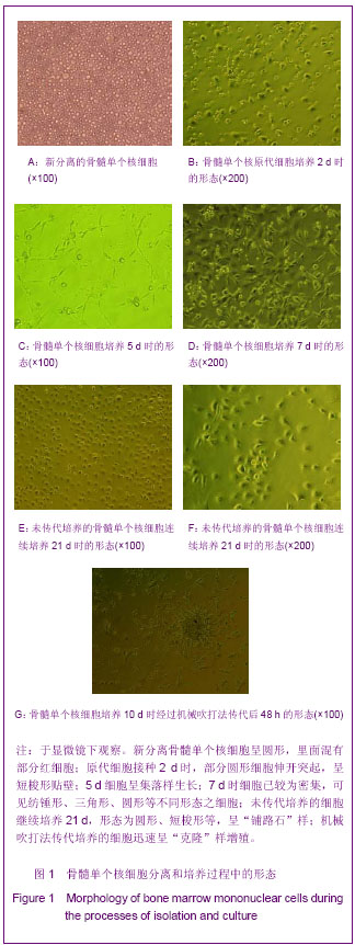
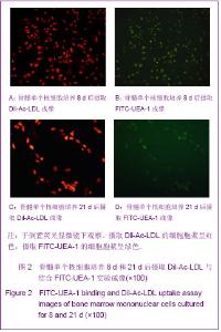
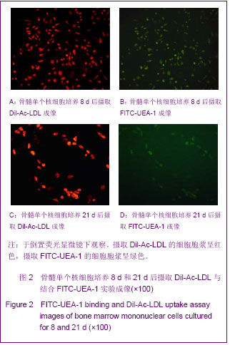
实验中不进行传代而持续培养的情况下,发现细胞生长存在2个生长高峰,第1个位于8 d前后,第2个生长高峰位于15 d左右。细胞的形态在生长的早期、晚期有一定差异,早期以纺锤形、三角形、圆形细胞多见,晚期以圆形、短梭形细胞多见。 2.2 摄取Dil-Ac-LDL与结合FITC-UEA-1实验结果 倒置荧光显微镜下观察,摄取Dil-Ac-LDL的细胞胞浆呈红色,摄取FITC-UEA-1的细胞胞浆呈绿色。骨髓单个核细胞培养8 d时,双阳性的细胞约占细胞总数的95%以上,见图2A、2B。骨髓单个核细胞培养21 d时,大部分细胞染色仍呈双阳性,见图2C、2D。说明骨髓单个核细胞培养8 d和21 d时均能够同时摄取Dil-Ac-LDL与FITC-UEA-1,为正在分化的内皮祖细胞。"
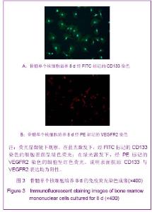
2.3 免疫荧光染色结果 骨髓单个核细胞培养8 d时,行免疫荧光化学法法检测细胞表面抗原表达情况。荧光显微镜下可见,在蓝光激发下,经FITC标记的CD133染色的细胞表面呈绿色荧光,见图3A;在绿光激发下,经PE标记的VEGFR2染色的细胞呈红色荧光,见图3B。说明表面抗原CD133与VEGFR2的表达均为阳性。"

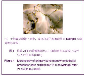
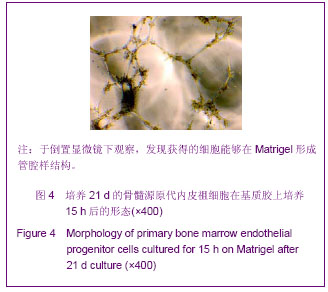
2.4 管腔形成实验结果 将培养21 d的骨髓源原代内皮祖细胞接种至包被了Matrigel的96孔板上后,15 h左右甚至更早即可见细胞拉长变形,分化为管腔状结构,见图4。"
| [1] Hur J, Yoon CH, Kim HS, et al. Characterization of two types of endothelial progenitor cells and their different contributions to neovasculogenesis. Arterioscler Thromb Vasc Biol. 2004; 24(2):288-293.[2] Dzau VJ, Gnecchi M, Pachori AS, et al. Therapeutic potential of endothelial progenitor cells in cardiovascular diseases. Hypertension. 2005;46(1):7-18.[3] Urbich C, Dimmeler S. Endothelial progenitor cells: Characterization and role in vascular biology. Circ Res. 2004; 95(4):343-353.[4] Asahara T, Mumhara T, Suilivan A, et al. Isolation of Putative Progenitor endothelial cells for angiogenesis. Science. 1997; 275 (5302):964-967.[5] Asahara T, Masuda H, Takahashi T, et al. Bone Marrow Origin of Endothelial Progenitor Cells Responsible for Postnatal Vasculogenesis in Physiological and Pathological Neovascularization. Circ Res. 1999;85(3):221-228.[6] Morgan R, Kreipke CW, Roberts G, et al. Neovascularization following traumatic brain injury:possible evidence for both angiogenesis and vasculogenesis. Neurol Res. 2007;29(4): 375-381.[7] Umemura T, Higashi Y. Endothelial progenitor cells:therapeutic target for cardiovascular diseases. J Pharmacol Sci. 2008;108(1):1-6.[8] Gao D, Nolan DJ, Mellick AS, et al. Endothelial progenitor cells control the angiogenic switch in mouse lung metastasis. Science. 2008;319(5860):195-198.[9] Su Y, Zheng L, Wang Q, et al. The PI3K/Akt pathway upregulates Id1 and integrin α4 to enhance recruitment of human ovarian cancer endothelial progenitor cells. BMC Cancer. 2010;10:459.[10] Moubarik C, Guillet B, Youssef B, et al. Transplanted late outgrowth endothelial progenitor cells as cell therapy product for stroke. Stem cell reviews. 2011;7(1):208-220.[11] Rouwkema J, Westerweel PE, de Boer J, et al. The use of endothelial progenitor cells for prevascularized bone tissue engineering.Tssue Eng Part A. 2009;15(8):2015-2027.[12] Asahara T. Cell therapy and gene therapy using endothelial progenitor cells for vascular regeneration. Handb Exp Pharmacol. 2007;(180):181-194.[13] Palladino M, Gatto l, Neri V, et al. Combined Therapy with Sonic Hedgehog Gene Transfer and Bone Marrow-Derived Endothelial Progenitor Cells Enhances Angiogenesis and Myogenesis in the Ischemic Skeletal Muscle. J Vasc Res. 2012;49(5):425-431.[14] Weber A, Pedrosa I, Kawamoto A, et al. Magnetic resonance mapping of transplanted endothelial progenitor cells for therapeutic neovascularization in ischemic heart disease. Eur J Cardiothorac Surg. 2004;26(1):137-143.[15] Khan SS, Solomon MA, McCoy JP Jr. Detection of circulating endothelial cells and endothelial progenitor cells by flow cytometry. Cytometry B Clin Cytom. 2005;64(1):l-8.[16] Medina RJ, O'Neill CL, Sweeney M, et al. Molecular analysis of endothelial progenitor cell (EPC) subtypes reveals two distinct cell populations with different identities. BMC Med Genomics. 2010;3:18.[17] Yuan JM, Guan SH, Dang RS, et al.Zhongguo Zuzhi Gongcheng Yanjiu yu Linchuang Kangfu. 2010;14(36): 6662-6666.袁建明,管松晖,党瑞山,等.绵羊骨髓来源内皮祖细胞的培养与鉴定[J].中国组织工程研究与临床康复,2010,14(36):6662-6666.[18] Asai J, Takenaka H, Li M, et al. Topical application of ex vivo expanded endothelial progenitor cells promotes vascularisation and wound healing in diabetic mice. Int Wound J.2012.[19] Zhao T, Li J, Chen AF. MicroRNA-34a induces endothelial progenitor cell senescence and impedes its angiogenesis via suppressing silent information regulator 1. Am J Physiol Endocrinol Metab. 2010;299(1):E110-E116.[20] Brunt KR, Hall SR, Ward CA, et al. Endothelial Progenitor Cell and Mesenchymal Stem Cell Isolation, Characterization, Viral Transduction. Methods Mol Med. 2007;139:197-210. [21] Chen J, Song M, Yu S, et at. Advanced glycation endproducts alter functions and promote apoptos in endothelial progenitor cells through receptor for advanced glycation endproducts mediate over pression of cell oxidant stress. Mol Cell Biochem. 2010;335(1-2):137-146.[22] li M. Bone Marrow-Derived Endothelial Progenitor Cells:Isolation and Characterization for Myocardial Repair. Methods Mol Biol. 2010;660:9-27.[23] Ahrens I, Domeij H, Topcic D, et al. Successful in vitro expansion an differentiation of cord blood derived CD34+ cells into early endothelial progenitor cells reveals highly differential gene expression. PLoS One. 2011; 6(8):e23210.[24] Thill M, Strunnikova NV, Berna MJ, et al. Late outgrowth endothelial progenitor cells in patients with age-related macular degeneration. Invest Ophthalmol Vis Sci. 2008; 49(6):2696-2708.[25] Umemura T, Higashi Y. Endothelial progenitor cells:therapeutic target for cardiovascular diseases. J Pharmacol Sci. 2008;108(1):1-6.[26] Garcia-Barros M, Paris F, Cordon-Cardo C, et al. Tumor Response to Radiotherapy Regulated by Endothelial Cell Apoptosis. Science. 2003;300(5622):1155-1159.[27] Ingram DA, Mead LE, Tanaka H, et al. Identification of a novel hierarchy of endothelial progenitor cells using human peripheral and umbilical cord blood. Blood. 2004;104(9): 2752-2760.[28] Werner N, Junk S, Laufs U, et al. Intravenous Transfusion of Endothelial Progenitor Cells Reduces Neointima Formation After Vascular Injury. Circ Res. 2003;93(2):e17-e24.[29] Cherqui S, Kurian SM, Schussler O, et al. Isolation and angiogenesis by endothelial progenitors in the fetal liver. Stem Cells. 2006;24(1):44-54.[30] Casamassimi A, Balestrieri ML, Fiorito C, et al. Comparison between total endothelial progenitor cell isolation versus enriched CD133+ culture. J Biochem. 2007;141(4):503-511.[31] Kahler CM, Wechselberger J, Hilbe W, et al. Peripheral infusion of rat bone marrow derived endothelial progenitor cells leads to homing in acute lung injury. Respir Res. 2007; 8(1):50.[32] Chen YH, Lin SJ, Lin F Y, et al. High glucose impairs early and late endothelial progenitor cells by modifying nitric oxide-related but not oxidative stress-mediated mechanism. Diabetes. 2007;56(6):1559-1568[33] Hirschi KK, Ingram DA, Yoder MC. Assessing identity,phenotype,and fate of endothelial progenitor cells. Arterioscle Thromb Vasc Biol. 2008;28(9):1584-1589.[34] Yoder MC. Defining human endothelial progenitor cells. J Thromb Haemost. 2009;7 Suppl 1:49-52.[35] Dimmeler S. Regulation of bone marrow-derived vascular progenitor cell mobilization and maintenance. Arterioscler Thromb Vasc Biol. 2010;30(6):1088-1093.[36] Leone AM, Valgimigli M, Giannico MB, et al. From bone marrow to the arterial wall:the ongoing tale of endothelial progenitor cells. Eur Heart. 2009;30(8):890-899.[37] Asahara T, Kawamoto A. Endothelial progenitor cells for postnatal vasculogenesis. Am J Physiol Cell Physiol. 2004; 287(3):C572-579.[38] Barsotti MC, Magera A, Armani C, et al. Fibrin acts as biomimetic niche inducing both differentiation and stem cell marker expression of early human endothelial progenitor cells. Cell Prolif. 2011;44(1):33-48.[39] Bleiziffer O, Hammon M, Naschberger E, et al. Endothelial progenitor cells are integrated in newly formed capillaries and alter adjacent fibrovascular tissue after subcutaneous implantation in a fibrin matrix. J Cell Mol Med. 2011;15: 2452-2461.[40] Shi S, He YZ, Song L, et al. Zhongguo Zuzhi Gongcheng Yanjiu yu Linchuang Kangfu. 2010;14(16):2879-2882. 施森,何延政,宋丽,等.血管内皮生长因子在鼠尾胶原凝胶诱导三维血管新生中的作用研究[J].中国组织工程研究与临床康复, 2010, 14(16):2879-2882. |
| [1] | Pu Rui, Chen Ziyang, Yuan Lingyan. Characteristics and effects of exosomes from different cell sources in cardioprotection [J]. Chinese Journal of Tissue Engineering Research, 2021, 25(在线): 1-. |
| [2] | Zhang Xiumei, Zhai Yunkai, Zhao Jie, Zhao Meng. Research hotspots of organoid models in recent 10 years: a search in domestic and foreign databases [J]. Chinese Journal of Tissue Engineering Research, 2021, 25(8): 1249-1255. |
| [3] | Wang Zhengdong, Huang Na, Chen Jingxian, Zheng Zuobing, Hu Xinyu, Li Mei, Su Xiao, Su Xuesen, Yan Nan. Inhibitory effects of sodium butyrate on microglial activation and expression of inflammatory factors induced by fluorosis [J]. Chinese Journal of Tissue Engineering Research, 2021, 25(7): 1075-1080. |
| [4] | Wang Xianyao, Guan Yalin, Liu Zhongshan. Strategies for improving the therapeutic efficacy of mesenchymal stem cells in the treatment of nonhealing wounds [J]. Chinese Journal of Tissue Engineering Research, 2021, 25(7): 1081-1087. |
| [5] | Liao Chengcheng, An Jiaxing, Tan Zhangxue, Wang Qian, Liu Jianguo. Therapeutic target and application prospects of oral squamous cell carcinoma stem cells [J]. Chinese Journal of Tissue Engineering Research, 2021, 25(7): 1096-1103. |
| [6] | Xie Wenjia, Xia Tianjiao, Zhou Qingyun, Liu Yujia, Gu Xiaoping. Role of microglia-mediated neuronal injury in neurodegenerative diseases [J]. Chinese Journal of Tissue Engineering Research, 2021, 25(7): 1109-1115. |
| [7] | Li Shanshan, Guo Xiaoxiao, You Ran, Yang Xiufen, Zhao Lu, Chen Xi, Wang Yanling. Photoreceptor cell replacement therapy for retinal degeneration diseases [J]. Chinese Journal of Tissue Engineering Research, 2021, 25(7): 1116-1121. |
| [8] | Jiao Hui, Zhang Yining, Song Yuqing, Lin Yu, Wang Xiuli. Advances in research and application of breast cancer organoids [J]. Chinese Journal of Tissue Engineering Research, 2021, 25(7): 1122-1128. |
| [9] | Wang Shiqi, Zhang Jinsheng. Effects of Chinese medicine on proliferation, differentiation and aging of bone marrow mesenchymal stem cells regulating ischemia-hypoxia microenvironment [J]. Chinese Journal of Tissue Engineering Research, 2021, 25(7): 1129-1134. |
| [10] | Zeng Yanhua, Hao Yanlei. In vitro culture and purification of Schwann cells: a systematic review [J]. Chinese Journal of Tissue Engineering Research, 2021, 25(7): 1135-1141. |
| [11] | Kong Desheng, He Jingjing, Feng Baofeng, Guo Ruiyun, Asiamah Ernest Amponsah, Lü Fei, Zhang Shuhan, Zhang Xiaolin, Ma Jun, Cui Huixian. Efficacy of mesenchymal stem cells in the spinal cord injury of large animal models: a meta-analysis [J]. Chinese Journal of Tissue Engineering Research, 2021, 25(7): 1142-1148. |
| [12] | Hou Jingying, Yu Menglei, Guo Tianzhu, Long Huibao, Wu Hao. Hypoxia preconditioning promotes bone marrow mesenchymal stem cells survival and vascularization through the activation of HIF-1α/MALAT1/VEGFA pathway [J]. Chinese Journal of Tissue Engineering Research, 2021, 25(7): 985-990. |
| [13] | Shi Yangyang, Qin Yingfei, Wu Fuling, He Xiao, Zhang Xuejing. Pretreatment of placental mesenchymal stem cells to prevent bronchiolitis in mice [J]. Chinese Journal of Tissue Engineering Research, 2021, 25(7): 991-995. |
| [14] | Liang Xueqi, Guo Lijiao, Chen Hejie, Wu Jie, Sun Yaqi, Xing Zhikun, Zou Hailiang, Chen Xueling, Wu Xiangwei. Alveolar echinococcosis protoscolices inhibits the differentiation of bone marrow mesenchymal stem cells into fibroblasts [J]. Chinese Journal of Tissue Engineering Research, 2021, 25(7): 996-1001. |
| [15] | Fan Quanbao, Luo Huina, Wang Bingyun, Chen Shengfeng, Cui Lianxu, Jiang Wenkang, Zhao Mingming, Wang Jingjing, Luo Dongzhang, Chen Zhisheng, Bai Yinshan, Liu Canying, Zhang Hui. Biological characteristics of canine adipose-derived mesenchymal stem cells cultured in hypoxia [J]. Chinese Journal of Tissue Engineering Research, 2021, 25(7): 1002-1007. |
| Viewed | ||||||
|
Full text |
|
|||||
|
Abstract |
|
|||||